
What You Can Save, Strengthen, or Restore After Enamel Damage
The deciding line is whether enamel is intact: early mineral loss may be strengthened through remineralization, but physically lost enamel cannot regrow.
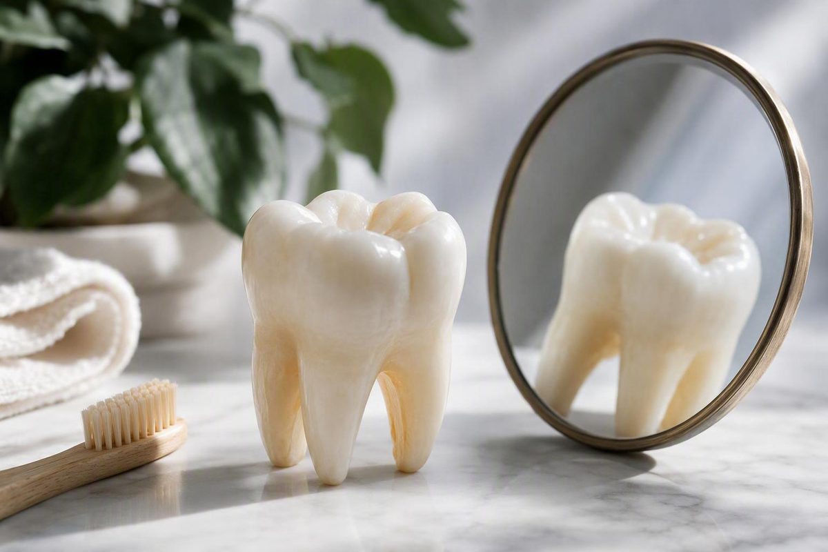
The short answer: lost enamel does not grow back
Physically lost natural enamel cannot currently regrow. However, enamel that is still present but has lost minerals may sometimes be strengthened through remineralization.
That distinction answers “Can enamel be replaced?” in two different ways:
- Natural enamel that has worn away, chipped off, or been lost to a cavity cannot be biologically replaced.
- An intact, early weakened area may be able to regain minerals before a hole or structural defect forms.
Enamel is the protective outer covering of the visible tooth crown. It shields the softer dentin and pulp inside the tooth from damage and everyday wear. Mature enamel lacks living cells capable of rebuilding structure after it has been physically lost, so it does not heal as skin or bone can. The Cleveland Clinic’s overview of tooth enamel similarly distinguishes protecting or strengthening enamel from making lost enamel grow back.
That does not mean nothing can be done. Care can pursue three practical goals:
- Strengthen enamel that remains. Early mineral loss may sometimes be arrested or remineralized.
- Slow or stop additional damage. This may mean reducing acid exposure, controlling plaque, addressing dry mouth or reflux, or managing grinding.
- Restore missing structure. A dentist can use composite resin, filling material, ceramic, or another restorative material to protect and reshape the tooth.
These goals are not interchangeable. Toothpaste may help with the first and contribute to the second, but it cannot accomplish the third. A filling or crown can replace missing structure, but it does not cause natural enamel to regenerate.
The relevant boundary is therefore not simply whether a tooth looks worn or discolored. It is whether the enamel remains physically intact and has only lost minerals, or whether part of the tooth is already missing. That distinction is not always visible in a mirror.
Decay Guide is an independent information publisher, not a dental practice. As explained on the site’s About Decay Guide page, it does not diagnose or treat patients. This article explains general possibilities but cannot determine what has happened to an individual tooth.
Remineralization is not the same as enamel replacement
Enamel undergoes repeated cycles of mineral loss and mineral return.
Demineralization is the loss of minerals, particularly calcium and phosphate, from existing enamel under acidic conditions. Acids may come directly from food, drinks, reflux, or vomiting. They may also be produced when bacteria in dental plaque process sugars and other fermentable carbohydrates.
Remineralization is the return of minerals to weakened enamel that remains in place. Saliva helps dilute and neutralize acids while supplying calcium and phosphate near the tooth surface. Fluoride supports mineral redeposition and can make the remaining enamel more resistant to subsequent acid exposure. This process strengthens existing enamel rather than recreating enamel that has disappeared, as the Penn Dental Family Practice explanation of enamel restoration emphasizes.
Remineralization may harden an early weakened area. It cannot:
- Fill a hole.
- Replace a missing edge.
- Reconstruct a chip.
- Restore the original contour of an eroded tooth.
- Cover dentin after the enamel over it has disappeared.
- Rebuild tooth structure removed during dental treatment.
The difference is comparable to reinforcing a wall that has become porous versus reconstructing a section of wall that is gone. Remineralization works within surviving enamel; restoration adds another material where tooth structure is absent.
Terminology to keep straight
- Regrowth: Production of new natural enamel by the body.
- Remineralization: Return of minerals to enamel that is still present.
- Restoration: Addition of a dental material to protect, fill, cover, or reshape a tooth.
- Enamel-like material: An engineered substance intended to reproduce selected properties or structures of enamel, rather than natural enamel produced spontaneously by the body.
Words such as repair, restore, and rebuild can blur these categories. A toothpaste label may use “enamel repair” to mean that the product supports remineralization. A dentist may say that a filling “restores” a tooth, meaning it restores form and function with a manufactured material. Neither use necessarily means that new natural enamel has formed.
When evaluating a claim, ask what the product or procedure physically does. Does it return minerals to existing enamel? Does it place a coating or restoration on the tooth? Or does it claim to produce new biological enamel? Those are materially different claims.
Fluoride is particularly important in this distinction. It can support remineralization, but it does not act as replacement enamel. An intact early weak spot and a visible hole may both be described casually as “enamel damage,” yet only the intact area may be within the practical scope of remineralization.
A stage-by-stage guide to what may still be reversible
A useful framework is to consider enamel damage in four broad stages. These are practical categories, not a method for self-diagnosis.
| Condition | What has happened | Can natural enamel return? | What home care can do | What professional care may do |
|---|---|---|---|---|
| Early mineral loss | An intact area has lost minerals but has not formed a hole | Missing enamel is not being replaced, but minerals may return to enamel that remains | Support remineralization, reduce frequent acid and sugar exposure, control plaque, and preserve saliva | Determine whether the surface is intact, provide preventive care, and monitor the area (Penn Dental Family Practice) |
| Established erosion or a chip | Part of the enamel’s thickness, edge, or contour is physically missing | No | Reduce ongoing wear and sensitivity triggers; protect remaining structure | Monitor, smooth, protect, bond, or otherwise restore the defect when appropriate (Colgate) |
| Formed cavity or visible hole | Tooth structure has broken down and a structural defect exists | No | Help prevent further disease, but not refill the defect | Assess and treat the damaged area, often with a filling or another restoration (Hayes Dental Group) |
| Extensive loss or a substantially weakened tooth | A large area is damaged, worn, fractured, or unable to tolerate normal forces reliably | No | Limit continuing contributors, but not restore the tooth’s original strength or shape | Consider broader protection, potentially including a crown, based on the examination (WebMD) |
Stage one: early mineral loss. An early lesion may appear chalky or different in color, but it can also be difficult to see. If the enamel surface remains intact, preventive care may shift the balance toward mineral return. “Reversible” in this context means that an early lesion may sometimes be arrested and rehardened—not that a new layer of enamel grows over it.
Stage two: established erosion or a chip. Once acid wear has removed part of the tooth’s contour, or trauma has broken off an edge, the missing portion will not return. The defect does not automatically require the same procedure in every case.
Stage three: a formed cavity or visible hole. A cavity is not merely enamel that needs more minerals. It is a structural defect. Toothpaste cannot occupy the missing space, treat damaged tissue, rebuild contact with an adjacent tooth, or reproduce the shape required for chewing. A formed cavity generally requires professional assessment and may require a filling or another restoration.
Stage four: extensive structural loss. When erosion, decay, fracture, or wear has substantially reduced a tooth’s strength, a small localized repair may not provide adequate protection. Broader coverage such as a crown may be considered, although the decision depends on how much sound tooth remains and the forces the tooth must withstand.
Dentists distinguish these situations through clinical examination. The purpose is to establish whether the surface is intact, whether decay extends beneath it, whether dentin is exposed, and whether a crack or mechanical problem is present.
This is why adding more “repair” toothpaste is not a dependable response to every white spot, dark mark, rough edge, or sensitive tooth. The appropriate response changes once mineral loss becomes structural loss.
What fluoride, saliva, and toothpaste can realistically do
Fluoride toothpaste can support remineralization of early mineral loss. It is not replacement enamel.
Saliva is part of the same protective system. It helps clear and neutralize acids while keeping calcium and phosphate available near the enamel surface. When acid attacks are limited and enough time passes between them, saliva can help shift the balance away from continuing mineral loss.
Staying hydrated supports normal saliva production. Chewing sugar-free gum may stimulate saliva for some people. These measures can support the mouth’s protective environment, but neither water nor gum reconstructs missing tooth structure.
Fluoride promotes mineral redeposition in weakened enamel and improves the acid resistance of the mineral that remains. Its usefulness depends on the target: an intact early lesion may benefit, while an eroded edge or cavity cannot be filled through fluoride exposure.
Some toothpastes contain hydroxyapatite, a calcium-phosphate mineral related to the mineral component of enamel. Available practice-based evidence presents it as a possible aid for early demineralization, but does not establish that hydroxyapatite toothpaste is equal or superior to fluoride across products, ages, and clinical situations. Formulation, concentration, study design, and the type of lesion all matter; one discussion of these limits appears in a dental-practice review of enamel repair.
A cautious interpretation is appropriate:
- Fluoride has consistent support for helping remineralize early weakened enamel.
- Hydroxyapatite may be intended to support mineral deposition at the enamel surface.
- Neither ingredient regrows a missing cusp, edge, or enamel layer.
- A toothpaste claim is not evidence that an established cavity can be treated at home.
No toothpaste can reconstruct the anatomy of a chip, restore a contour removed by erosion, cover substantially exposed dentin, or fill a cavity. Even if a product leaves mineral deposits on a surface, that is not equivalent to recreating the organized natural enamel that was lost.
Treat phrases such as enamel repair, rebuilds enamel, and restores enamel as descriptions of strengthening existing enamel unless the manufacturer clearly defines and supports another mechanism. Look beyond the headline to see whether the claim is limited to remineralization, surface hardness, sensitivity, or laboratory findings.
Home remedies require the same skepticism. Lemon juice and other acids add acid exposure rather than restoring enamel. Improper use of abrasive powders such as baking soda may add mechanical wear. Brushing harder does not drive minerals back into teeth. Guidance summarized by Hayes Dental Group likewise advises against acidic or improperly used abrasive do-it-yourself remedies.
A reasonable general routine centers on fluoride toothpaste, gentle brushing, cleaning between the teeth, hydration, and reducing repeated acid and sugar exposure. Individual fluoride instructions vary with age, cavity risk, water supply, medical history, and existing dental conditions, so product strength and treatment schedules should be chosen with appropriate professional guidance rather than prescribed generically here.
How dentists restore a tooth when enamel is missing
When enamel is missing, dentistry can protect or restore the tooth with a non-enamel material. This may re-establish shape, appearance, contact, chewing function, or protection, but it is not biological enamel regrowth.
The most conservative suitable option is not the same for every tooth. A defect’s depth, position, and cause matter, as do symptoms, appearance, bite forces, and the amount of sound structure remaining.
| Treatment | Main purpose | Coverage | Is natural enamel regenerated? | Important limitation |
|---|---|---|---|---|
| Sealant | Create a protective barrier over a selected surface | A thin coating, commonly over pits or grooves | No | Protects a surface but does not rebuild a missing contour or strengthen a severely damaged tooth |
| Filling | Replace structure lost to a cavity after the damaged area is treated | A localized area within the tooth | No | Suitability depends on the defect and the amount of sound tooth remaining |
| Bonding | Add tooth-colored composite resin to reshape or repair selected damage | A localized surface, edge, or contour | No | May chip, wear, or stain and may be unsuitable where forces or damage are extensive |
| Veneer | Cover the visible front surface for selected structural or cosmetic concerns | Primarily the front surface | No | Traditional placement generally removes enamel and is irreversible |
| Crown | Provide more extensive coverage and protection | Most or all of the visible crown | No | Requires more tooth preparation and is not the most conservative answer for every defect |
A sealant bonds over enamel to form a protective barrier. It may be useful on selected surfaces, particularly where grooves are vulnerable, but it is an imperfect substitute rather than replacement enamel. It does not recreate a chipped edge or restore a deeply eroded chewing surface.
A filling replaces tooth structure after decay has created a cavity and the affected area has been treated. The material occupies and seals the prepared defect. It can restore local shape and function, but it does not turn into natural enamel.
Dental bonding adds tooth-colored composite resin directly to the tooth. It may be considered for selected minor chips, worn edges, contour irregularities, or limited areas of damage. Bonding can preserve more existing tooth structure than broader coverage, but suitability depends on the location of the damage and the forces the repair will receive.
A veneer is a shell placed mainly over the front surface of a tooth. It can mask discoloration or alter contour and may cover selected enamel defects. Traditional veneer placement generally requires removal of some existing enamel so the shell can fit. That makes the procedure irreversible: the covering may restore appearance or form, but the natural enamel removed during preparation does not return. A comparison of bonding and traditional veneers describes this trade-off between localized resin and a front-surface shell requiring enamel preparation.
A crown provides more extensive coverage. It may be considered when erosion, fracture, decay, or a large existing restoration leaves a tooth substantially weakened. It is therefore not a universal upgrade from bonding or a filling.
Treatment selection is conditional. A small front-tooth chip and a heavily worn back tooth may both involve enamel loss, but they face different functional demands.
If reflux, dry mouth, grinding, repeated acid exposure, or another contributor continues, restoring the visible defect alone may not stop further damage.
There is no useful universal ranking of sealants, fillings, bonding, veneers, and crowns. They solve different problems and require different amounts of tooth preparation. Costs and longevity also vary with material, technique, tooth position, bite, habits, and maintenance, so one fixed figure or guarantee would be misleading.
Why enamel is being lost matters as much as the repair
Restoring the surface without addressing the cause can leave the same tooth—and nearby teeth—vulnerable to continued damage.
Dental erosion and tooth decay are not identical. Erosion is chemical wear caused by nonbacterial acids, including dietary acids and stomach acid from reflux or vomiting. Tooth decay involves acids produced when plaque bacteria process sugars and other fermentable carbohydrates. The processes can coexist.
Frequency matters. Repeated sugary or starchy snacks can likewise support repeated plaque-related acid production.
Possible contributors include:
- Frequent acidic drinks or foods.
- Repeated sugary or starchy snacks.
- Slowly sipping or grazing over extended periods.
- Acid reflux.
- Recurrent vomiting.
- Dry mouth.
- Grinding or clenching.
- Aggressive brushing.
- Repeated friction or contact.
- Ordinary wear over time.
Reflux and vomiting deserve attention beyond the tooth surface because they can repeatedly expose teeth to stomach acid. A restoration may repair a defect, but it cannot prevent another acid episode.
Dry mouth creates a different problem. Reduced saliva can weaken acid neutralization, clearance, and mineral availability. It may be associated with dehydration, medication use, or health conditions. Drinking water may support hydration, but persistent dry mouth warrants assessment rather than an assumption that more brushing will solve it.
Mechanical forces can compound chemical weakening. Grinding and clenching place repeated loads on tooth surfaces and edges. Aggressive brushing may add abrasion, especially where enamel has already been softened. These causes and their interactions are summarized in guidance from Felton Dentistry.
Cause-linked protective measures include:
- Reduce the frequency of acidic and sugary exposures rather than focusing only on one food or drink.
- Use fluoride toothpaste as part of routine oral care.
- Brush gently with a soft-bristled brush.
- Clean between teeth to reduce plaque where a toothbrush does not reach effectively.
- Stay hydrated.
- Avoid acidic substances as whitening or “repair” remedies.
- Discuss persistent reflux, vomiting, dry mouth, grinding, or clenching with an appropriate professional.
- Ask whether a habit, medication, or health condition may be contributing.
Brushing immediately after an acidic exposure may add abrasion while the tooth surface is temporarily softened. Published consumer guidance does not give one universal waiting period: some sources recommend at least 30 minutes, while others advise an hour. The practical point is to avoid scrubbing immediately, rinse with water if appropriate, and resume gentle brushing after the immediate acidic period has passed; one of the longer waiting recommendations appears in Pronamel’s acid-erosion guidance.
These measures preserve enamel that remains. They do not reconstruct an eroded surface, refill a cavity, or make a broken edge grow back.
Signs that need a dental assessment rather than more toothpaste
Possible signs of enamel wear or structural damage include:
- Persistent sensitivity to cold, heat, sweets, touch, or brushing.
- Yellowing or another change in tooth color.
- Rough or increasingly translucent edges.
- Smooth, shiny, flattened, or worn areas.
- Cupping or small indentations in chewing surfaces.
- Chips or cracks.
- A visible hole.
- Repeated cavities.
- Unexplained changes in tooth shape or bite.
Yellowing can occur when thinning or missing enamel allows more of the naturally yellower dentin underneath to show through. That differs from a removable surface stain, although both can occur together. Whitening can change color, but it does not add back lost tooth structure or cover exposed dentin. Sensitivity, discoloration, chips, cracks, shiny surfaces, and cupping are among the changes associated with erosion in the WebMD guide to enamel erosion.
None of these signs diagnoses enamel loss by itself. Sensitivity can have several causes. A dark or yellow area may reflect staining, visible dentin, decay, an existing restoration, or another change. Cracks vary in depth and significance, and enamel loss can progress without obvious pain.
Professional evaluation is appropriate for:
- Sensitivity that persists, worsens, or affects eating and drinking.
- A visible hole or suspected cavity.
- A new chip or crack.
- Pain when biting.
- An area that appears to expose dentin.
- Unexplained changes in color, shape, texture, or tooth length.
- Damage that continues despite changes in home care.
Toothpaste cannot stabilize a major fracture, assess deeper tissue, or replace a missing section of tooth. Erosion and structural damage can increase vulnerability to sensitivity, cavities, and infection, as explained in Colgate’s overview of enamel restoration.
An examination helps distinguish early demineralization from erosion, decay, fracture, grinding-related wear, staining, sensitivity from another source, or a combination of problems. That distinction determines whether the priority is remineralization, control of the cause, restoration, or another form of care.
Are scientists close to regrowing enamel?
Research into enamel-like materials is scientifically important, but it should not be confused with spontaneous biological regrowth of natural enamel.
One University of Washington project described a lozenge containing an engineered peptide derived from amelogenin, a protein involved in enamel formation, together with calcium and phosphorus. The proposed mechanism was for the peptide to bind to damaged tooth surfaces and organize minerals into enamel-like layers.
At the stage reported by the university, the technology had been tested on extracted teeth and in animals, and the team was preparing for human clinical trials. The University of Washington School of Dentistry’s account of the peptide lozenge also reported the developers’ proposed dosing and claims about deposition, whitening, compatibility with low-concentration fluoride, and expected safety.
Those statements should be read as claims from the development team at that reported stage, not as independently confirmed consumer guidance. The cited report does not establish:
- Completed human efficacy trials.
- Long-term durability under chewing and repeated acid exposure.
- Regulatory approval.
- Commercial availability.
- Routine use in dental practices.
- Permanent sensitivity relief.
- Proven safety and effectiveness across adults and children.
Even if an engineered system successfully deposits an enamel-like mineral layer, that would not necessarily mean that the body had regenerated natural enamel with the same developmental structure. Material deposition, biological regrowth, and conventional restoration are distinct concepts.
Future research could change available treatment. When evaluating a headline about “regrowing enamel,” ask:
- Were the results obtained in a laboratory, in animals, or in human patients?
- Was the research peer reviewed?
- Was the treatment compared with existing care?
- How long did the deposited material remain attached and functional?
- Did it withstand chewing, temperature changes, and repeated acid exposure?
- Were adverse effects assessed?
- Has an appropriate regulator approved the product?
- Is it available through routine clinical care?
For now, the practical conclusion from this evidence remains unchanged: it does not establish a proven, approved, routinely available treatment that regrows missing natural enamel.
Frequently asked questions
Can enamel grow back naturally after it has worn away?
No. Once natural enamel has been physically lost through erosion, wear, chipping, decay, or tooth preparation, the body cannot grow it back. Mature enamel lacks living cells capable of rebuilding the missing structure, according to the Cleveland Clinic.
An intact but weakened area may sometimes regain minerals. That is remineralization, not regrowth. The key question is whether the enamel structure is still present.
Can fluoride or hydroxyapatite toothpaste replace lost enamel?
No. Fluoride toothpaste can support remineralization and improve the acid resistance of remaining enamel. Hydroxyapatite toothpaste may also be intended to support mineral deposition in early weakened areas, but the evidence considered here does not establish that it equals or outperforms fluoride.
Neither type of toothpaste can fill a cavity, reconstruct a chip, restore an eroded contour, or cover substantially exposed dentin.
Does yellow dentin showing through mean enamel has been lost?
It may indicate that enamel has become thinner or is missing, allowing more of the naturally yellow dentin to show. But yellowing is not diagnostic by itself. Surface stains, lighting, natural tooth color, restorations, and other dental changes can also affect appearance.
If a color change is new, uneven, accompanied by sensitivity, or associated with a change in shape, a dental assessment can help identify the cause. Whitening may alter color, but it cannot replace missing enamel.
Do bonding, veneers, fillings, or crowns count as replacement enamel?
They are dental restorations, not regenerated natural enamel.
Bonding adds composite resin. A filling replaces structure within a treated cavity. A veneer covers mainly the front surface of a tooth and, when traditionally placed, generally requires some enamel removal. A crown provides more extensive coverage. These treatments can restore shape, appearance, protection, or function, but their materials remain distinct from natural enamel.
Is the enamel-regrowing peptide lozenge proven or commercially available?
The available university report describes an experimental technology that was preparing for human clinical trials after testing on extracted teeth and animals. It does not establish completed human efficacy trials, regulatory approval, routine commercial availability, or long-term durability.
Claims about how much material the lozenge deposits, how often it would be used, its whitening effects, and its expected safety came from the development team. They should not be treated as confirmed clinical outcomes without completed human research and regulatory review. The reported development status is described in the University of Washington account.
The practical distinction to remember is simple: weakened enamel may sometimes regain minerals, but missing enamel does not naturally return. The next step depends on whether the goal is to remineralize an intact early lesion, stop an ongoing cause such as acid exposure or dry mouth, or restore lost structure with a dental material.
Persistent sensitivity, pain, a visible hole, a chip, a crack, swelling, or worsening changes deserve professional assessment. This article provides general information, not a diagnosis of an individual tooth.