
When Teeth Form With Too Little Enamel
A small, stable defect without decay or symptoms may be monitored. Vulnerable structure may need sealants, fillings or coverage. Enamel cannot regrow.
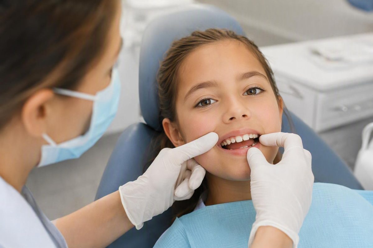
What hypoplastic dental enamel means
Hypoplastic dental enamel, also called enamel hypoplasia, is a developmental defect of enamel quantity. Too little enamel formed while the affected tooth was developing beneath the gum. The shortage was therefore present when the tooth erupted; it did not begin because of poor brushing or because normal enamel later wore away. The Cleveland Clinic’s overview of enamel hypoplasia describes the enamel as unusually thin or missing from part of one or more teeth.
Enamel is the mineral-rich outer covering of the visible tooth. It protects the underlying dentin and pulp, including sensitive living tissue, from chewing forces, temperature changes, acids, and biofilm-related disease. Although enamel is highly mineralized, it can still be damaged. A tooth may have reduced protection because too little enamel developed initially, because enamel was lost after eruption, or because both processes occurred.
The amount of missing enamel varies. A hypoplastic defect may appear as:
- A small pit or depression
- One or more grooves
- A rough or irregular area
- A generally thin enamel covering
- A defined patch where enamel is absent
- More extensive loss of normal tooth shape
The defect can involve part of one tooth, the entire crown of one tooth, several teeth, or a broader portion of the dentition. Primary teeth, permanent teeth, or both may be affected because their enamel develops during different but partly overlapping periods.
That developmental origin is the distinction that resolves much of the confusion around this condition. Hypoplasia means the enamel was insufficient at eruption. Erosion, grinding, abrasion, and other forms of wear remove enamel after eruption. A tooth can also begin with too little enamel and later lose additional structure through decay, acid exposure, wear, or fracture.
Enamel that never formed cannot naturally grow back. Existing mineralized tissue may sometimes be protected or supported with preventive care, but this is not the same as recreating the missing enamel layer. When lost structure needs to be replaced, dentists use restorative materials.
This article provides general information and cannot determine whether a particular mark, groove, color change, or symptom is enamel hypoplasia. Decay Guide is an independent information publisher, not a dental practice, and does not diagnose or provide individualized dental treatment.
What hypoplastic enamel can look and feel like
The appearance and effects of hypoplastic enamel depend on how much enamel is missing, where the defect is located, and what has happened to the tooth since eruption. Some defects are small and remain stable. Others expose dentin or create irregular areas where plaque and food are difficult to remove.
Appearance. Pits are small, localized depressions. Grooves or lines may cross part of the crown, while broader defects can make the surface look rough, ridged, uneven, or misshapen. In more extensive cases, enamel may be thin over much of the tooth or absent from a visible area.
Affected teeth may also show white, cream, yellow, or brown changes. Color alone cannot identify hypoplasia. Staining, early decay, fluorosis, mineral-quality defects, exposed dentin, and natural differences in tooth color can produce similar findings.
A yellow appearance may occur when naturally yellow dentin is easier to see through an unusually thin enamel covering. Where enamel is absent, dentin may be directly exposed. Neither observation means that every yellow tooth has hypoplasia.
Symptoms. Possible findings include discomfort while chewing, unusual wear, rough edges, chipping, and repeated cavities around a vulnerable area. Small defects may be discovered only during a dental examination.
Reduced enamel leaves less separation between the mouth and the more sensitive inner tooth. Pits and grooves may also retain plaque, while thin or absent areas offer less protection against acids and chewing forces. Sensitivity, decay, erosion, wear, and structural deterioration are therefore possible, although they are not inevitable. A dentist-authored overview of hypoplastic teeth likewise identifies sensitivity, staining, excessive wear, cavities, and altered tooth shape among the possible findings.
Later complications. The original developmental defect should be separated from damage that happens afterward. Hypoplasia does not spread across the tooth after eruption: the amount of enamel that failed to form is fixed. The affected tooth may nevertheless develop:
- Decay in or around a pit or groove
- Acid erosion of the remaining surface
- Mechanical wear from chewing or grinding
- Chipping or fracture
- Further loss around a restoration
- Persistent sensitivity or pain
- Changes in tooth shape, contact, or bite
Visible defects can also create appearance concerns, particularly on front teeth. More extensive structural loss may make it harder to preserve the tooth’s normal contour or maintain contact with neighboring and opposing teeth. Cosmetic and functional concerns should be considered separately: a visible mark does not necessarily mean the tooth is unstable, while a less noticeable defect may still require protection.
Dental assessment is appropriate when a newly erupted tooth has pits, grooves, or visibly missing structure; when dentin appears exposed; or when there is persistent sensitivity, recurrent decay, chipping, or rapid breakdown. These are reasons for examination, not a self-diagnostic checklist.
Hypoplasia versus other enamel changes
Several enamel problems can produce spots, sensitivity, roughness, or structural loss. The key distinction is not simply how a tooth looks, but what happened to the enamel and when.
| Condition or change | Nature of the problem | When it develops | What appearance alone cannot establish |
|---|---|---|---|
| Enamel hypoplasia | Too little enamel formed; enamel quantity is deficient | Before eruption | A pit, groove, or color change does not prove hypoplasia |
| Hypomineralization | Enamel formed but has deficient mineral quality | During tooth development; breakdown may become evident after eruption | Discoloration or crumbling does not identify the exact mineral defect |
| Erosion | Acids dissolve enamel that was present | After eruption | A smooth or thinned area does not reveal the acid source |
| Mechanical wear | Enamel is lost through grinding, abrasion, or other physical forces | After eruption | A wear pattern does not by itself identify its cause |
| Fluorosis | A developmental enamel change associated with fluoride exposure during formation | Before eruption | White or brown areas cannot be labeled fluorosis without considering history and distribution |
| Early decay | Mineral is lost through a biofilm-mediated disease process | After eruption | A white, brown, or rough spot is not automatically a cavity |
| Amelogenesis imperfecta | An inherited group of disorders affecting enamel development | Before eruption | Widespread abnormalities do not establish an inherited diagnosis |
The most important comparison is with hypomineralization. Hypoplasia concerns enamel quantity: physically too little enamel formed. Hypomineralization concerns quality: enamel formed, but its mineral characteristics are deficient. The two can resemble one another, and more than one developmental defect may sometimes be present.
Hypoplastic enamel should not automatically be described as soft. Pronounced softness, rapid crumbling, or breakdown soon after eruption may suggest a mineral-quality defect or another condition requiring professional differentiation. The clinically useful distinction remains quantity versus quality, as summarized in this comparison of hypoplasia and hypomineralization.
Hypoplasia also differs from erosion and mechanical wear. In hypoplasia, the shortage exists when the crown emerges. Erosion removes enamel through acid exposure after eruption, while grinding and abrasion remove it mechanically. A groove documented when a tooth first erupts therefore suggests a different sequence from a smooth, thinning area that appears and enlarges later.
These categories are not mutually exclusive. A hypoplastic tooth can subsequently be eroded, worn, decayed, chipped, or fractured. By the time someone seeks care, the original developmental outline may be partly obscured by later damage or previous restorations.
Color is particularly unreliable as a diagnostic shortcut. White, yellow, and brown marks can occur with hypoplasia, but they can also reflect fluorosis, hypomineralization, early decay, staining, exposed dentin, or other enamel changes. Even a pit is not specific enough to establish the cause without considering its depth, shape, distribution, and relationship to surrounding enamel.
Amelogenesis imperfecta is an inherited group of enamel-development disorders. Some forms produce reduced enamel quantity and can therefore have a hypoplastic appearance. Enamel hypoplasia, however, is a descriptive defect that may have inherited or noninherited contributors. It is not a synonym for amelogenesis imperfecta, and an isolated defect should not be assumed to represent a genetic disorder.
Molar-incisor hypomineralization is another distinct mineral-quality disorder involving a characteristic pattern of permanent teeth. Its name may arise when children have sensitive, discolored, or breaking-down molars or incisors, but visible breakdown alone does not make it hypoplasia or establish that diagnosis.
Photographs and symptom lists cannot confirm which enamel disorder is present. A dentist must assess whether tissue was missing from eruption, formed with deficient mineral quality, or was removed later—and whether several processes now affect the same tooth.
Why only certain teeth may be affected
The pattern of affected teeth can provide clues about when enamel formation was disrupted. It usually cannot identify a precise illness, deficiency, exposure, or event.
A simple developmental timeline is more useful than an exhaustive tooth-by-tooth chart:
- Before birth into infancy: Enamel formation for primary teeth begins before birth and continues during infancy.
- Infancy through early childhood: Enamel formation for permanent teeth begins in infancy as different permanent crowns develop.
- Up to approximately age eight: Some later-developing permanent teeth are still forming enamel, so a childhood disturbance may affect only the teeth active at that stage. This broad timeline is described in a clinician-authored pediatric discussion of enamel development.
These periods overlap, but individual teeth are not all at the same developmental stage at once. A disturbance can affect whichever teeth—or whichever parts of their crowns—were forming enamel at that time.
Enamel within one crown also forms in stages. A limited disturbance may therefore leave a line, groove, pit, or band instead of affecting the whole tooth. If several crowns were forming simultaneously, comparable portions of multiple teeth may be involved.
Hypoplastic primary teeth do not automatically mean that permanent teeth will be affected. The two dentitions have related but distinct developmental schedules. A disruption may occur while primary teeth are vulnerable but end before the relevant permanent enamel forms, or vice versa.
A localized defect in one tooth may suggest a local developmental event. Turner’s hypoplasia is a term used for a localized defect, often in a permanent tooth whose developing crown was disrupted by trauma or infection involving its primary predecessor. This pattern is described in the available dental literature, but a single-tooth defect alone cannot prove that a particular injury or infection caused it.
Symmetrical or widespread involvement may prompt a broader review. A dentist may ask about family patterns, pregnancy and birth history, childhood development, significant illnesses, nutrition, medication or other exposures, and previous dental trauma. Widespread defects can make inherited or systemic contributors more plausible, but enamel appearance cannot diagnose a genetic syndrome, nutritional deficiency, or medical condition.
Distribution is therefore a clue rather than proof. It may help estimate when enamel-producing cells were affected, but it generally cannot reconstruct an exact cause retrospectively.
Causes, risk factors, and cases with no clear explanation
Enamel hypoplasia develops when enamel-matrix formation is disrupted while the tooth crown is developing. Specialized enamel-producing cells create this matrix before eruption; if formation is incomplete, the finished tooth has too little enamel.
Potential explanations are best grouped rather than presented as a list of equally established causes.
Inherited enamel disorders. Amelogenesis imperfecta can include enamel-quantity defects and commonly affects multiple teeth. Other inherited conditions may also involve enamel development. These possibilities matter when defects are widespread or follow a family pattern, but inherited disorders do not explain every case.
Local developmental events. Trauma or infection near a developing tooth may disrupt one crown or one portion of it. A permanent tooth developing beneath an injured or infected primary tooth may be affected even when the rest of the dentition forms normally. The defect’s location and the dental history can support or weaken this explanation, but they cannot prove causation by themselves.
Reported systemic or environmental associations. Premature birth, low birth weight, malnutrition or nutritional deficiency, infections, inflammation, selected systemic conditions, prenatal factors, and certain exposures have been reported as possible contributors or associations. Available patient-education evidence recognizes both health-related and environmental factors but does not establish that each reported factor directly causes hypoplasia in an individual.
These associations require caution because their support is uneven. Retrospective conclusions are complicated by:
- When the illness, exposure, or nutritional problem occurred
- Its dose, duration, or severity
- Which teeth were forming enamel at the time
- Other health and developmental factors
- Whether the observed defect is truly hypoplasia rather than another enamel condition
Fluoride illustrates the diagnostic difficulty. Excessive fluoride exposure during enamel development is conventionally associated with fluorosis, which can also produce visible enamel changes. A discolored tooth should not be attributed to fluoride—or labeled hypoplastic—without distinguishing among possible conditions.
Unexplained cases. Sometimes no precise cause is identified. The term idiopathic means that the available clinical and historical information has not revealed an explanation. That uncertainty does not make the defect unreal; it means the developmental event cannot be reconstructed confidently.
Crucially, hypoplastic enamel is not evidence that a parent or patient failed to brush properly. Brushing happens after eruption, whereas the enamel shortage formed beforehand. Poor plaque control can contribute to later decay around a defect, but it did not create the original shortage.
Enamel appearance alone cannot diagnose malnutrition, celiac disease, a vitamin deficiency, or an inherited syndrome. Such decisions require an appropriate clinical history and professional assessment.
How a dentist evaluates suspected enamel hypoplasia
Diagnosis is a clinical process rather than a single test. A dentist first examines the surface and determines what kind of structural change is present.
The examination may consider:
- Pits, depressions, grooves, or rough areas
- Unusually thin or absent enamel
- Exposed dentin
- Active or previously treated decay
- Erosion and mechanical wear
- Chips, cracks, or fractures
- Sensitivity or pain
- Changes in tooth shape, contact, or bite
Mapping the distribution is important. The dentist considers which teeth are affected and which portions of their crowns show defects. A localized pit in one tooth, corresponding bands across several teeth, and widespread thin enamel represent different patterns, although none establishes a cause on its own.
Timing provides another piece of evidence. Useful questions include when the teeth erupted, whether the marks were visible immediately, and whether the area has changed since then. A defect observed at eruption supports a developmental origin. Progressive loss that appeared later raises the possibility of erosion, wear, decay, post-eruptive breakdown, or a combination of processes.
The history may cover pregnancy and birth, childhood development, significant illnesses, dental infection or trauma, family enamel patterns, medication or other exposures, and previous dental treatment. The purpose is to determine whether the history fits the clinical pattern—not to assign blame or force a causal explanation.
An image alone cannot prove hypoplasia or identify its cause. The available overview of clinical assessment for enamel irregularities describes a combination of examination, history, symptoms, and imaging where appropriate.
Before treatment, the dentist may need to distinguish hypoplasia from hypomineralization, fluorosis, early decay, staining, erosion, and mechanical wear.
Five useful questions to take to an appointment are:
- What diagnosis best fits this tooth, and what alternatives are being considered?
- How much sound enamel and dentin remain?
- Are complications such as decay, erosion, fracture, or bite changes already present?
- What protection or restoration is proposed, and what is it intended to achieve?
- How will the tooth and any restoration be monitored?
Treatment follows the severity of the defect
Treatment is a progression based on the defect’s extent and location, the condition of the remaining tissue, symptoms, decay, function, and the likelihood of further breakdown. The available evidence does not establish one best treatment for every hypoplastic tooth.
The main goals are to reduce sensitivity, prevent or treat decay, preserve sound tooth structure, maintain tooth shape and bite, restore comfortable function, and address appearance when appropriate.
1. Monitoring and prevention
A small, stable defect without decay or significant symptoms may be monitored rather than restored immediately. A dentist may chart or photograph it, provide preventive guidance, and reassess it according to the tooth’s risk.
Monitoring is still an active management choice. It allows the dental team to look for new decay, sensitivity, wear, chipping, or structural change before deciding whether restoration is necessary.
2. Protecting the tissue that remains
For vulnerable pits or fissures, sensitivity, or concern about decay, a dentist may consider topical fluoride, desensitizing measures, remineralizing agents, or sealants. These options have different purposes:
- Fluoride and remineralizing products may support mineralized tissue that remains.
- Desensitizing measures may reduce symptoms.
- A sealant may cover a retentive pit or fissure and make it easier to protect.
None of these approaches can create the missing thickness of developmental enamel. Remineralization acts on existing mineralized tissue; it is not biological replacement of an absent layer.
3. Repairing localized loss or decay
Bonding or a tooth-colored filling may be considered when a defined area needs rebuilding or decay requires treatment. These restorations can replace missing structure and restore contour.
A practical limitation is that bonding may be less predictable when restoration margins depend on defective enamel. The pediatric treatment overview from Hurst Pediatric Dentistry notes that tooth-colored fillings may adhere less reliably to affected enamel. The location of sound tissue and the need for follow-up therefore matter.
4. Providing partial or full coverage
Coverage may protect vulnerable surfaces and restore the contour needed for chewing and bite stability. Whether it is appropriate depends on the affected tooth, the amount of sound tissue, symptoms, age, growth, and the overall treatment plan. The evidence supplied here does not support a universal crown threshold, material choice, or durability estimate.
5. Considering appearance-oriented care
Veneers and other cosmetic procedures appear among the treatment options described in patient-education sources, but they are not routine solutions for every hypoplastic tooth. Cosmetic treatment does not remove the need to assess decay, sensitivity, remaining tooth structure, and function.
Professional whitening is also listed as an appearance-oriented option in some dental-practice material, but the supplied evidence does not establish when it is suitable for hypoplastic enamel. It changes color rather than replacing missing structure, so any decision should follow a dental assessment rather than self-diagnosis from discoloration alone.
6. Extraction in selected severe cases
Extraction may occasionally be considered when a severely compromised tooth cannot be predictably preserved. That decision depends on the tooth’s role, symptoms, age, growth, neighboring teeth, and replacement planning. It should not be understood as the usual outcome of enamel hypoplasia.
Across this treatment ladder, the plan may be influenced by:
- Whether the tooth is primary or permanent
- The person’s age and remaining growth
- The amount and location of sound enamel
- Dentin exposure and sensitivity
- Existing decay or fracture
- Tooth shape, function, and bite
- Appearance priorities
- Ability to keep the area clean
- The condition of previous restorations
The available evidence does not establish one universally superior restorative material, a mandatory treatment sequence, or precise lifespan figures. Treatment should be matched to the actual tooth rather than selected from the diagnosis label alone.
Protecting affected teeth after they erupt
Home care cannot reverse the developmental shortage, but it can reduce additional pressure from plaque, acids, and mechanical stress.
Brush gently twice daily with fluoride toothpaste. This general protective measure helps control plaque and exposes remaining tooth tissue to fluoride; it does not cure missing enamel.
Clean between teeth daily using a method suited to age, dexterity, tooth spacing, and professional guidance. The goal is to clean areas a toothbrush does not reach without injuring the gums or vulnerable tooth structure.
Reducing frequent exposure to sugary, sticky, and acidic foods or drinks may lower the repeated decay and erosion challenges faced by a less protected tooth. Rinsing with water after acidic intake is a limited practical measure, not a way to neutralize every risk or replace normal oral hygiene.
Grinding, clenching, chewing hard objects, and other mechanical stresses should be discussed with a dentist if wear or chipping is occurring. A dentist can determine whether the pattern is mechanical and whether any protective intervention is appropriate; the available evidence does not support recommending an appliance for every affected tooth.
Dental monitoring should be based on risk rather than one schedule for everyone. A stable shallow pit and a painful tooth with repeated breakdown do not require identical follow-up. Reviews allow the dentist to look for decay, erosion, fracture, dentin exposure, sensitivity, and restoration problems.
Do not start fluoride supplements, high-concentration fluoride products, calcium or vitamin supplements, or remineralizing agents solely because a tooth looks abnormal. The product, concentration, and need depend on the diagnosis and individual circumstances.
Arrange prompt dental assessment for:
- Persistent pain or sensitivity
- Visible or suspected exposed dentin
- A newly broken or chipped area
- Rapid change in tooth shape or surface
- Difficulty eating
- Recurrent decay around the same tooth
Diligent brushing can control plaque, but it cannot replace enamel that failed to form—a broader distinction also discussed in What Daily Brushing Cannot Fix. The developmental shortage remains, but later complications are not inevitable. Decay, erosion, sensitivity, fractures, and restoration problems may often be reduced, monitored, or treated once the tooth’s actual risks are understood.
Can hypoplastic dental enamel grow back?
No. Enamel that never formed cannot naturally regenerate because the enamel-producing cells are no longer available to rebuild the erupted crown. Fluoride and remineralizing measures may help protect mineralized tissue that remains, while bonding, fillings, or crowns may replace or cover missing structure with restorative material.
Does enamel hypoplasia get worse after a tooth erupts?
The developmental defect itself does not spread after eruption. The tooth begins with a fixed shortage of enamel. It may nevertheless experience later decay, erosion, wear, chipping, fracture, or other structural loss. The tooth can therefore appear to worsen even though the original hypoplasia is not expanding.
Does hypoplasia in baby teeth mean permanent teeth will also be affected?
Not necessarily. Primary and permanent teeth form during overlapping but distinct periods. A disturbance may affect primary enamel without continuing into the relevant period of permanent-tooth development, or it may affect only the permanent teeth forming at that time. The pattern offers timing clues but does not predict the other dentition with certainty.
Is enamel hypoplasia the same as amelogenesis imperfecta?
No. Enamel hypoplasia is a descriptive defect in which too little enamel formed. Amelogenesis imperfecta is an inherited group of enamel disorders, some forms of which can produce hypoplastic enamel. Hypoplasia can also occur without amelogenesis imperfecta after a local developmental event, in association with other factors, or with no identifiable explanation.
Can fluoride or remineralizing products replace missing enamel?
No. These products may support or protect enamel and other mineralized tooth tissue that remains, but they cannot recreate enamel thickness that never developed. Where structure is missing, a dentist may monitor the area, protect it, or replace it with restorative material depending on the defect and its consequences.
The central distinction remains: hypoplastic enamel was insufficient when the tooth formed, while decay, erosion, wear, and fractures are problems that can happen afterward. Missing enamel cannot be regenerated, but deterioration is not inevitable. A dentist can distinguish hypoplasia from similar-looking conditions, assess how much protection remains, and match monitoring or treatment to the tooth’s actual needs. Pits, exposed dentin, persistent sensitivity, recurrent decay, or rapid breakdown warrant dental assessment rather than diagnosis from appearance alone.