
The 5 Stages of Cavity Formation: How Tooth Decay Develops Over Time
A true hole will not rebuild on its own. Before one forms, fluoride and less frequent sugar exposure may help early damage stall or partly repair.
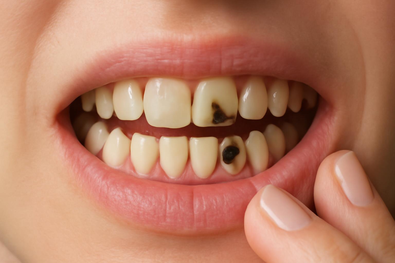
Cavities do not usually appear all at once. What people call “a cavity” is the visible result of a longer process in which plaque bacteria use sugars and starches as fuel, produce acids, and gradually pull minerals out of the tooth until the surface gives way. If nothing interrupts that cycle, the damage can move from enamel into dentin, then into the pulp, and in advanced cases beyond the tooth itself.
The common five-stage model is best understood as an educational map, not a stopwatch. Different patient-facing dental sources start counting at slightly different points, but they generally describe the same arc: early mineral loss, enamel breakdown, faster spread in dentin, pulp involvement, and then deeper infection if the problem is left untreated. Thinking of cavity formation this way helps explain two things at once: why decay can stay quiet for a long time, and why early detection matters so much.
What Causes Cavities: The Role of Plaque Bacteria and Acids
Cavity formation starts with a repeating chemical cycle. Some of the bacteria that normally live in the mouth use sugars and starches from food and drinks as fuel. As they digest those carbohydrates, they produce acids. Those acids sit against the teeth inside plaque, the sticky bacterial film that builds up on tooth surfaces. The result is a continual “tug-of-war” between mineral loss from acid and repair from saliva and fluoride, with decay developing when acid attacks happen too often for repair to keep up. NIDCR explains this process and the role of saliva and fluoride in early repair.
That is why cavity risk is shaped less by one isolated treat than by exposure pattern. Decay needs a tooth surface, bacteria living in plaque, and fermentable carbohydrates for those bacteria to use. Frequent snacking, grazing, or sipping sweet drinks over long periods gives bacteria repeated chances to make acid and gives teeth less time to recover between attacks. A pediatric dental overview describes this three-part setup and the effect of repeated sugar exposure on acid production.
Enamel is often described as the hardest tissue in the body, but “hard” does not mean immune to chemistry. Enamel is highly mineralized, and acids do not need to crack it in a dramatic way to damage it. They dissolve minerals little by little.
This also explains why people sometimes feel baffled after getting a cavity despite brushing regularly. Brushing is essential, but it does not change tooth anatomy, saliva flow, snack frequency, or whether plaque is repeatedly exposed to sugar in hard-to-clean areas. Deep grooves, tight contacts between teeth, exposed roots, and dry mouth can all keep the decay process going even when someone is making a real effort.
Stage 1: Demineralization and White Spot Lesions
The first stage of cavity formation is demineralization. At this point, acids have started dissolving minerals out of enamel, but the tooth has not yet developed a permanent hole. The earliest visible sign is often a white, chalky, dull, or matte-looking patch rather than a brown or black spot. That appearance reflects mineral loss beneath or within enamel that still looks mostly intact from the outside.
This stage matters because it is the point at which the process is most often described as reversible. If the damage is limited to early mineral loss, saliva can help redeposit minerals, and fluoride can strengthen that repair process. In practical terms, the goal is to change the mouth back from a frequent-acid environment to a repair-friendly one: better plaque control, fewer repeated sugar exposures, and enough time between eating and drinking episodes for the tooth surface to recover.
Most people do not feel anything at stage 1. There is usually no pain, no visible hole, and no dependable home test. Early lesions are also easy to miss when they sit near the gumline, on the back teeth, or in areas that are awkward to see in a bathroom mirror. A five-stage dental guide notes that white spots are easy to miss, especially on back teeth, and that early enamel changes can be subtle.
A helpful way to think about demineralization is not “the cavity is definitely coming,” but “the tooth is under repeated attack.” Some early lesions progress, some stall, and some partly repair. The direction depends on whether plaque keeps maturing in place, how often acids are being produced, how much fluoride is available, and whether saliva can do its buffering job. Stage 1 is real damage, but it is still damage with room for recovery.
Stage 2: Enamel Decay and Initial Cavities
Stage 2 begins when the enamel is no longer only weakened but structurally broken. This is the point where a true cavity opening forms in the enamel. A small pit, rough spot, or brownish area may appear, though many people still feel little or nothing because the damage remains confined to the outer layer. Patient education sources commonly describe this as the stage where small cavities form and restorative treatment becomes more likely because the enamel has actually cavitated. Sisko Dentistry’s stage guide describes stage 2 as enamel breakdown with small cavities forming in the enamel layer.
This is where everyday language becomes slippery. Some people use “cavity” to mean any early tooth decay, including a white-spot lesion. Others use it only once a real hole exists. Clinically, that distinction matters. Early demineralization may still be managed conservatively. Lost tooth structure does not rebuild its original shape on its own.
One description often used in patient education is that enamel can weaken from beneath the surface before the outer shell finally collapses. That helps explain why a small opening can sometimes sit over a wider zone of damaged enamel. It is a simplified explanation, but it captures the basic idea that the visible surface can understate the structural weakening already present. Ponte Vedra Complete Dentistry describes enamel breakdown as progressing from the underside outward before the surface ultimately gives way.
The practical takeaway is not that every stained groove needs an immediate filling, but that true cavitation is different from early mineral loss. Once enamel has caved in, the conversation usually shifts from remineralization alone to whether the area needs a restoration to stop food trapping, plaque retention, and deeper spread. That is a judgment call made on examination, not something most people can sort out confidently just by looking in a mirror.
Stage 3: Dentin Decay and Emerging Sensitivity
Stage 3 starts when decay breaks through enamel and reaches dentin, the layer beneath it. This is the stage many people first notice because dentin is softer than enamel, less resistant to acid, and easier for decay to spread through. Dentin also contains tiny tubules that communicate with the inside of the tooth, which helps explain why sensitivity becomes more common once decay gets this deep. Colgate’s tooth decay guide explains that dentin is softer than enamel, that decay often progresses faster there, and that dentinal tubules help explain increased sensitivity.
The symptoms at this stage are more familiar to patients: cold drinks start to sting, sweets cause a quick ache, or chewing on one side becomes uncomfortable. A visible hole may look darker or larger. Food may catch repeatedly in one spot. But the experience is still variable. Some dentin-stage cavities are quite symptomatic, while others remain surprisingly quiet until they are already deep.
Dentin decay also creates a bigger mismatch between what is visible and what is involved. That is one reason self-inspection is unreliable for judging how advanced a cavity is.
At this depth, professional treatment is commonly needed. Patient-facing stage guides usually describe fillings as standard care at this point, with larger defects sometimes requiring bigger restorations such as inlays or onlays depending on how much tooth structure has been lost.
Stage 4: Pulp Involvement and Infection
Stage 4 is when decay reaches the pulp, the soft inner tissue of the tooth. The pulp contains nerves and blood vessels, so once bacteria and inflammation involve that tissue, the problem is no longer confined to hard tooth structure. Many patient resources describe this phase as pulp damage, pulp infection, or pulpitis depending on the exact clinical picture. Cleveland Clinic explains that deeper decay can reach the pulp and describes the pulp as the part of the tooth containing nerves and blood vessels.
By this stage, the symptoms often change in character as well as intensity. Instead of a quick zing from cold, pain may linger, throb, wake a person at night, or flare without an obvious trigger. Nearby gum tissue can look irritated or swollen, and some teeth darken as the internal tissues become more affected. A five-stage cavity guide describes stage 4 symptoms as throbbing pain with possible swelling when bacteria reach the pulp.
Once the pulp is involved, treatment generally becomes more invasive because the issue is not just a damaged outer surface anymore. Patient education sources commonly frame this as the stage where root canal treatment may be needed to preserve the tooth, while extraction becomes the fallback if the tooth cannot be restored predictably.
One practical lesson from this stage is that pain is not a dependable early warning system. A tooth can move through the enamel stages with few or no symptoms, and some people do not feel a cavity until it reaches dentin or pulp. Cleveland Clinic notes that you might not feel a cavity until it reaches the dentin or pulp.
Stage 5: Abscess Formation and Severe Complications
Stage 5 is the end-stage picture of untreated progression: infection extends beyond the tooth and may form an abscess, a pocket of pus associated with bacterial invasion near the root. At that point, the problem is no longer just a defect in the tooth surface. It has become a deeper dental infection with the potential for serious pain, surrounding tissue involvement, and eventual tooth loss if not managed. Healthline’s stage summary describes an abscess as a pocket of pus at the tooth root caused by bacterial invasion.
Symptoms at this stage can include intense toothache, swelling in the gums or face, tenderness when biting, unpleasant-tasting drainage, and sometimes fever. Patient education sources often describe the pain as radiating into the jaw or face rather than staying confined to one pinpoint area.
This is the stage at which prompt dental evaluation becomes especially important. Mayo Clinic notes that untreated cavities can lead to infection and tooth loss, and advises seeing a dentist as soon as possible for toothache or mouth pain. Mayo Clinic explains that untreated cavities can progress to deeper infection and tooth loss and advises prompt dental care for toothache or mouth pain.
It is also worth keeping the timeline in perspective. The dramatic symptoms of stage 5 are late, not first, chapters in the same process.
Types of Cavities and Progression Factors
Not all cavities start in the same place, and location changes both how they behave and how easy they are to notice. The three classic types are smooth-surface cavities, pit-and-fissure cavities, and root cavities. Smooth-surface cavities occur on flatter outer surfaces and can also involve the areas between teeth. Pit-and-fissure cavities develop in the grooves and pits of chewing surfaces, especially molars and premolars. Root cavities form on exposed root surfaces, which becomes more relevant after gum recession. Mayo Clinic illustrates these three cavity types and where they occur.
Those locations matter because they change both retention and detectability. Grooves on molars are efficient at trapping plaque and food. Between-the-teeth lesions can stay invisible from a straight-on view. Root cavities present a different problem: once the root surface is exposed, the tooth no longer has the same outer anatomy it had on the crown. So the same underlying acid-bacteria process can look quite different depending on where it begins.
Patient education sources usually describe the total process as taking anywhere from weeks to years, with speed influenced by diet, oral hygiene, saliva flow, fluoride exposure, and the location of the lesion. They also tend to agree that progression is usually faster once decay has moved beyond enamel and when it starts in plaque-retentive areas.
Age changes the pattern, too. Cavities are commonly described as especially common in children, teenagers, and older adults. In children, baby teeth are more susceptible in part because their enamel is thinner.
The practical result is that two people with similar brushing habits can still have very different cavity patterns. One person may struggle mainly with grooves on molars, another with areas between teeth, and another with root surfaces after gum recession. “I brush twice a day” tells only a small part of the story.
Brushing Limitations and General Prevention Insights
Brushing matters because plaque is the bacterial film in which cavity-causing acids are produced. But brushing alone does not control every part of cavity risk. Decay still depends on how often sugars and starches are consumed, whether saliva flow is normal, how much fluoride protection is present, and whether plaque keeps maturing in difficult areas of the mouth.
A toothbrush also cannot solve every problem created by time and tooth anatomy. Plaque that is not removed can harden into tartar, and once that happens it is no longer something home brushing can simply scrub away. Cavities also commonly start in places that are harder to keep clean or even see, such as grooves, root surfaces, and the contact areas between teeth.
For the earliest stage, prevention and partial reversal are mostly about changing conditions rather than “brushing harder.” Lower sugar frequency means fewer acid attacks. Fluoride helps enamel resist mineral loss and supports remineralization. Saliva buffers acids and supplies minerals.
Regular dental checkups matter partly because the earliest stages can be silent and visually subtle. A dentist may spot a lesion that has not yet caused pain, or notice a pattern of risk before a tooth actually cavitates. That is one reason waiting for symptoms is such a poor strategy: by the time a cavity hurts consistently, the disease is usually no longer in its easiest-to-manage stage.
For a broader non-clinical discussion of why effort and outcomes do not always match, see our guide to what daily brushing cannot fix. And as explained on our about page, this article is general educational information, not a diagnosis or treatment plan for your own teeth.
Can early white spots from demineralization be reversed?
Often, yes. Early white-spot lesions are widely described as the stage where enamel has lost minerals but has not yet collapsed into a permanent hole. At that point, fluoride, improved plaque control, and less frequent sugar exposure may help the lesion stall or partly remineralize.
The key limit is that reversibility applies to mineral loss, not to a cavitated surface. Once enamel has actually broken and a real hole exists, home remineralization does not rebuild the missing shape of the tooth.
How long does it typically take for a cavity to form through all stages?
There is no single timeline that fits everyone. Patient education sources usually describe cavity development as taking anywhere from weeks to years overall, with speed influenced by lesion location, diet, oral hygiene, saliva flow, and whether decay is still limited to enamel or has already reached deeper layers.
The safest takeaway is not to rely on a clock. Early lesions can stay quiet for a long time, while deeper lesions can become much more active once dentin or pulp is involved.
What are the main types of cavities and where do they occur?
The main three are pit-and-fissure cavities, smooth-surface cavities, and root cavities. Pit-and-fissure cavities occur in the grooves of chewing surfaces. Smooth-surface cavities occur on flatter sides of teeth, including areas that can involve the spaces between teeth. Root cavities occur on root surfaces that have become exposed.
That classification matters because each type tends to be found differently. Pit-and-fissure decay hides in anatomy, smooth-surface decay between teeth can be hard to see directly, and root decay becomes more relevant when gum recession exposes the root.
Why is dentin decay faster and more painful than enamel decay?
Because dentin is a softer, less acid-resistant layer than enamel, and it contains tubules that communicate with the inside of the tooth. Once decay reaches dentin, it can spread more quickly and is more likely to trigger sensitivity to cold, heat, sweets, or pressure.
That is why a cavity can seem to change character quickly at stage 3. What was once a quiet spot may start acting like a tooth problem you can actually feel.
Does brushing alone prevent all cavity stages?
No. Brushing is one of the most useful daily habits because it helps remove and disrupt plaque, but cavity formation also depends on sugar frequency, saliva, fluoride exposure, and whether plaque is repeatedly retained in vulnerable areas.
That is why prevention is broader than brushing alone. Interdental cleaning, fluoride exposure, lower snacking frequency, and routine professional evaluation all help determine whether early lesions stall or keep progressing.