
How a Tooth Changes From Early Mineral Loss to Deep Infection
A four-stage guide groups abscesses under advanced pulp infection. You cannot diagnose a stage from a photo, spot color or symptom checklist.
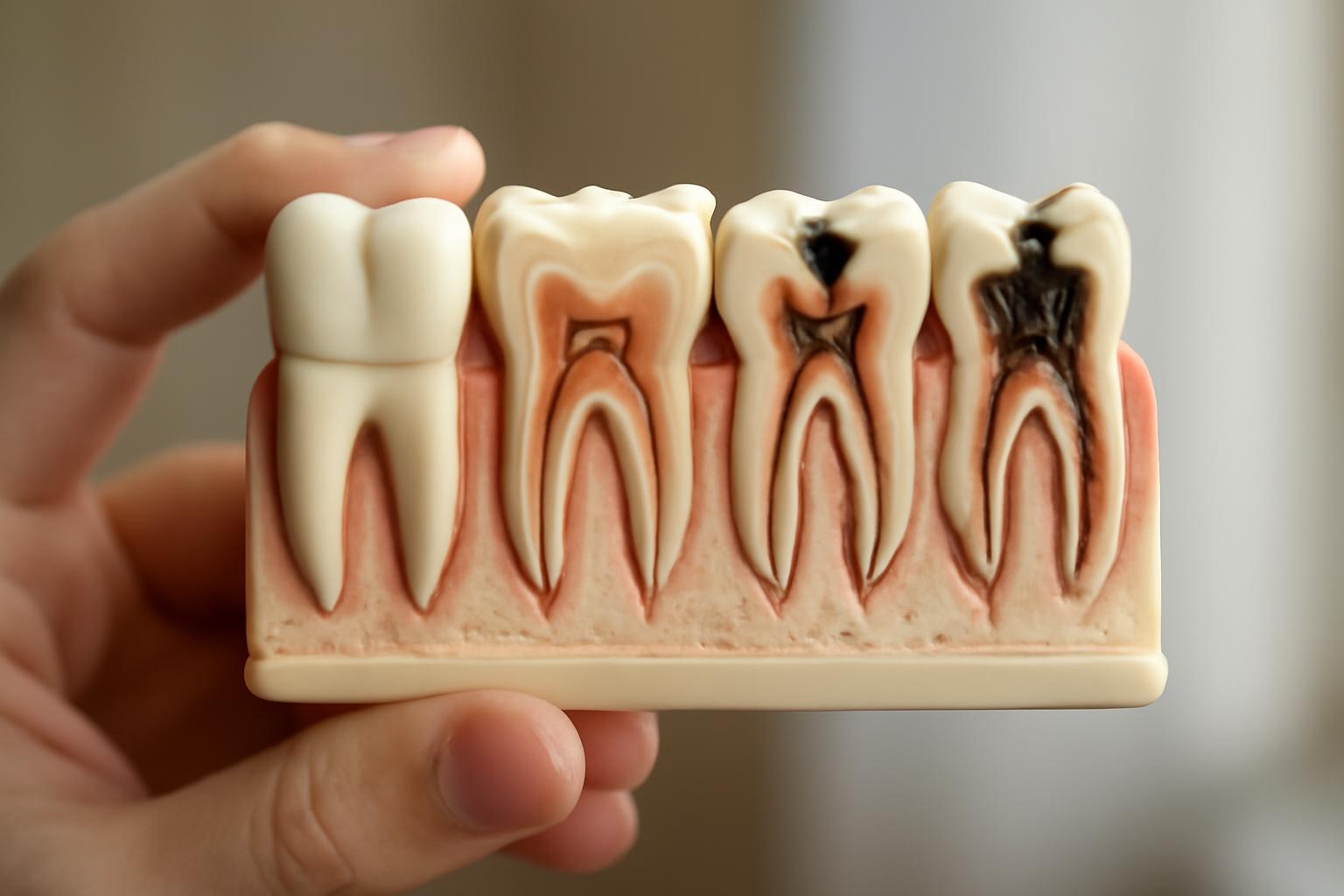
Are There Four or Five Stages of Tooth Decay?
Many patient guides use five stages of tooth decay:
- Demineralization
- Enamel cavitation
- Dentin decay
- Pulp involvement
- Abscess formation
This is a useful educational framework, not a universally standardized clinical staging system. Cleveland Clinic uses a comparable five-stage model progressing from demineralization to an abscessed tooth. Its overview describes the anatomy, possible symptoms and treatment options at each point.
One four-stage patient guide, for example, groups abscesses under advanced pulp infection. That framework illustrates why stage counts can differ even when the underlying sequence is similar.
Regardless of the number used, the biological progression is broadly consistent: mineral loss begins in enamel; continued damage can create a hole and reach dentin; deeper disease can affect the pulp; and untreated infection may extend beyond the tooth.
Three anatomical terms make this process easier to understand:
- Enamel is the hard outer covering of the tooth.
- Dentin is the softer layer beneath enamel.
- Pulp is the central tissue containing nerves and blood vessels.
The boundaries are not always as neat as a diagram suggests. A lesion may have a small surface opening while spreading more broadly underneath. Pulp irritation, pulp infection and an abscess are also related but distinct conditions. Pain, sensitivity or color cannot reliably separate them.
The most useful dividing line is often not between stages three and four or four and five. It is the boundary between non-cavitated mineral loss, where the enamel surface remains intact, and cavitation, where tooth structure has physically broken down. Early mineral loss may sometimes be arrested or remineralized. Missing tooth structure cannot grow back.
The following comparison is a general educational summary rather than a tool for diagnosing an individual tooth. The five-stage framework and its conditional treatment options are consistent with Cleveland Clinic’s patient guidance.
| Educational stage | Affected structure | Possible appearance | Possible symptoms | Reversibility | Common care considerations | General urgency |
|---|---|---|---|---|---|---|
| 1. Demineralization | Enamel minerals; surface may remain intact | White or chalky area, or no obvious change | Often none | May be arrested or remineralized before cavitation | Fluoride-based care, plaque control and reduced sugar frequency | Routine dental assessment |
| 2. Enamel cavitation | Enamel has physically broken down | Small opening, pit or discoloration | May remain painless; sensitivity is possible | Lost structure is not reversible | Professional evaluation; a filling or another restoration may be considered | Routine or prompt assessment, depending on symptoms |
| 3. Dentin decay | Softer dentin beneath enamel | Visible cavity or darker area; internal damage may exceed the apparent opening | Sensitivity, discomfort or pain may occur, but symptoms vary | Not reversible through remineralization alone | Filling, crown or another restoration may be considered after assessing remaining structure | Prompt assessment is sensible |
| 4. Pulp involvement | Central tissue containing nerves and blood vessels | Deep cavity, dark discoloration, gum redness or swelling may occur | Persistent or severe pain is possible, but pain does not prove the stage | Missing structure is not reversible | Root canal therapy and restoration may save some teeth; extraction may be considered if the tooth is not restorable | Prompt care; greater urgency if infection signs appear |
| 5. Abscess formation | Infection near the pulp, root or surrounding tissues | Swelling, pus or a gum lesion may be visible | Severe or radiating pain, unpleasant taste, fever or swollen neck lymph nodes may occur | Not reversible | Treatment must address the infection and its source; options depend on whether the tooth can be restored | Urgent professional assessment |
A stage label is therefore a map of the process—not a diagnosis based on a photograph, spot color or symptom checklist. A dentist must assess the lesion’s location and depth, whether the surface is intact, the condition of the pulp, the amount of remaining tooth structure and any evidence of infection.
How the Decay Process Begins—and Where Reversibility Ends
Tooth decay begins with repeated chemical cycles. Bacteria in dental plaque use sugars and starches from foods and drinks and produce acids that remove minerals from enamel. Teeth also undergo mineral replacement: saliva supplies minerals, while fluoride can help replace lost minerals and make enamel more resistant to acid. The National Institute of Dental and Craniofacial Research describes this as a continuing balance between mineral loss and replacement. Its explanation distinguishes early mineral loss from a permanent cavity.
Demineralization means that enamel is losing minerals. It does not necessarily mean that a physical hole is present. If the mineral loss is limited and the enamel surface remains intact, changing the conditions around the tooth may allow minerals to return to weakened enamel.
Repeated acid exposure can shift the balance toward continuing loss. Over time, the weakened enamel may collapse or break down, creating a physical opening. That is cavitation.
Color is not a dependable substitute for this distinction. Early mineral loss may appear as a white or chalky patch, but white areas can have other causes. Brown, gray or black marks may represent decay, staining or another change. Color alone cannot establish whether decay is active, how far it extends or whether the surface has cavitated.
Potentially reversible versus repairable
Potentially reversible: Before cavitation, an early enamel lesion may sometimes be arrested or remineralized with fluoride, saliva, effective plaque control and changes in the frequency of sugar exposure.
Repairable: After a hole forms, the missing enamel does not regenerate. A dentist may need to manage the decayed tissue and restore the lost structure. Fluoride and better home care remain useful, but they cannot rebuild the hole.
This distinction clarifies what “healing a cavity” can and cannot mean. People sometimes use cavity for any early sign of decay. Clinically, however, a physical cavity is a structural defect. Selected areas of early mineral loss can improve without drilling; an established hole cannot be brushed closed.
Fluoride, saliva, brushing and dietary changes support a healthier mineral balance, but they do not replace examination of a suspected defect.
Stages 1 and 2: Demineralization and Enamel Cavitation
Stage 1: Demineralization
In the earliest part of the process, repeated acid exposure removes minerals from enamel faster than they are replaced. The surface may remain intact even though the enamel below it has weakened.
A chalky or opaque white area is one possible sign. Early decay, however, commonly causes no symptoms and may not produce a change that a person can recognize. The National Institute of Dental and Craniofacial Research states that early decay can be stopped or reversed before a cavity forms and that saliva and fluoride can help enamel replace minerals. Its patient guidance also notes that early decay usually has no symptoms.
A white spot is not a home diagnosis. A dental examination is needed to make that distinction.
If a lesion is non-cavitated, care may focus on changing the conditions that caused the mineral imbalance. Depending on the examination and the person’s risk, possible measures include:
- Using fluoride toothpaste
- Professionally applied fluoride when appropriate
- Improving plaque removal
- Cleaning difficult-to-reach areas effectively
- Reducing how often sugary foods and drinks contact the teeth
- Addressing persistent dry mouth
- Monitoring the area over time
The aim is to arrest the process and support remineralization—not to replace structure that has not yet been lost.
Stage 2: Enamel cavitation
If mineral loss continues, weakened enamel can eventually break down. A small opening, pit or hole may form. This is the practical transition from potentially reversible mineral loss to permanent structural damage.
An enamel cavity can remain painless. Some people notice a defect, discoloration or sensitivity, while others notice nothing. The absence of discomfort does not show that the surface is intact.
Once a physical hole exists, saliva and fluoride cannot reconstruct the enamel’s original shape. Professional evaluation and usually structural repair are needed. A filling is one common option: the dentist manages the decayed area and replaces missing structure with restorative material.
That does not mean every area called “stage two” automatically receives a filling. A clinician must first establish whether a cavity is present and assess its size, location, accessibility and activity.
The transition may also be difficult to see at home. Cavities between teeth, beneath contact points or in deep grooves do not always appear as obvious holes. Conversely, a dark groove does not prove that enamel has cavitated. Surface integrity and lesion extent matter more than color alone.
Stages 3 to 5: Dentin Decay, Pulp Involvement and Abscess
Stage 3: Dentin decay
Dentin lies directly beneath enamel. Because it is softer and less resistant to acid, decay may progress more readily after reaching it.
Possible findings include:
- Sensitivity to sweet, hot or cold foods and drinks
- Discomfort when chewing or biting
- A visible opening or pit
- Brown or darker discoloration
- Intermittent toothache
- No noticeable symptoms
These findings do not confirm dentin involvement by themselves. Sensitivity has several possible causes, and a tooth with substantial decay may not hurt.
A cavity’s surface opening can also underestimate its internal extent. Enamel may retain a relatively small opening while damage spreads through the softer dentin underneath. This helps explain why a tooth that looks only mildly affected may require a larger restoration after the full lesion is assessed.
Structurally damaged dentin cannot be restored by fluoride alone. Depending on the extent of decay and the strength of the remaining tooth, care may involve a filling, crown or another restoration.
Stage 4: Pulp involvement
The pulp occupies the center of the tooth and contains nerves and blood vessels. When decay reaches or closely approaches this space, bacterial irritation and swelling can produce pressure and pain.
Possible findings include:
- Persistent, spontaneous or severe toothache
- Gum redness or localized swelling
- Pain when biting
- Dark discoloration
- A deep visible cavity
Pain intensity is not a dependable staging rule. A painful tooth does not automatically have pulp infection, and an extensively damaged tooth is not necessarily painful. Clinical assessment is needed to distinguish a deep cavity, pulp disease and other possible sources of dental pain.
If the pulp is sufficiently inflamed, damaged or infected, root canal therapy may be considered. This treatment addresses tissue inside the tooth so that the tooth can potentially be restored and retained. Suitability depends on the pulp condition, infection, remaining sound structure and whether a durable restoration is possible.
Stage 5: Abscess formation
An abscess is a collection of pus associated with infection. In the five-stage framework, it may develop after bacteria invade deeply and infection extends toward or beyond the root.
Possible warning signs include:
- Severe or radiating pain
- Swelling of the gum or face
- Pus or drainage
- An unpleasant or foul taste
- Fever
- Swollen lymph nodes in the neck
An abscess may affect surrounding tissue or bone and requires prompt professional treatment. Healthline’s medically reviewed five-stage overview describes severe or radiating pain, swelling, fever and swollen neck lymph nodes among possible advanced signs. It also warns that infection can spread beyond the affected tooth.
An abscess is not simply a very large cavity. It is an infection-related complication. Treatment must address both the infection and its source. Some teeth can be treated with root canal therapy and restored; others may not have enough healthy, supported structure to be retained.
The underlying tooth and surrounding tissues still require assessment.
Why Symptoms Cannot Tell You the Stage
Early tooth decay commonly causes no symptoms. Even more substantial damage may be silent, so the absence of pain does not rule out a cavity or establish that decay is minor.
Symptoms overlap because they are affected by more than lesion depth. Location, size, number of cavities and proximity to the pulp can all influence what a person feels. Mayo Clinic lists toothache, sensitivity, visible pits, staining and pain when biting as possible symptoms while emphasizing that a beginning cavity may cause none. Its symptom overview treats these findings as possibilities, not fixed stage markers.
Sensitivity to sweet, hot or cold foods can occur with tooth decay, including after decay reaches dentin. It does not prove dentin involvement. Sensitivity can arise from other dental conditions, and not every dentin lesion produces it.
Appearance has similar limits. A white, brown, gray or black area may justify an examination, but it cannot independently confirm:
- Whether the mark is decay
- Whether the process is active
- Whether the surface is intact
- How deeply the lesion extends
- Whether the pulp is affected
A visible pit is more suggestive of structural loss, but its internal extent cannot be estimated reliably from a mirror or photograph. Lighting, angle, staining and natural tooth anatomy can all change how an area appears.
When appropriate, radiographs can reveal decay that is difficult to see directly, particularly between teeth, and help assess its location or depth. NHS inform states that dentists can usually identify tooth decay and related problems through examination or an X-ray. Its guidance also notes that tooth decay may cause no pain.
Myth versus fact
Myth: “No pain means no decay.” Fact: Early decay is often painless, and some larger lesions also remain quiet.
Myth: “Every dark spot is a cavity.” Fact: Dark areas can have several causes. Color alone does not confirm active decay.
Myth: “Sensitivity reveals the exact stage.” Fact: Sensitivity may occur with decay, but it cannot show precisely which tissue is affected.
What a person notices can raise suspicion and help explain why care is being sought. Determining whether a cavity is present—and how deep or active it is—requires professional confirmation.
How Treatment Usually Changes as Decay Deepens
Treatment is clearer when organized by goal rather than by assigning one procedure automatically to every stage.
1. Remineralize or arrest early enamel changes
For a non-cavitated lesion, the goal is to stop continued mineral loss and support mineral replacement. Possible measures include fluoride-based care, improved plaque control and reducing the frequency of sugary foods and drinks.
These measures are most relevant while the enamel surface remains intact. The dentist may also consider where the lesion is located, whether it can be cleaned and the person’s wider cavity risk.
2. Restore missing structure
After cavitation, the objective changes. Minerals cannot recreate the tooth’s original shape, so the missing structure must be managed professionally. A common approach is to remove or otherwise manage decayed tissue and restore the defect with a filling.
A filling is not selected merely because a guide calls the lesion “stage two” or “stage three.” The dentist must evaluate the cavity’s size and position, the amount of healthy structure remaining and whether the tooth can withstand normal biting forces.
3. Protect a weakened tooth
A larger defect may leave too little strong tooth structure for a straightforward filling. A crown or another restoration may then be considered to protect and rebuild the tooth.
There is no universal visual threshold that tells a patient when a crown is required. Two cavities that appear similar can differ in location, internal spread and the amount of supported enamel and dentin that remains.
4. Treat diseased pulp while trying to retain the tooth
When the pulp is sufficiently damaged or infected, root canal therapy may be a tooth-saving option. The treatment addresses tissue inside the tooth, after which the tooth must be restored appropriately.
5. Address a tooth that cannot be restored
Extraction may be considered when decay, fracture, infection or loss of supporting structure means that a tooth cannot be restored. It is not inevitable for every deeply decayed or abscessed tooth. Some can be treated with root canal therapy and a suitable restoration; others cannot.
In broad terms, a dentist assesses:
- The lesion’s size, location and activity
- The amount and quality of remaining tooth structure
- Whether the remaining enamel and dentin can support a restoration
- The condition of the pulp
- The presence and extent of infection
- Cracks or other structural problems
- Whether the tooth is restorable
- The tooth’s function and the wider condition of the mouth
NHS inform describes this general progression in treatment: early disease may be managed with dietary measures and fluoride, while more advanced decay may require a filling or crown; pulp involvement may require root canal treatment, and a tooth that cannot be restored may need removal. Its treatment guidance emphasizes that care depends on how advanced the decay is.
When Tooth Decay Needs Prompt or Urgent Care
The following framework is general education, not individualized advice. Symptoms overlap, and only a dental professional who examines the tooth can determine what is happening.
Routine assessment
Arrange a dental assessment for:
- A possible white or chalky spot
- New discoloration
- A suspicious groove or pit
- A suspected cavity without significant pain
“Routine” does not mean unimportant. Early decay may be silent, and treatment is generally less extensive before a cavity becomes larger.
Prompt assessment
Do not ignore:
- Toothache
- Persistent or recurring sensitivity
- Pain when biting
- A visible hole
- Worsening discomfort
These signs do not reveal the stage, but they justify timely evaluation. Mayo Clinic advises seeing a dentist as soon as possible for toothache or mouth pain and notes that cavities may be developing even when the mouth feels fine. Its guidance supports assessment based on symptoms rather than attempted self-staging.
Urgent assessment
Seek urgent professional assessment for possible infection when there is:
- Facial or gum swelling
- Fever
- Pus or drainage
- A foul or unpleasant taste alongside other infection signs
- Severe or radiating pain
- Swollen lymph nodes in the neck
These findings may occur with an abscess or infection extending beyond the tooth. An abscess can involve surrounding tissues or bone and requires prompt treatment.
Do not assume that an infected tooth has healed because its pain decreases. Reduced pain does not establish that the source of infection has gone, so definitive assessment is still needed.
Decay Guide is an informational publisher, not a dental practice, and cannot examine, diagnose or stage an individual tooth. The site’s About page explains the limits of its educational role.
How Fast Decay Progresses and How to Slow It
There is no reliable universal timeline from a white spot to dentin decay, pulp involvement or an abscess. Progression varies with factors such as:
- The frequency of sugar and starch exposure
- Plaque accumulation and removal
- Fluoride exposure
- Saliva flow
- Persistent dry mouth
- Tooth shape and the depth of pits or grooves
- Lesion location
- Age and individual risk factors
- Existing restorations and access for cleaning
Frequency matters because each acid-producing episode pushes the tooth toward mineral loss. Repeated exposure can restart that process before saliva and fluoride have fully supported mineral replacement. Sipping a sugary drink over an extended period or repeatedly snacking therefore creates a different exposure pattern from less frequent contact.
No one should assume that a cavity will reach the pulp after a fixed number of months or remain harmless for a certain number of years. Assessment is safer than countdown-based self-monitoring.
Useful risk-reduction measures include:
- Brushing thoroughly with fluoride toothpaste
- Cleaning effectively between teeth
- Reducing how often sugary foods and drinks contact the teeth
- Seeking professional advice about persistent dry mouth
- Discussing professionally applied fluoride when appropriate
- Attending dental visits on a schedule based on individual risk
Dental sealants may also be considered for vulnerable pits and grooves. They form a barrier over areas where food and bacteria can become trapped. NIDCR explains that sealants protect these grooves and that fluoride toothpaste, reduced sugar frequency and professional dental care can help prevent cavities. Its prevention guide also explains how saliva and fluoride support mineral replacement.
A fixed six-month recall interval is not right for every person. Timing should be agreed with a dental professional according to cavity risk, existing disease, age, health factors and previous findings.
It cannot replace repair of tooth structure that is already missing.
It is also important not to confuse progression stages with cavity locations:
- Smooth-surface cavities occur on relatively flat tooth surfaces.
- Root cavities affect exposed root surfaces.
- Pit-and-fissure cavities occur in grooves, often on chewing surfaces.
These categories describe where decay occurs, not how advanced it is. A cavity in any of these locations can be shallow or deep.
Frequently Asked Questions
Can an early white spot heal without a filling?
Possibly—if the spot represents non-cavitated mineral loss and the enamel surface remains intact. Fluoride, saliva, effective plaque control and reduced sugar frequency may support remineralization or arrest the process before a permanent hole forms.
A white spot cannot be assumed to be decay from appearance alone. It may represent another enamel change, and a dentist must determine whether the surface is intact and whether the area appears active. Once a physical cavity has formed, remineralization cannot recreate the missing structure.
Can a cavity be advanced even if the tooth does not hurt?
Yes. Early cavities often cause no symptoms, and pain does not increase according to a reliable stage-by-stage formula. A lesion may also be difficult to notice when it is between teeth or spreading beneath a relatively small enamel opening.
No pain does not prove that decay is absent or shallow. Examination—and sometimes radiographs—is needed to assess its extent.
Can fluoride or better brushing reverse a physical cavity?
No. Fluoride and effective brushing can improve conditions around the tooth, reduce continuing mineral loss and support remineralization before cavitation. They cannot rebuild enamel or dentin that has already been physically lost. Colgate’s patient guide likewise distinguishes potentially reversible demineralization from an enamel cavity that requires professional restoration. Its stage overview explains this transition.
Once a hole exists, professional evaluation is needed. A filling or another restoration may be considered depending on the defect and the remaining tooth structure. Preventive care remains important afterward, but prevention and structural repair have different goals.
Can an abscessed or severely decayed tooth be saved?
Sometimes. Root canal therapy followed by an appropriate restoration may save a tooth when the affected tissue can be treated and enough sound structure remains. In other cases, extensive decay, fracture, poor support or insufficient restorable structure makes extraction more appropriate. A dental-practice overview included in the evidence for this guide similarly notes that some severely decayed teeth may be saved with root canal treatment and a crown, while others require removal. Its discussion presents these as conditional options rather than guarantees.
An abscess does not automatically mean extraction, but root canal therapy cannot guarantee that every tooth will be retained. The decision requires examination of the tooth, pulp, roots, surrounding tissues and remaining structure.
How does a dentist determine how deep tooth decay is?
A dentist considers the tooth’s appearance, surface integrity, lesion location, symptoms and surrounding tissues. The examination may reveal a visible defect, staining or another change requiring closer assessment.
NIDCR notes that dental examinations may identify staining, holes or altered areas and that X-rays can also show decay. Its diagnostic overview explains why professional findings are needed to plan treatment.
Symptoms and online photographs cannot provide the same information.
The central distinction throughout the stages of tooth decay is reversible versus repairable. Early mineral loss may sometimes be arrested before a hole forms, but missing tooth structure does not grow back. Because decay can remain painless and tooth color is unreliable, a suspected cavity should be assessed rather than self-staged. Swelling, fever, pus, severe or radiating pain, or swollen neck lymph nodes call for prompt or urgent professional care.
Decay Guide provides general education rather than diagnosis or personalized treatment advice. Its content is written by a health writer and has not been reviewed by a clinician, so it cannot replace a dental examination. Decay Guide’s editorial disclosure documents these limitations.