
How a Tooth Changes From Early Mineral Loss to Deep Infection
Intact early mineral loss may sometimes be remineralized; permanent structural breakdown cannot be repaired through home care alone.
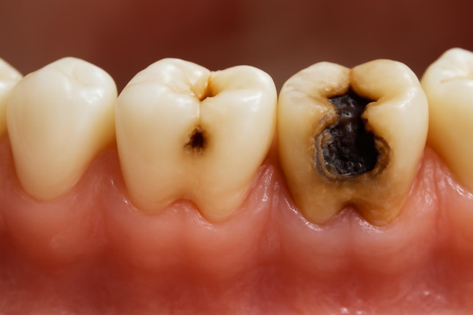
The five-stage framework—and why some guides list only four stages
A practical way to understand the tooth decay stages is to follow damage as it moves from the tooth’s surface toward its center and, in advanced cases, into surrounding tissues:
- Demineralization: enamel loses minerals, but its surface may remain intact.
- Enamel decay: continued damage breaks down enamel and may form a hole.
- Dentin decay: decay reaches the softer layer beneath enamel.
- Pulp damage: the inner tissue containing nerves and blood vessels becomes inflamed, damaged, or infected.
- Dental abscess: infection is associated with a pus-filled pocket and may extend beyond the tooth.
This five-stage sequence is a useful patient-education framework, not a universally established clinical classification. Cleveland Clinic uses the five-part model above (Cleveland Clinic’s overview of cavities). Some four-stage consumer guides combine pulp involvement and abscess-related disease under one advanced “pulp infection” stage (an example of the four-stage model).
The different counts therefore do not necessarily represent disagreement about the direction in which decay progresses. They often reflect where a publisher chooses to divide the advanced part of the process.
The most useful boundary is not the stage number. It is the distinction between:
- Early, non-cavitated mineral loss: Enamel has been weakened, but the surface remains sufficiently intact. Under suitable conditions, minerals may be replaced and the process may sometimes be stopped or reversed.
- Cavitation: Enamel or deeper tooth structure has broken down into a true opening or hole. Missing structure does not regrow through brushing, fluoride, or dietary changes and generally requires professional repair.
The table summarizes the five-stage model. “Possible” matters throughout: appearances and symptoms vary, and none of these observations can confirm a stage on its own.
| Stage | Affected tissue | Possible appearance | Possible symptoms | Reversible? | Diagnostic limits | Typical next step |
|---|---|---|---|---|---|---|
| 1. Demineralization | Enamel minerals; surface may remain intact | White or chalky area | Often none | Sometimes, if early and non-cavitated | White spots can have causes other than active decay | Dental assessment, fluoride support, plaque control, and risk-factor management |
| 2. Enamel decay | Enamel structure | Light-brown or dark area, pit, or visible or hidden opening | May remain painless | Mineral loss may be reversible before cavitation; missing structure is not | Stains and natural pits can resemble decay | Professional assessment; restoration may be needed if a hole has formed |
| 3. Dentin decay | Dentin beneath enamel | A larger or darker area may be present, but damage can also be hidden | Possible hot or cold sensitivity; sometimes no symptoms | Structural loss does not regrow | Sensitivity alone cannot establish the cause, depth, or treatment | Filling, crown, or another approach depending on the tooth |
| 4. Pulp damage | Inner tissue containing nerves and blood vessels | Deep cavity, discoloration, or no obvious external sign | Pain, lingering sensitivity, swelling, or few symptoms | Deep structural loss is not reversible through home care; pulp condition requires diagnosis | Symptoms alone cannot establish whether the pulp can be preserved | Assessment for pulp-preserving care, root canal treatment, restoration, or extraction |
| 5. Abscess | Infection associated with deep tooth structures and surrounding tissue | Gum, facial, or jaw swelling; sometimes limited visible change | Severe or radiating pain, swelling, fever, or swollen neck nodes | No home-care reversal | Symptoms cannot reveal the infection’s full extent | Prompt professional care |
A dark mark may be staining. A white area may have a cause other than active mineral loss. A significant cavity may not hurt, while sensitivity can arise from conditions unrelated to decay. This guide explains common patterns rather than providing a way to diagnose a tooth at home.
Editorial disclosure: Decay Guide is an informational publisher, not a dental practice. It does not diagnose or treat patients (Decay Guide’s non-clinical scope).
Its content is written by a health writer, has not been reviewed by a clinician, and does not replace a dental examination (Decay Guide’s authorship and review disclosure).
How decay moves from the tooth surface inward
Tooth decay begins with a repeated chemical process rather than with a hole appearing all at once. Certain bacteria in dental plaque use sugars and starches from foods and drinks and produce acids that remove minerals from enamel. When repeated mineral loss outweighs mineral replacement, enamel becomes progressively weaker.
This balance changes throughout the day:
- Demineralization occurs when acids draw minerals out of enamel.
- Remineralization occurs when minerals are deposited back into weakened enamel.
- Saliva supplies calcium and phosphate that support mineral replacement.
- Fluoride can reduce mineral loss and support remineralization of early damaged enamel.
The National Institute of Dental and Craniofacial Research describes this as an ongoing “tug-of-war”: teeth lose and regain minerals, but frequent exposure to sugary or starchy foods and drinks can shift the balance toward continuing damage (NIDCR’s explanation of the tooth decay process).
A simplified cross-section helps show what changes as decay moves inward:
                     TOOTH CROWN
              ┌─────────────────────┐
              │       ENAMEL        │  Hard outer covering
              │  ┌───────────────┐  │
              │  │    DENTIN     │  │  Softer supporting tissue
              │  │  ┌─────────┐  │  │
              │  │  │  PULP   │  │  │  Nerves and blood vessels
              │  │  └────┬────┘  │  │
              └──┴───────┼───────┴──┘
                         │
                    ROOT CANAL
                         │
                      ROOT TIP
                         ▼
              Surrounding ligament,
                   bone, and tissue
Educational diagram only; proportions are simplified, and the image cannot be used to diagnose decay.
Enamel does not contain the nerve-and-blood-vessel tissue found in the pulp. Mineral loss and decay confined to enamel can therefore remain symptomless. The absence of toothache does not establish that a tooth is free of clinically important decay.
Beneath enamel lies dentin. Dentin is softer than enamel and contains microscopic tubules leading toward the inner pulp. Once decay reaches dentin, it may progress more readily, and temperature sensitivity may become more likely. Even then, a cavity can remain painless.
If decay reaches the pulp, inflammation or infection can affect tissue confined within the tooth. Pain, sensitivity, swelling, or discoloration may occur, but symptoms vary. Infection associated with deep decay can eventually extend into tissues around the root and contribute to an abscess.
A lesion does not always move from one stage to the next within a fixed number of days, months, or years. The stage model describes the general direction of progression, not its speed in a particular tooth.
Stage 1: Demineralization before a cavity forms
Stage 1 affects enamel. Minerals are being lost, but the outer surface may still be intact. This matters because sufficiently early, non-cavitated damage may still be able to gain minerals.
A possible early sign is a small white or chalky area that looks duller or more opaque than nearby enamel. Not every white spot is active decay, however. A mirror or photograph cannot establish the cause, show whether the surface is intact, or determine whether the change is active.
This stage may cause no pain or other noticeable symptoms. Early changes are therefore often found during a dental examination rather than because a person feels something wrong.
If the lesion remains sufficiently early and non-cavitated, care may focus on shifting the local environment toward remineralization. Measures can include:
- fluoride from toothpaste or professionally selected treatment;
- saliva-supported replacement of calcium and phosphate;
- more effective plaque removal;
- fewer frequent exposures to sugary or starchy foods and drinks; and
- management of individual factors that keep the tooth under repeated acid challenge.
“Reverse” has a limited meaning here. It means that minerals may return to early damaged enamel, strengthening it and potentially stopping progression. It does not mean that the body can regrow a missing piece of enamel or dentin. Mayo Clinic similarly limits potential reversal to the earliest stage and distinguishes it from established cavities that require restorative care (Mayo Clinic’s cavity diagnosis and treatment guide).
Educational illustration: intact white spot versus formed cavity
```text INTACT EARLY LESION FORMED CAVITY
Enamel surface Enamel surface ───────────────── ───────╲ ╱─────── pale mineral-loss area ╲╱ with an intact surface opening
May be remineralizable Missing structure does if sufficiently early not regrow through home care ```
This is a conceptual illustration, not a diagnostic image. A dentist must determine whether the surface is intact and whether a visible mark represents decay.
Do not scrape, pick, or probe a suspicious area to see whether it feels soft or open. Professional assessment is the appropriate way to distinguish an early lesion from staining, an enamel variation, or an established cavity.
Stages 2 and 3: Enamel breakdown and decay into dentin
Stage 2: enamel decay
If mineral loss continues, enamel can weaken until its structure breaks down. The result may be a small opening at the surface, although some lesions remain difficult to see because they occur between teeth, within grooves, or beneath a modest-looking surface change.
Possible clues include:
- a light-brown or darker area;
- a visible pit;
- a rough-looking defect; or
- an obvious hole.
None is proof by itself. Teeth naturally have pits and grooves, and discoloration may be staining rather than decay. Conversely, damage beneath the surface may be more extensive than its external appearance suggests.
Once a true hole has formed, the missing structure does not regrow. Fluoride can still help protect the remaining enamel and reduce future mineral loss, but it does not rebuild the missing wall of a cavity.
Enamel decay can remain painless. A person may therefore have a formed cavity without realizing it, particularly when it lies between teeth or in a hard-to-see groove.
Stage 3: dentin decay
Dentin lies beneath enamel and makes up much of the tooth’s structure. Because it is softer than enamel, decay may progress more quickly after reaching it. Its microscopic tubules communicate toward the pulp, helping explain why hot or cold foods and drinks may trigger sensitivity.
Sensitivity is not a dependable staging tool. A dentin cavity may produce noticeable sensitivity, mild symptoms, or no symptoms at all.
Care at stages 2 and 3 depends on more than the tissue name. A smaller formed cavity may be suitable for a filling. If extensive decay has weakened the tooth or removed a large amount of supporting structure, a crown may be considered. Treatment cannot be assigned from the stage label alone (Penn Dental’s comparison of fillings and root canal treatment).
Relevant factors can include:
- whether the lesion is cavitated and active;
- its size, depth, and location;
- how much strong tooth structure remains;
- whether an existing filling or crown is involved;
- the tooth’s function and risk of fracture; and
- the condition of the pulp.
The stage describes biological progression. It does not prescribe one procedure for every tooth.
Stages 4 and 5: Pulp damage and dental abscess
Stage 4: pulp damage
The pulp is the inner soft tissue containing nerves and blood vessels. When deep decay approaches or reaches it, the pulp may become inflamed, damaged, infected, or no longer vital.
Possible findings include:
- substantial or spontaneous tooth pain;
- lingering sensitivity after hot or cold exposure;
- pain when biting;
- redness or swelling near the tooth;
- tooth discoloration; or
- few or reduced symptoms if pulp tissue has lost vitality.
These findings overlap with other dental conditions. Symptoms alone cannot establish how deeply decay extends, whether the pulp is infected, or which treatment is appropriate.
If pulp tissue is damaged or infected but the tooth remains restorable, root canal treatment may be considered. This involves removing diseased pulp and cleaning and filling the canal spaces. The tooth may then need a suitable restoration to protect its remaining structure.
Extraction may be considered when decay or fracture has left too little sound structure for predictable restoration. It is not an automatic consequence of reaching a stage called “pulp damage.” The dentist must assess both pulp condition and restorability.
Stage 5: dental abscess
A dental abscess is a pus-filled pocket associated with infection. In advanced tooth decay, infection may extend through deep tooth structures and into tissues around the root.
Possible warning signs include:
- severe or radiating tooth pain;
- swelling of the gum, face, or jaw;
- fever; and
- swollen lymph nodes in the neck.
A suspected abscess requires prompt professional care because infection can spread into surrounding tissues. Distant spread, such as spread to the bloodstream or brain, is described as rare, but the risk of progression is why an abscess should not simply be watched at home (Cleveland Clinic’s guidance on cavity complications).
Severe or radiating pain, facial or jaw swelling, fever, or swollen neck nodes warrants prompt evaluation. Home care cannot determine the extent of the infection or remove the source within an affected tooth.
How dentists find and assess decay
Early decay can be difficult to see, especially when it begins between teeth or beneath a seemingly small surface mark. It may also cause no symptoms. Dentists therefore do not rely on pain or appearance alone.
Assessment may include:
- Symptom history: when pain or sensitivity began, what triggers it, how long it lasts, whether swelling has occurred, and whether symptoms have changed.
- Examination: inspection of the teeth, gums, existing restorations, and surrounding tissues.
- Professional probing when appropriate: checking suspicious areas for clinical signs such as softened tooth structure. Readers should not attempt this themselves.
- Dental X-rays when appropriate: imaging can reveal decay that is difficult or impossible to identify through casual visual inspection.
Mayo Clinic describes diagnosis using pain and sensitivity history, examination, professional probing, and dental X-rays (Mayo Clinic’s explanation of cavity diagnosis).
No isolated clue can reliably determine depth or treatment:
| Isolated clue | Why it is not enough |
|---|---|
| White spot | May represent early mineral loss, an inactive change, or a non-decay enamel difference |
| Dark mark | Could be decay, staining, or discoloration from another cause |
| Visible pit | Could be natural anatomy, a stain, or a cavity |
| Cold sensitivity | Has several possible causes and does not establish lesion depth |
| Toothache | May result from decay or another dental condition |
| Severe symptoms | Cannot establish whether a filling, crown, root canal, or another treatment is needed |
| No pain | Does not rule out enamel or dentin decay |
Treatment selection may depend on lesion depth, size, location, activity, remaining tooth strength, pulp condition, and whether the tooth can be restored predictably.
A practical care pathway is:
- No symptoms: Continue routine dental examinations because early or hidden decay may be painless.
- New hole, persistent sensitivity, or tooth pain: Arrange a dental appointment rather than waiting for symptoms to become severe.
- Severe or radiating pain, facial or jaw swelling, fever, or swollen neck nodes: Seek prompt professional evaluation for possible advanced infection.
The absence of an obvious hole in a photograph does not exclude decay between teeth or beneath an existing restoration.
How treatment changes as damage becomes deeper
Treatment is better understood as an individualized ladder than as a one-to-one stage chart. The aim changes as damage progresses: first to improve the mineral balance, then to replace missing structure, protect a weakened tooth, address diseased pulp, or remove a tooth that cannot be restored.
| Option | General situation in which it may be considered | What it addresses | Why examination is necessary |
|---|---|---|---|
| Fluoride and risk-factor management | Very early, non-cavitated enamel mineral loss | Supports remineralization and reduces continuing mineral loss | An examiner must distinguish an intact early lesion from a formed cavity or another enamel change |
| Filling | A formed cavity limited enough for direct restoration | Removes decayed tissue and replaces missing structure | Size, depth, location, access, and remaining tooth strength affect suitability |
| Crown | Extensive decay or a tooth weakened by substantial structural loss | Covers and protects the remaining tooth | A crown may be unnecessary for a small lesion and insufficient if the tooth cannot be restored |
| Root canal treatment | Damaged or infected pulp in a tooth that remains restorable | Removes diseased pulp and cleans and fills the canal system | Symptoms alone cannot establish pulp condition or restorability |
| Extraction | Severe damage leaves the tooth unable to be predictably restored | Removes the affected tooth | The decision requires assessment of structure, infection, function, and alternatives |
Fluoride and risk-factor management: For sufficiently early, non-cavitated enamel damage, care may focus on strengthening enamel and reducing the factors driving mineral loss. This can involve fluoride, improved plaque removal, fewer frequent sugar or starch exposures, and monitoring to determine whether the lesion has stopped progressing.
Filling: Once a restorable hole has formed, a dentist may remove decayed tissue and rebuild the area with restorative material. A direct filling is more likely to be suitable when enough strong tooth remains to support it.
Crown: A crown is a custom-fitted covering. It may be considered when decay is extensive, a large amount of structure must be replaced, or the remaining tooth is too weakened for a smaller filling to offer suitable protection.
Root canal treatment: If the pulp is damaged or infected and the tooth can be saved, treatment may involve removing the affected pulp, cleaning and shaping the canals, and filling the internal space. The tooth is commonly protected with an appropriate restoration afterward.
Extraction: If severe decay, fracture, or structural loss means the tooth cannot be predictably restored, extraction may be considered. Advanced decay does not automatically make a tooth non-restorable; that conclusion requires clinical assessment and often imaging.
Overlapping symptoms cannot tell someone whether they need a filling, crown, root canal, extraction, or another approach. Two cavities described with the same stage name may differ substantially in size, location, remaining support, pulp health, and restorability.
Reducing the chance that early decay progresses
Prevention is risk reduction, not a guarantee that decay will never occur. Daily care can reduce plaque accumulation and shift the balance toward mineral replacement, but individual risk varies.
Useful measures include:
- Brush twice daily with fluoride toothpaste. This combines mechanical plaque removal with regular fluoride exposure.
- Clean between teeth regularly. Depending on spacing and dexterity, an appropriate method may be floss, an interdental brush, or another professionally recommended aid.
- Reduce frequent sugary or starchy exposures. Each exposure can support another period of acid production. Fewer eating and sipping episodes leave more time for saliva- and fluoride-supported mineral replacement.
- Attend regular dental examinations. Early decay may be painless, and professional assessment can identify lesions before they require more extensive treatment.
- Consider sealants when appropriate. Sealants form a protective barrier over pits and grooves in molars where food and bacteria can become trapped.
These measures reflect NIDCR guidance recommending brushing with fluoride toothpaste twice daily, limiting repeated snack exposures, obtaining regular dental care, and using sealants to protect vulnerable grooves when appropriate (NIDCR’s cavity-prevention guidance).
It also helps to separate cavity location from decay stage:
- Smooth-surface cavities occur on smooth tooth surfaces, including areas between teeth.
- Pit-and-fissure cavities occur in grooves on chewing surfaces.
- Root cavities occur on exposed root surfaces.
These terms describe where decay occurs, not how advanced it is.
For parents and caregivers, primary teeth may be more susceptible to progression because their enamel is thinner. That does not establish a universal timetable or a single treatment rule for children. Prevention, monitoring, fluoride use, sealants, and treatment still require age-appropriate professional assessment (Healthline’s medically reviewed overview of tooth decay stages).
The practical limit of home care remains clear: brushing, fluoride, plaque control, and eating-pattern changes can help prevent decay and may arrest or remineralize sufficiently early mineral loss. They cannot rebuild a formed hole.
The central distinction is simple but important. Early mineral loss with an intact enamel surface may sometimes be remineralized; permanent structural breakdown cannot be repaired through home care alone. Symptoms do not map neatly onto the tooth decay stages, and significant disease may be painless. A dental examination—not a color chart, photograph, or symptom checklist—determines what a particular tooth needs.
Which stage of tooth decay can be reversed?
The earliest stage—demineralization—may sometimes be stopped or reversed if the enamel surface remains sufficiently intact and the lesion has not become a formed cavity. Saliva, fluoride, improved plaque control, and fewer frequent sugar or starch exposures can support mineral replacement.
“Reversed” means remineralizing early damaged enamel. It does not mean regrowing enamel or dentin after a hole has formed.
Can you have a cavity without pain?
Yes. Decay limited to enamel may cause no symptoms, and some dentin lesions can also remain painless. Cavities between teeth or within grooves may be difficult to see.
Pain is not a dependable staging tool. Routine examinations matter because waiting for pain can allow a silent lesion to become more extensive.
Does every white or dark spot on a tooth mean decay?
No. A white or chalky area can be associated with early demineralization, but enamel differences and inactive changes may look similar. A dark mark may be decay, staining, or another form of discoloration.
Appearance alone cannot establish whether a lesion is active, whether the surface has broken down, or how deeply it extends. A dentist may need to combine examination findings with X-rays.
How does a dentist decide between a filling, crown, root canal, and extraction?
The decision depends on the cavity’s depth, size, and location; the amount of strong tooth structure remaining; the condition of the pulp; and whether the tooth can be predictably restored.
A smaller formed cavity may be suitable for a filling. A tooth weakened by extensive structural loss may need a crown. Root canal treatment may be considered when the pulp is damaged or infected but the tooth remains restorable. Extraction may be considered when severe damage leaves too little structure to save predictably. Symptoms alone cannot make that distinction.
Which symptoms can indicate a dental abscess?
Possible symptoms include severe or radiating tooth pain, gum or facial swelling, jaw swelling, fever, and swollen lymph nodes in the neck. Not everyone has every symptom, and these signs cannot confirm an abscess without professional assessment.
A suspected abscess requires prompt professional care because infection may spread into surrounding tissues.