
Why Tooth Decay Has No Single Countdown
Acid attacks can begin quickly, but a physical cavity develops through a variable process that cannot be dated from color, sensation, or appearance.
The short answer: cavity formation is gradual, but the clock varies
How long does a cavity take to form? There is no reliable universal timetable. Tooth decay is generally gradual rather than an overnight event, but the supplied evidence does not establish a research-backed average that can predict when a particular tooth will develop a physical hole.
Some dental-practice articles describe cavity development in terms ranging from weeks to years. At most, these estimates support a cautious generalization that the process may take months or longer—not a clinical deadline. The articles use inconsistent starting and ending points and do not cite longitudinal research establishing their ranges as standard timelines (see one example and its limitations).
The word cavity can refer to at least three different endpoints:
- The start of mineral loss: Acids produced by plaque bacteria begin removing minerals from enamel.
- A detectable lesion: A dentist identifies demineralization or decay during an examination, sometimes with the help of an X-ray.
- Surface cavitation: Weakened tooth structure breaks down, leaving a physical defect or hole.
These events do not occur at the same moment. Acid activity and microscopic mineral loss can begin soon after bacteria encounter sugars or other fermentable carbohydrates, but an acid challenge after eating is not evidence that a permanent hole has formed within minutes. Saliva can neutralize acids and supply minerals involved in enamel repair; repeated imbalance between loss and repair is what allows decay to progress (review the explanation of acid activity, saliva, and variable progression).
The time required for decay to pass through enamel and reach dentin is a separate question. The time needed to involve the pulp, cause infection, or contribute to an abscess is even less predictable. These outcomes depend on factors such as lesion location and activity, tooth structure, saliva, fluoride exposure, diet, plaque control, existing dental work, and whether treatment interrupts the process.
The most useful questions are therefore not “How many weeks do I have?” but “Is the surface still intact?” and “Is the lesion active?” Early mineral loss beneath an intact surface may sometimes be arrested or remineralized. Once the surface has collapsed into a hole, prevention cannot rebuild the missing tooth structure.
What counts as a cavity: acid attack, early lesion, or physical hole?
Plaque is a bacterial film that forms over teeth. When plaque bacteria use sugars from food and drink, they produce acids that can break down the tooth surface and contribute to cavities, according to NHS inform’s tooth-decay guidance.
A single acid challenge is not identical to a cavity. The decisive issue is whether repeated acid production keeps mineral loss ahead of repair. Frequent sugar exposure, persistent plaque, reduced saliva, limited fluoride exposure, and vulnerable tooth surfaces can all shift that balance toward continued demineralization.
An early non-cavitated lesion is an area of mineral loss where the outer surface may still be intact. Subsurface changes can alter the appearance of enamel and may produce a chalky or opaque white area. That appearance can be a warning sign, but it cannot by itself establish whether the lesion is active, how deep it is, or whether it needs a filling.
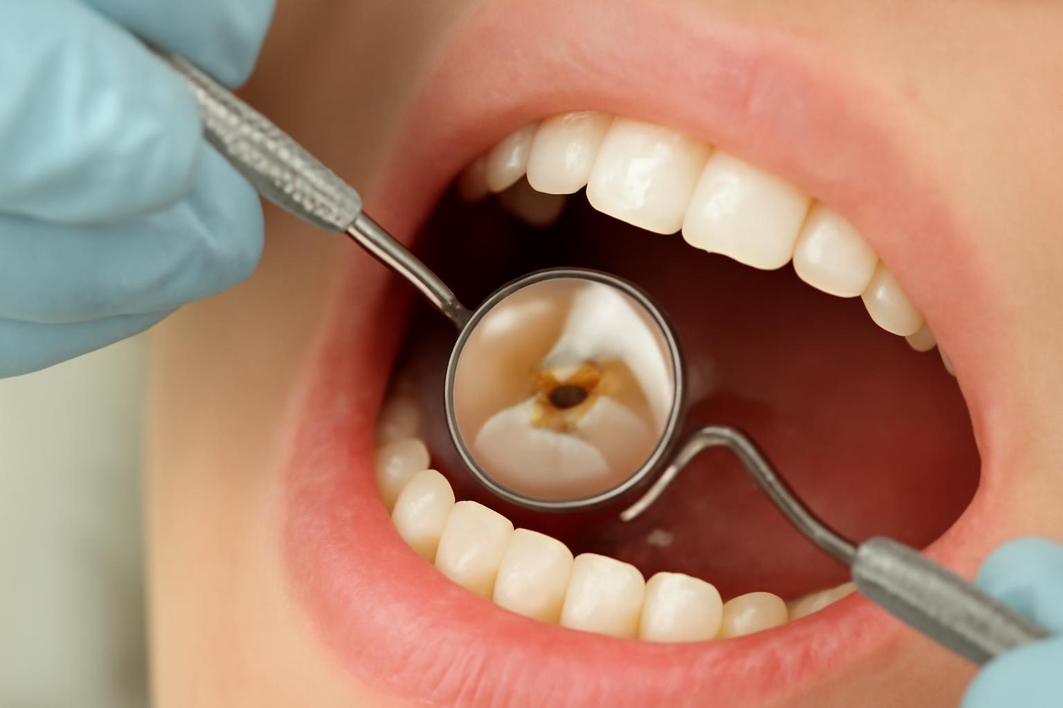
A cavitated lesion has undergone structural breakdown. The surface has collapsed or developed a physical defect—the “cavity” or hole meant in ordinary conversation. Cleveland Clinic defines cavities as holes or areas of tooth decay caused by acids wearing down enamel and notes that they can form on either the crown or root of a tooth (see the medically reviewed overview).
Sources do not always use cavity so narrowly. Some call initial demineralization an “early cavity,” while others reserve the term for structural loss. This difference helps explain apparently conflicting timelines:
- Minutes may refer to acid production or the beginning of mineral loss after eating.
- Weeks or months may refer to persistent demineralization or a detectable lesion.
- Months or longer may refer to structural breakdown or movement into deeper tooth layers.
- Years may describe a slow lesion that remains confined, stable, or intermittently active.
These are descriptions of different biological or clinical endpoints, not a reliable schedule.
Color does not resolve the ambiguity. A white area can have causes other than active decay, while a brown, grey, or black area may represent staining or decay at an uncertain stage. A dentist considers the location, surface integrity, texture, change over time, overall cavity risk, and imaging when appropriate.
The stages of decay—and why they should not be treated as a fixed schedule
Tooth decay is often described as a sequence from enamel mineral loss to deeper infection. That sequence helps explain what can happen, but it should not be converted into a stage-by-stage calendar. A medically reviewed clinical overview describes demineralization, enamel decay, dentin decay, pulp involvement, and abscess as progressive stages while emphasizing that early disease may be painless and that decay accelerates after reaching softer dentin (review the stages and treatment distinctions).
| Stage | What is happening | Possible visibility or symptoms | Reversibility | Usual management |
|---|---|---|---|---|
| 1. Enamel demineralization | Acids remove minerals from enamel while the outer surface may remain intact. | Often no pain. A chalky or opaque white area is possible but is not diagnostic by itself. | Mineral loss may sometimes be arrested or remineralized while the surface remains intact. | Fluoride, effective plaque removal, less frequent sugar exposure, saliva support, and professional monitoring. |
| 2. Enamel breakdown or cavitation | Weakened enamel loses structural integrity and develops a defect or hole. | A pit, rough edge, visible hole, food trapping, or sensitivity may occur; symptoms may also be absent. | Preventive measures cannot reconstruct missing tooth structure. | Dental assessment and restorative care when indicated by the defect’s size and location. |
| 3. Dentin involvement | Decay passes through enamel into the softer dentin underneath. | Sensitivity or discomfort may occur, although symptoms vary. | Lost structure cannot be restored through remineralization alone. | Restorative treatment is generally required. |
| 4. Pulp involvement | Decay or infection reaches the central tissue containing nerves and blood vessels. | Toothache, prolonged sensitivity, or signs of inflammation or infection may develop. | Not reversible through home care. | More extensive treatment may be required, potentially including root canal treatment. |
| 5. Abscess or spread beyond the tooth | Infection may extend through the root into surrounding tissues. | Pain, swelling, inflammation, drainage, or an unpleasant taste may occur. | Not reversible through preventive measures. | Prompt professional assessment; treatment depends on the tooth and extent of infection. |
Decay generally advances more quickly after reaching dentin because dentin is softer than enamel. If the process reaches the pulp, pain or infection may develop, and untreated infection can contribute to an abscess.
“Stage” does not mean “scheduled stop.” A non-cavitated lesion may remineralize, arrest, remain stable, or progress as conditions in the mouth change.
Location also matters. Decay in a deep groove, between teeth, around existing dental work, or on an exposed root may behave differently from a lesion on an accessible smooth enamel surface. Consequently, estimates assigning a fixed number of months to enamel decay or a set number of years to dentin involvement should not be treated as clinical standards.
Why one cavity may progress faster than another
Cavity progression depends on the balance between repeated mineral loss and opportunities for repair. Risk factors rarely act alone: several unfavorable conditions together may favor progression, while protective conditions may help an early lesion slow or arrest.
| Conditions that may favor faster progression | Conditions that may favor slower progression or arrest |
|---|---|
| Frequent sugary snacks, sweets, or drinks | Fewer eating and drinking occasions involving sugar |
| Repeated sipping or grazing | More recovery time between sugar exposures |
| Plaque remaining in difficult-to-clean areas | Effective brushing and interdental cleaning |
| Dry mouth or reduced saliva flow | Adequate saliva and professional management of persistent dryness |
| Limited fluoride exposure | Regular fluoride toothpaste use and professionally advised fluoride care |
| Deep pits, fissures, exposed roots, or worn enamel | Accessible, cleansable surfaces and individualized prevention |
| Plaque-retentive areas around dental work | Monitoring restorations and cleaning vulnerable margins |
| A lesion already cavitated or extending into dentin | A non-cavitated lesion with an intact surface |
Sugar frequency and recovery time
Each exposure to fermentable carbohydrates gives plaque bacteria another opportunity to produce acid. Frequency therefore matters as well as the total amount consumed. Repeated sipping or grazing can reduce the time available for saliva to neutralize acids and support mineral return.
This does not mean that one sweet item immediately creates a hole. It means that frequent exposure can repeatedly tilt the environment toward mineral loss. No simple formula can translate a person’s snack pattern into a cavity-formation date.
Plaque removal
Brushing and interdental cleaning reduce bacterial plaque in areas where acid-producing activity can persist.
Regular brushing is protective, but it is not a guarantee against decay. Deep grooves, tightly contacting teeth, exposed roots, worn enamel, and margins around fillings or crowns may remain vulnerable. Technique, fluoride exposure, sugar frequency, saliva flow, and existing disease still matter.
Saliva and dry mouth
Saliva washes the mouth, helps neutralize acids, and supplies minerals involved in enamel repair. Reduced saliva flow removes part of that natural protection. Persistent dry mouth may be related to medicines, health conditions, or other factors and warrants discussion with a dentist or healthcare professional rather than reliance on water alone.
Commercial dental guidance consistently identifies frequent sugar exposure, dry mouth, deep grooves, enamel wear, inconsistent interdental cleaning, and existing dental work as factors that may affect progression, while also emphasizing that no single timeline follows from them (see the risk-factor overview).
Fluoride
Fluoride strengthens enamel’s resistance to acid and supports remineralization during the non-cavitated stage. It can shift the balance toward repair, but it is not a universal cure. Its usefulness depends partly on whether the surface remains intact and whether repeated causes of mineral loss are also addressed.
Location, anatomy, and individual differences
Deep pits and fissures can retain plaque and be difficult to clean. Enamel wear, tooth position, existing fillings or crowns, and inaccessible contact areas can also alter local risk.
Diet, age, health, saliva, fluoride exposure, enamel characteristics, and cavity history add further variation. These factors do not support a dependable child-versus-adult rate or a numerical timeline for any particular cavity type.
A bounded comparison illustrates the point: frequent sugar exposure combined with dry mouth, limited fluoride, and ineffective plaque removal may favor faster progression than less frequent sugar exposure, adequate saliva, fluoride exposure, and effective cleaning. That comparison indicates direction only. It cannot tell either person how many weeks a lesion will take to cavitate.
The reversible window: when early decay may be stopped
Can a cavity heal without a filling? The answer depends on what cavity means and whether the surface remains intact.
Early enamel mineral loss may sometimes be arrested or remineralized. Fluoride can strengthen enamel, while effective plaque removal, fewer frequent sugar exposures, adequate saliva, and dental monitoring can improve the conditions for repair. This is why identifying a non-cavitated lesion early matters.
Once the surface has collapsed into a hole, however, fluoride, brushing, and dietary changes cannot reconstruct the missing shape of the tooth. Those measures remain useful for protecting surrounding tooth structure and reducing further disease, but they do not replace restoration of lost structure. Decay extending into dentin also generally requires restorative care.
Public patient guidance describes treatment according to how advanced the decay is:
- Preventive advice and fluoride for suitable early lesions
- A filling or crown for more advanced structural decay
- Root canal treatment when decay has reached the pulp and the tooth can be treated
- Extraction when the tooth cannot appropriately be restored
These are broad treatment categories, not instructions for self-treatment. NHS inform explains the progression from preventive care to fillings, crowns, root canal treatment, or extraction, but only an examining dentist can decide what is appropriate for a particular tooth.
A dentist may consider silver diamine fluoride in selected situations, including some cavities in baby teeth. It may help stop progression, but it does not rebuild missing structure and should not be treated as suitable for every patient or lesion (see the practice guidance discussing this limited use).
Nor does the date when a spot was first noticed establish whether it is active.
Clinical assessment may include visual and surface findings, the lesion’s location, cavity-risk history, changes over time, and imaging where appropriate. Detection also does not automatically mean that a filling is needed: an intact early lesion may be managed differently from a cavitated or dentin-level lesion.
Symptoms cannot tell you how old a cavity is
Early tooth decay may be painless, so waiting for toothache can allow disease to progress unnoticed. Conversely, sensitivity or pain does not prove that decay is the cause.
Possible signs include:
- Persistent or recurring sensitivity to hot, cold, or sweet foods and drinks
- Toothache
- Discomfort when biting or chewing
- A white, brown, grey, or black area
- A rough spot or edge that catches floss
- A pit or visible hole
- Food repeatedly lodging in one location
- Bad breath
- An unpleasant taste
None of these signs is a timestamp.
Deeper decay may be more likely to cause symptoms, but pain still cannot specify when the process began or precisely how far it has advanced. Some early lesions are sensitive, while some deeper lesions cause little warning. The absence of pain does not establish that a tooth is healthy.
A dental examination can assess the location and surface. When appropriate, X-rays may reveal decay between teeth or in other areas that are difficult to inspect directly. Dental-practice guidance likewise notes that examinations and imaging may identify decay before symptoms appear, while listing sensitivity, discoloration, pits, holes, and chewing discomfort as possible—not diagnostic—signs (review the detection overview).
How to reduce repeated acid attacks and slow early disease
Prevention is most useful before the surface has cavitated. The measures below can reduce repeated acid challenges and support enamel repair, but they cannot restore tooth structure that has already been lost.
Brush twice daily with fluoride toothpaste
Brush accessible tooth surfaces twice a day with fluoride toothpaste. The goals are to disrupt plaque and maintain regular fluoride contact with enamel. Technique matters because rushed or incomplete brushing may miss back teeth, inner surfaces, grooves, and areas near the gumline.
Fluoride supports enamel resistance and remineralization, but brushing cannot reach every vulnerable location and does not guarantee that an existing lesion will stop.
Clean between teeth daily
Use floss or another interdental cleaning method appropriate for the spaces between your teeth. Contact areas can retain plaque even when visible tooth surfaces appear clean.
A rough area that repeatedly catches or shreds floss can have several causes. Rather than assuming it proves decay, mention it during a dental assessment so the area can be examined.
Reduce how often teeth encounter sugar
Focus on frequency as well as quantity. Repeated sipping, sucking sweets, or grazing on sugary snacks creates recurring opportunities for plaque bacteria to produce acid. Reducing the number of exposures allows more recovery time between challenges.
This does not require treating every carbohydrate as equally harmful or trying to calculate an exact “safe” interval. The practical goal is to avoid repeated sugar exposure throughout the day.
Choose water and take persistent dry mouth seriously
Water is a practical alternative to sugary drinks and can help clear loose food debris. It does not correct the underlying cause of chronic dry mouth.
If your mouth regularly feels dry, discuss it with a dentist or healthcare professional. Assessment may be particularly useful when the change is persistent, began after a new medicine, or occurs alongside increasing tooth decay.
Use monitoring based on individual risk
There is no universal checkup interval appropriate for every person. NHS guidance states that adult recall intervals can vary from three months to two years depending on individual needs; this is a range for professional planning, not a schedule readers should choose for themselves. Monitoring frequency depends on factors such as cavity history, current lesions, dry mouth, diet, fluoride exposure, dental work, age, and health.
For a separate visual explanation, see Decay Guide’s How Cavities Form Stages: 5 Progressive Tooth Decay Stages Explained. That first-party article is supplementary educational material, not independent clinical validation.
When to arrange a dental examination—and what the timeline cannot decide
Arrange a dental examination if you notice:
- Persistent or recurring sensitivity
- Toothache or worsening pain
- Discomfort when chewing or biting
- A visible pit or hole
- A rough area that repeatedly catches floss
- A spot that appears to grow, spread, or darken
- Recurrent food trapping around one tooth
- A damaged filling or a change around existing dental work
Possible infection or an abscess warrants prompt professional assessment, particularly when pain occurs with swelling, inflammation, drainage, or an unpleasant taste. Untreated decay can reach the pulp and contribute to an abscess or infection beyond the tooth, so an online timeline should not be used to justify waiting.
The supplied evidence does not provide a supported checklist for distinguishing every dental emergency from a problem requiring prompt dental care. A dentist or local urgent-care service should therefore triage severe or worsening symptoms rather than a reader relying on this article to determine urgency.
A treatment decision depends on examination findings: whether the surface is intact, the lesion’s depth and location, signs of activity, symptoms, changes over time, overall cavity risk, and sometimes imaging. The number of weeks since a spot was first noticed is only background information. It cannot determine whether the lesion is active, arrested, cavitated, or close to the pulp.
The supplied public-health and medically reviewed sources strongly support the mechanism of tooth decay, its broad stage sequence, the possibility of painless early disease, and the distinction between potentially remineralizable mineral loss and missing tooth structure. They do not establish a research-backed average formation time that can be applied to an individual tooth. Practice articles that offer week-, month-, or year-level estimates use inconsistent endpoints and should not be converted into personal diagnostic thresholds.
Decay Guide is an informational publisher, not a dental practice. Its content is written by a health writer rather than a dentist, has not been reviewed by a clinician, and cannot replace an examination, as disclosed in its editorial information.
Frequently asked questions
Can a cavity form overnight?
A permanent physical hole does not usually form overnight. Acid production and microscopic enamel mineral loss can begin quickly after sugar exposure, but those chemical changes are not the same as structural collapse.
A newly noticed defect deserves assessment, but the date of discovery does not reveal when the decay began.
Can a permanent cavity really form within a few weeks?
Some dental-practice articles place “a few weeks” at the lower end of broad cavity-development ranges. They do not consistently define whether this means early demineralization, a detectable lesion, or a cavitated hole, and they do not provide sufficient longitudinal evidence to establish that a permanent hole routinely forms within weeks.
Unfavorable conditions can favor progression, but the supplied evidence does not support assigning a week-based cavitation estimate to a particular tooth. Surface integrity and lesion depth must be assessed directly.
Can fluoride reverse a cavity before a filling is needed?
Fluoride may help arrest or remineralize early mineral loss while the enamel surface remains intact. It strengthens enamel’s resistance to acid and supports mineral repair, particularly when combined with effective plaque control and fewer frequent sugar exposures.
Fluoride cannot rebuild a hole or replace missing tooth structure. If a lesion is cavitated or has entered dentin, restorative treatment is generally needed. A dentist must determine which side of that boundary the lesion occupies.
Do cavities form faster in children than in adults?
However, the supplied evidence does not establish a dependable child-versus-adult formation rate.
Claims that a child’s cavity forms within a specified number of months while an adult’s always takes longer are too broad for personal prediction. Saliva, fluoride, sugar frequency, plaque control, tooth location, enamel characteristics, and existing disease are more useful considerations than age alone.
Can a dentist detect tooth decay before it hurts?
Yes. Early decay can be painless, and a dentist may identify changes during an examination before a person notices a hole or toothache. X-rays may also be appropriate for detecting decay in locations that are difficult to see directly, such as between teeth.
Detection does not automatically mean that a filling is required. The dentist considers whether the surface is intact, whether the lesion appears active, how deep it is, where it is located, and whether preventive management or monitoring is reasonable.
The practical conclusion is to replace the idea of a precise countdown with a clinical threshold. Acid attacks can begin quickly, but a physical cavity develops through a variable process that cannot be dated from color, sensation, or appearance. The opportunity for non-restorative management is greatest while the enamel surface remains intact. Once structure is lost or decay reaches dentin, professional treatment is generally needed. Prevention and timely examination are therefore more useful than waiting for pain or trying to calculate whether a tooth remains “safe” for another few weeks.