
Why Tooth Decay Has No Single Countdown
Permanent damage may emerge over months, while many cavities take years. Early mineral loss may still be repairable. A suspected hole needs assessment.
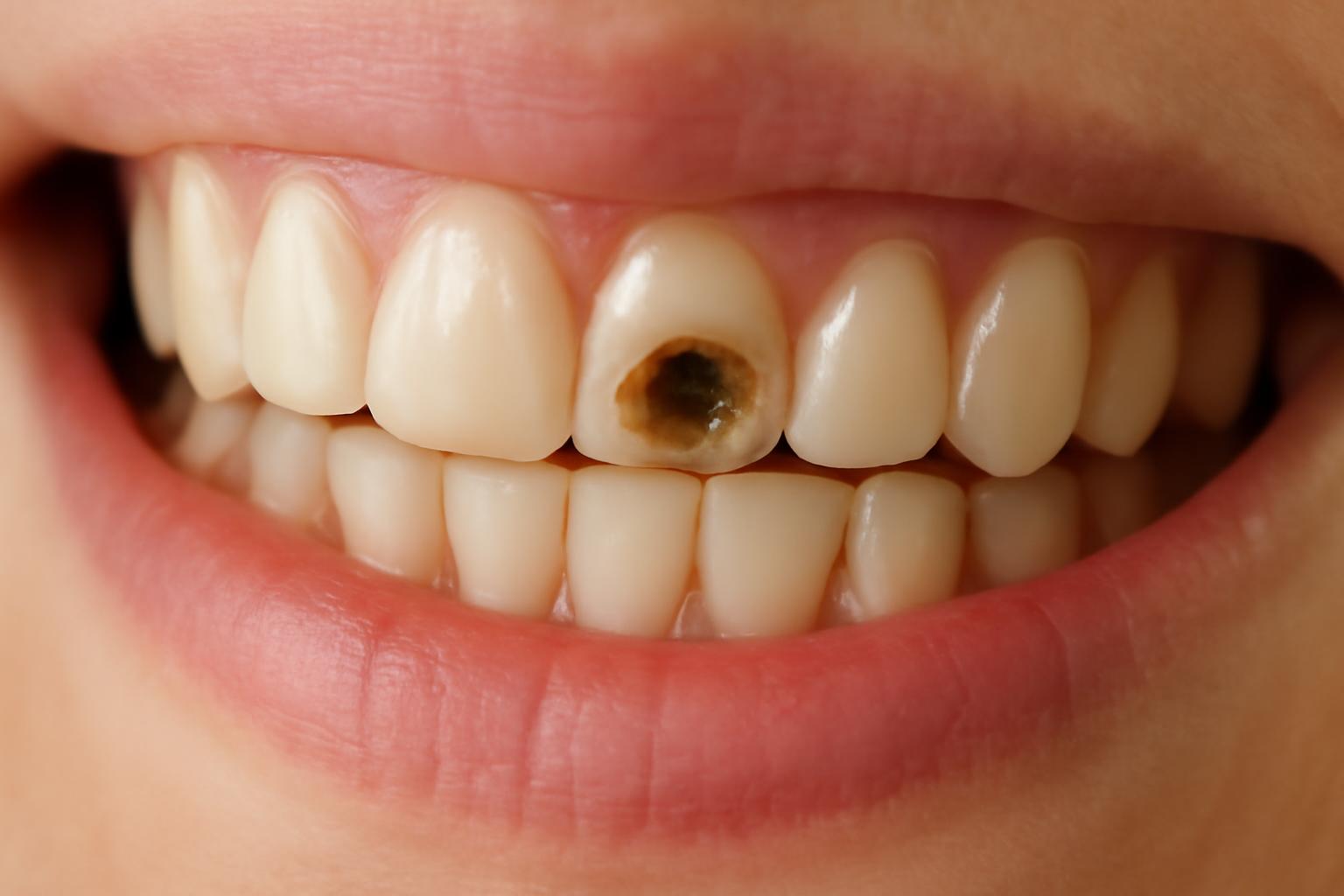
The short answer: months to years, not a predictable clock
A cavity may develop over months, while many develop over years. This is a broad orientation from a medically reviewed consumer-health source—not a minimum, average, maximum, or prediction for an individual tooth. Healthline’s medically reviewed overview describes this months-to-years range and emphasizes that timing varies.
Authoritative public-health sources describe tooth decay as a process that unfolds over time, but they do not provide a universal formation time. The CDC explains cavity formation as an imbalance between acid damage and enamel repair rather than a fixed countdown. It does not specify a standard number of days, months, or years in which a cavity must form. The CDC explains the balance between enamel damage and repair.
Quick answer
- A permanent cavity does not ordinarily form overnight.
- It may form over months under unfavorable conditions, while many cavities take years.
- That range is an approximate secondary estimate, not a government-derived average or a prediction for a particular tooth.
- Early mineral loss is not necessarily a permanent hole and may still be repairable.
- A persistent tooth change, suspected hole, or new symptom should be assessed rather than monitored according to a general timeline.
Published estimates extending from weeks to years mostly come from dental-practice articles. Some of those pages also assign durations to individual stages, but the estimates are inconsistent and generally are not supported there by cited clinical studies. They should not be treated as established rules or used as permission to postpone an examination.
Part of the confusion comes from the word cavity. In a narrower clinical sense, it refers to a hole or permanently damaged area in a tooth. Cleveland Clinic distinguishes early demineralization from a formed hole and describes cavities as holes or areas of decay on tooth surfaces. Its medically reviewed cavity guide explains that distinction.
That difference changes the answer. Acid activity can begin after exposure to food or drink, and microscopic mineral loss can occur before anything is visible. Neither event means that a permanent cavity has already formed. The important transition is from an intact surface that may still be remineralized to irreversible loss of tooth structure.
“Not overnight” should not be mistaken for “safe to ignore.” Decay can be painless, hidden between teeth, or progressing on a vulnerable surface. A general timeline cannot reveal how deep or advanced a particular lesion is.
Three different clocks: acid activity, mineral loss, and a permanent cavity
When people ask how long it takes for a cavity to form, they often combine three different events:
- Bacteria produce acid after exposure to food or drink.
- Enamel loses minerals during repeated acid challenges.
- Enough tooth structure is destroyed to create permanent cavitation.
These events are related, but they are not interchangeable.
Bacteria in dental plaque use sugars and starches from food and drinks. They produce acids that remove minerals from enamel. This activity can begin well before a person has a visible hole, pain, or anything that would ordinarily be called a completed cavity.
Teeth also do not move continuously toward destruction after every meal. They undergo repeated periods of mineral loss and repair. Saliva helps clear acid and supplies minerals, while fluoride can reduce mineral loss and help replace minerals during early damage. The National Institute of Dental and Craniofacial Research describes this as a continuing “tug-of-war” throughout the day. Its tooth-decay guide explains demineralization, remineralization, and the role of fluoride.
A simple balance sheet helps illustrate the process:
| Factors shifting the balance toward damage | Factors shifting the balance toward repair |
|---|---|
| Repeated sugary or starchy exposures | Time between exposures |
| Acid-producing plaque retained on teeth | Effective plaque removal |
| Reduced saliva protection | Adequate saliva flow |
| Hard-to-clean grooves or exposed roots | Fluoride exposure |
| Continued mineral loss | Early detection and risk-based care |
One snack or drink can contribute to an acid challenge, but it does not automatically create a hole. The long-term pattern matters: Are damaging episodes frequent and sustained, or does the tooth repeatedly have enough protection and time to recover?
This is why the claim that “a cavity forms within 20 minutes of eating” is misleading. A dental-practice article may use a short post-meal estimate for the beginning of acid-related mineral loss, but that is not the same event as cavitation. An intact tooth does not ordinarily become a formed hole within those minutes.
Repeatedly sipping a sugary drink through the day can create a different exposure pattern from consuming it once with a meal. This does not establish a universally safe number of snacks or drinks; it means recurring challenges can leave less opportunity for saliva and fluoride-supported repair.
The practical answer therefore involves three clocks:
- Acid activity: may begin after bacteria receive fermentable carbohydrates.
- Mineral loss: develops through recurring episodes and may remain microscopic or appear as a surface change.
- Permanent cavity formation: occurs when continued damage destroys tooth structure beyond what remineralization can rebuild.
Only the third represents irreversible structural loss. Confusing it with the first makes cavity formation sound much more immediate than the evidence supports.
From a white spot to deeper decay: what changes at each stage
A stage-based framework is more useful than assigning fixed dates. It shows what has changed within the tooth and why treatment possibilities change, without pretending that every stage lasts a predictable amount of time.
1. Demineralization
The earliest stage is mineral loss from enamel. An affected area may look white, opaque, or chalky while the outer surface remains intact. Under more favorable conditions, minerals may return to the enamel and the lesion may stop progressing or partially reverse.
A new or persistent change deserves professional assessment, but it should not automatically be labeled a cavity.
2. Enamel breakdown
If mineral loss continues, enamel becomes more porous and weak. Eventually, the surface may collapse or tooth structure may be lost. This is the key transition from potentially reversible mineral loss to permanent damage.
Fluoride can support mineral replacement while an early lesion remains intact, but it cannot recreate missing enamel in the original shape of the tooth. NIDCR describes a formed cavity as permanent damage that must be repaired by a dentist. Its guide distinguishes reversible early decay from permanent cavitation.
3. Dentin involvement
Dentin lies beneath enamel. It is softer and less resistant to acid, so decay may advance faster after reaching this layer. Sensitivity may become more likely, although some people still have few or no noticeable symptoms.
Dentin involvement also changes management. Plaque control, fluoride, and dietary changes remain important for reducing further damage, but decay that has extended into dentin generally requires restorative care rather than toothpaste or brushing alone. Cleveland Clinic notes both the faster progression in dentin and the usual need to restore a cavity once a hole has formed. Its cavity guide describes progression and treatment across the tooth’s layers.
4. Pulp involvement
The pulp is the inner part of the tooth containing nerves, blood vessels, and connective tissue. Deeper decay can irritate or infect it.
Treatment may involve root-canal treatment or, when a tooth cannot be restored, extraction. There is no dependable timetable for how long decay takes to travel from enamel to the pulp.
5. Abscess
An abscess is a later infectious complication that can occur when decay reaches the pulp and infection extends beyond it. Significant pain, swelling, or a suspected abscess warrants prompt professional assessment rather than observation based on a general cavity timeline.
These stages form a sequence, not a schedule. A lesion may remain stable, stop progressing, or continue deeper. Its course depends on surface integrity, location, plaque retention, exposure pattern, saliva, fluoride, and whether the conditions driving decay change.
For a more detailed visual and conceptual walk-through, see How Cavities Form Stages: 5 Progressive Tooth Decay Stages Explained.
Why one cavity may progress faster than another
No risk factor adds or subtracts a fixed number of weeks. Each one changes the balance between damage and repair.
Frequent sugary or starchy exposure
Plaque bacteria can use fermentable carbohydrates, including sugars and starches, to produce acid. Frequent snacks, repeated sips, and prolonged exposure can create recurring acid challenges before a tooth has had much opportunity to recover.
This does not justify a universal numerical limit for snacks or drinks. The relevant pattern includes what is consumed, how often teeth are exposed, how much plaque remains on the surface, and which protective factors are present.
Plaque removal
Brushing and cleaning between teeth disrupt plaque and reduce the amount retained on tooth surfaces. Technique and access matter: someone may brush routinely while grooves, contact areas, appliance-adjacent surfaces, or the gumline remain difficult to clean.
Plaque removal is preventive, but it should not be confused with repairing a formed cavity. Better cleaning can reduce continued damage and help manage an intact early lesion; it cannot rebuild a missing section of tooth.
Fluoride exposure
Fluoride shifts the balance toward repair by reducing mineral loss and helping replace minerals during the earliest stage. Fluoride toothpaste therefore remains important even for people who already brush consistently.
Its ability to reverse damage depends on the condition of the surface. Fluoride may support an intact early lesion and help protect nearby surfaces, but it does not act as a structural filling once a hole exists.
Saliva and dry mouth
Saliva clears acid and supplies minerals involved in enamel repair. Reduced saliva protection can therefore increase cavity risk. The CDC identifies reduced saliva, exposed roots, braces or other oral appliances, and difficulty brushing among factors associated with a higher chance of cavities. The CDC’s risk-factor guidance summarizes these conditions.
Persistent dry mouth may create a less favorable repair environment even when a person’s diet has not changed.
Tooth location and anatomy
Decay does not behave identically on every surface:
- Pits and fissures on chewing surfaces can retain plaque in narrow grooves, allowing decay to progress more quickly.
- Areas between teeth can be difficult to see and clean directly.
The surface involved influences both risk and management. A general estimate cannot account for all these anatomical differences.
Braces, appliances, and cleaning barriers
Physical, visual, or cognitive difficulties may also make effective brushing harder. These circumstances do not establish a timeline, but they can shift the balance toward damage when plaque remains around vulnerable surfaces.
Previous decay and surface condition
The condition and location of the affected surface matter as much as general habits.
Practical higher-risk checklist
A tooth may warrant closer attention when one or more of the following applies:
- Frequent sugary or starchy snacking or sipping
- Inadequate fluoride exposure
- Reduced saliva or persistent dry mouth
- Deep grooves, crowded contacts, or other hard-to-clean surfaces
- Exposed tooth roots
- Braces or other oral appliances
- Difficulty brushing or cleaning between teeth
- A personal history of cavities
This is a risk checklist, not a speed calculator. It cannot show whether a mark is decay, how deep a lesion is, or how long it has been present. Claims that genetics, tartar, “weak enamel,” or a particular bacterial profile alter the timeline by a known amount go beyond the supplied evidence.
Do tooth type, cavity location, and age change the pace?
Decay does not progress identically on every tooth or surface. Location affects plaque retention, access to saliva and fluoride, ease of cleaning, and the type of tissue exposed.
A smooth-surface lesion develops on a relatively flat side of a tooth. It may progress more slowly, particularly when the surface is accessible to cleaning and protective factors.
A pit-and-fissure lesion develops in the grooves of a chewing surface. Narrow anatomy can shelter plaque and make complete cleaning more difficult.
A root-surface lesion occurs on an exposed root. Root surfaces do not have the same enamel covering as tooth crowns, so their vulnerability and treatment considerations differ.
Decay can also develop around or beneath an existing restoration. A filling repairs lost structure but does not make the tooth immune to future decay. If plaque and repeated acid-producing conditions continue around a restoration margin, another lesion may occur. There is no standard recurrence timetable.
Age alone does not determine speed. Children, adults, and older people can all develop cavities, but their mix of tooth structure, saliva, exposure, cleaning access, fluoride, and lesion location may differ.
Baby teeth have thinner enamel than permanent teeth, making faster progression biologically plausible once decay is active. However, the available guidance does not establish a dependable child-versus-adult timeline. A dental-practice article claims cavities may form within a few months in young children and take up to a year in adults, but it does not cite studies supporting those limits. That practice article provides the enamel comparison and age estimates as generalizations.
Those figures should not be used to decide how long to watch a child’s tooth. A child with a persistent suspicious spot, visible breakdown, pain, swelling, or difficulty eating needs assessment based on the current condition—not an assumed number of months.
The same caution applies to adults. An older person with exposed roots and reduced saliva may have a different risk profile from a younger adult with intact enamel and normal saliva flow. Conversely, being young does not mean every lesion will progress rapidly. Tissue, location, exposure, and protection are more informative than age by itself.
Why a cavity can grow without hurting
Early decay may cause no pain, sensitivity, or obvious hole. A person can have active mineral loss—or even a formed cavity—without knowing it.
The absence of pain does not prove that a tooth is healthy or reveal how long a lesion has been present. Pain and sensitivity may become more likely as decay enlarges or approaches the pulp, but symptoms cannot reliably determine depth.
Possible signs include:
- A persistent white, brown, or black area
- Sensitivity to sweets, cold, or heat
- Pain when biting
- A rough area, pit, or visible hole
- Toothache or lingering pain
- Swelling or tenderness near a tooth
These findings are reasons to seek assessment, not diagnostic proof. Mayo Clinic notes that beginning cavities may have no symptoms and lists toothache, sensitivity, visible pits, staining, and pain when biting among possible later signs. Its patient guide explains the range of possible cavity symptoms.
A dental examination can assess:
- Whether the surface is intact or has broken down
- Which tooth and surface are involved
- Whether plaque is retained in the area
- The apparent extent of damage
- Whether a restoration is already present
- Whether previous records show a change
When clinically appropriate, dental radiographs can reveal decay between teeth or below a surface that does not show an obvious hole. Imaging is not required on one universal schedule; its timing should reflect individual risk, symptoms, examination findings, age, and professional judgment.
When early decay may be stopped—and when a filling is usually needed
“Reversing a cavity” can mean two different things, and separating them prevents false expectations.
The first meaning is remineralizing early damage while the enamel surface remains intact. Saliva and fluoride can help replace minerals, while improved plaque removal and less frequent sugary or starchy exposure can reduce continuing acid challenges. Under favorable conditions, an early lesion may stop progressing or partially reverse.
The second meaning would be regrowing tooth structure after a hole has formed. Routine brushing, toothpaste, saliva, and fluoride cannot do that.
Not every early lesion behaves the same way. The outcome depends on surface integrity, depth, location, plaque control, fluoride exposure, saliva, and whether the exposure pattern changes. A person looking in a mirror cannot reliably determine all those features.
Once decay has permanently destroyed tooth structure, restoration is commonly required after professional assessment. A limited cavity may be treated with a filling. More extensive structural loss may require a larger restoration or crown. Pulp involvement may lead to consideration of root-canal treatment, while some teeth cannot be restored.
Confirmed decay extending into dentin generally crosses the threshold from remineralization alone to restorative care. Dentin is softer and less resistant to acid than enamel, and missing tooth structure cannot be replaced by improved brushing.
Consumer guidance may appear to disagree about whether every “small cavity” needs an immediate filling. Part of the disagreement is terminology: one source may call an intact early lesion a small cavity, while another reserves “cavity” for a formed hole. Management also depends on surface integrity, depth, location, evidence of progression, and whether the area can be monitored reliably.
The useful rules are:
- Intact early mineral loss may sometimes be arrested or reversed.
- Not every early lesion will reverse.
- Missing tooth structure does not regrow.
- Dentin involvement commonly requires restoration.
- Management depends on professional assessment of the surface, depth, location, and change over time.
The earlier the process is identified, the more opportunity there may be to control it with preventive measures and limited treatment. Here, “early” refers to the lesion’s condition—not necessarily to how recently someone noticed it.
What to do if you think a cavity is forming
Do not try to date a possible cavity from its color, symptoms, or the date of your last checkup. Use the change to decide what kind of assessment is appropriate.
Arrange a dental examination
A routine or reasonably prompt appointment is appropriate for:
- An unexplained white, brown, or black area that persists
- New sensitivity to sweets, heat, or cold
- A rough area or suspected pit
- Food repeatedly catching in the same place
- A suspected hole
- A change around a filling, crown, or other restoration
How quickly an appointment is needed depends on the persistence and severity of the finding. A painless spot may still warrant assessment even if it does not require the same response as worsening pain or swelling.
Seek prompt care for significant symptoms
Do not wait for a predicted cavity timeline if you have toothache, worsening or persistent pain, swelling, or signs of a possible abscess. These concerns warrant professional assessment rather than home monitoring. Mayo Clinic advises seeing a dentist as soon as possible for toothache or mouth pain. Its cavity guidance also explains that untreated decay can affect deeper tooth layers.
Improve the conditions around the tooth
While arranging care, general preventive measures include:
- Brush thoroughly with fluoride toothpaste.
- Clean between teeth using an appropriate method.
- Reduce repeated sugary or starchy snacks and drinks.
- Avoid prolonged sipping or grazing when possible.
- Drink water and maintain hydration.
- Clean carefully around braces, retainers, or other appliances.
- Discuss persistent dry mouth with an appropriate health professional.
These steps may reduce further acid challenges and support repair of intact early damage. They do not confirm whether a cavity is present, and they cannot replace treatment for missing tooth structure.
Use an individualized follow-up schedule
A cavity can develop or progress between routine dental visits, particularly when risk factors are present. A recent checkup does not guarantee that every surface will remain unchanged until the next scheduled appointment.
There is no need to impose one examination or radiograph interval on everyone. Follow-up should reflect personal cavity history, current findings, symptoms, exposed roots, saliva protection, appliances, cleaning difficulty, age, and professional judgment. Imaging should be used when clinically appropriate rather than automatically according to a universal calendar.
Frequently asked questions
Can a cavity form overnight?
A permanent cavity does not ordinarily form overnight. Tooth decay develops through repeated acid-driven mineral loss and, if damage continues to outpace repair, eventual breakdown of tooth structure.
A person may suddenly notice pain, a rough edge, or a visible hole, but that does not establish that the underlying decay began that day. The appropriate response is to assess the tooth’s current condition rather than calculate when the process started.
Can a cavity form between dental checkups?
Yes. A cavity can develop, or an existing early lesion can progress, between checkups—especially when repeated acid exposure, reduced saliva, inadequate fluoride, plaque-retentive anatomy, exposed roots, or cleaning difficulties are present.
A previous normal examination is useful information, but it is not a guarantee against later change. If you notice a persistent mark, sensitivity, pain, or a suspected hole, contact a dental professional rather than automatically waiting for the next routine visit.
How long does it take for a cavity to reach the nerve?
There is no dependable timetable. The rate depends on where the lesion started, the surface involved, whether it has reached dentin, the exposure pattern, saliva, fluoride, plaque removal, and whether progression continues.
Decay may advance faster after reaching dentin because dentin is softer and less acid-resistant than enamel. That fact cannot be converted into a reliable number of weeks or months for reaching the pulp.
A deep cavity may produce few symptoms, while another dental problem may be painful. Persistent or worsening pain should be assessed promptly.
Can a dentist tell how long a cavity has been there?
Usually not with precision. A dentist can evaluate its location, whether the surface has broken down, its apparent extent, and whether it has changed compared with earlier examinations or radiographs.
Do cavities form faster in baby teeth?
They may progress faster in some circumstances because baby teeth have thinner enamel than permanent teeth. That makes faster movement through the outer layer plausible, but it does not establish a universal pediatric timeline.
A child’s actual risk also depends on exposure frequency, plaque removal, fluoride, saliva, tooth anatomy, and lesion location. Do not use age-based estimates to monitor a suspicious baby tooth at home. Persistent discoloration, visible breakdown, pain, swelling, or difficulty eating warrants assessment based on the child’s current condition.
The bottom line
The useful answer is not a countdown. Cavities develop when repeated mineral loss outpaces repair. Permanent damage may emerge over months, while many cavities take years, but no universal timeline can predict what will happen to a particular tooth.
Early demineralization may still be stopped or partially reversed while the surface remains intact. Once tooth structure has been permanently lost, brushing and fluoride cannot regrow it, and professional assessment is needed. Use risk factors and symptoms as reasons to seek care—not as tools for dating or diagnosing a cavity.
Decay Guide is an informational publisher, not a dental practice. Its content is written by a health writer, has not been reviewed by a clinician, and does not replace an examination, diagnosis, or individualized professional advice. Decay Guide’s terms explain its authorship and clinical-review limitations.