
Why Back Teeth Develop Cavities—and What the Stage of Decay Changes
Early mineral loss in intact enamel may remineralize; once a hole forms, missing structure cannot return. Pain or color cannot reliably distinguish the two.
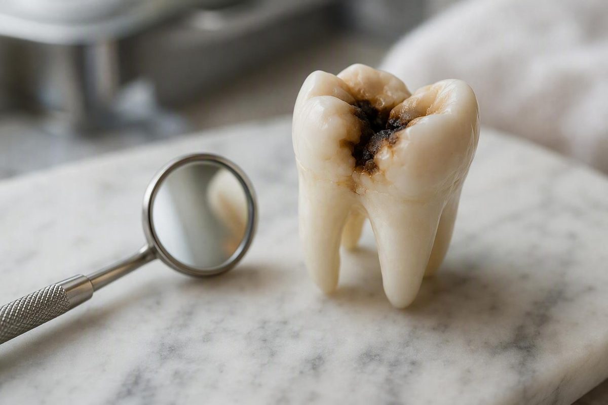
Molar tooth decay is shaped by the anatomy of the back teeth, cleaning access, bacterial plaque, repeated exposure to sugars and starches, fluoride, and saliva. Molars can develop cavities even when someone brushes regularly because narrow pits and fissures may retain plaque and food beyond the reach of toothbrush bristles. Decay is not proof that someone simply failed to brush.
The most important distinction is the stage of damage. Early mineral loss in enamel, before the surface has broken, may sometimes be arrested or remineralized. Once decay has produced an opening or hole, however, the missing tooth structure cannot be brushed or remineralized back into place. An established cavity generally needs professional repair.
Appearance and symptoms cannot reliably establish that stage. A decayed molar may be painless, a dark groove may be stained rather than decayed, and damage between teeth may not be visible in a mirror. A dentist must determine whether decay is present, how far it extends, and what treatment—if any—the tooth requires.
What molar tooth decay is—and why back teeth are vulnerable
Tooth decay is a process in which acids produced by bacteria in dental plaque remove minerals from enamel, the tooth’s hard outer covering. If mineral loss continues, the surface can break down. A cavity is permanent structural damage that has developed into an opening or hole.
Molars are built for grinding food. Their broad chewing surfaces are not uniformly flat: they contain natural depressions called pits and branching grooves called fissures. These features make chewing efficient, but they can also retain plaque and food. Some fissures are so narrow or deep that toothbrush bristles cannot clean their full depth, even when the visible chewing surface is brushed carefully. The National Institute of Dental and Craniofacial Research explains how molar grooves can trap food and bacteria and contribute to the tooth-decay process.
That does not make brushing ineffective. Careful brushing disrupts plaque on accessible surfaces and reduces risk. It does mean that diligent brushing cannot guarantee that every anatomical recess has been reached. Access can be particularly difficult around a partly erupted tooth or at the back of the dental arch.
Not every molar cavity begins in a visible groove. Decay can occur in several locations:
- Pit-and-fissure decay develops on the chewing surface, where plaque collects in natural pits and grooves.
- Smooth-surface decay develops on the smoother sides of a tooth.
- Between-tooth decay develops where adjacent teeth meet, an area toothbrush bristles do not clean directly.
- Root-surface decay affects an exposed root rather than the enamel-covered crown.
Premolars also have pits and fissures and can develop chewing-surface decay. Molars remain a particular concern because their larger, more complex surfaces sit farther back in the mouth and can be difficult to see and clean.
Cavity risk ultimately reflects a balance. Acid-producing plaque and frequent exposure to fermentable carbohydrates push the balance toward mineral loss. Saliva, fluoride, effective cleaning, and time between dietary exposures support protection and mineral replacement. Anatomy is one factor within that larger process—not a guarantee that a molar will decay.
How decay progresses from mineral loss to a cavity
Plaque is a bacterial film that develops on teeth. When bacteria within it use sugars and starches from food and drinks, they produce acids. Those acids remove minerals from enamel. Saliva helps neutralize acids and supplies calcium and phosphate that can replace some of the minerals lost from an intact surface.
This creates a repeated cycle of mineral loss and replacement. If protective conditions are strong enough, the surface can remain intact. If acid attacks happen frequently and mineral loss repeatedly exceeds replacement, the enamel becomes progressively weaker.
Fluoride changes that balance in several helpful ways. It reduces mineral loss, supports mineral replacement in early damaged areas, and makes enamel more resistant to later acid exposure. It does not regenerate tooth structure after a physical hole has formed.
A simplified progression looks like this:
- Mineral loss or a white-spot change. Enamel loses minerals, but its surface may still be intact.
- Enamel breakdown. Continuing damage causes the surface to collapse, producing a cavity.
- Dentin involvement. Decay passes through enamel into the softer tissue beneath it.
- Pulp damage. Decay reaches or irritates the central tissue containing nerves and blood vessels.
- Possible abscess. Infection may develop around the tooth or its root.
The boundary between the first two stages changes management. A non-cavitated enamel lesion may sometimes be arrested or remineralized through improved plaque control, less frequent sugar exposure, fluoride-based measures, and professional monitoring. A formed hole represents missing structure and generally requires a restoration. That is why “early decay can be reversed” does not mean every cavity can heal without a filling.
Once decay reaches dentin, it may progress more readily because dentin is softer than enamel. There is no reliable universal timetable, however.
If decay approaches or enters the pulp, inflammation or infection may follow. Pain becomes more likely at deeper stages, but it is not inevitable and does not provide a precise measure of depth. Cleveland Clinic distinguishes early demineralization from an established cavity and describes progression through enamel, dentin, pulp damage, and possible abscess as separate stages of tooth decay.
Possible signs—and why a painless molar may still be decayed
Molar tooth decay can produce symptoms, but no single symptom confirms a cavity. Possible signs include:
- Toothache or an unexplained ache around a back tooth
- Sensitivity to sweet, hot, or cold foods and drinks
- Pain when biting or chewing
- A visible pit or opening
- A white, brown, grey, or black change on the tooth
- Persistent bad breath or an unpleasant taste
- Redness or swelling near the tooth
- Facial swelling if an infection has developed
Several of these findings have other possible explanations. Biting pain may be associated with decay, but it can also occur with other dental problems. Bad breath and an unpleasant taste are nonspecific.
Early decay may produce no discomfort at all. A lesion can also be concealed within a deep fissure, between neighboring molars, or beneath a surface that appears intact. The absence of pain or a visible hole therefore does not rule out decay. Mayo Clinic notes that beginning cavities may be symptom-free, while larger lesions can cause sensitivity, toothache, visible pits, discoloration, or biting pain as decay progresses.
Conversely, severe discomfort does not prove that a large cavity is present. A painful tooth may have a crack or another problem without an obvious cavity, while an extensively decayed tooth may remain relatively quiet.
What does a black line in a molar groove mean?
A black or brown line can be a surface stain, the appearance of a deep fissure, or tooth decay. Color alone cannot distinguish among those possibilities. A mark that has been present for a long time should not automatically be assumed harmless, while a newly noticed line should not automatically be labeled a cavity. Stains and cavities can look similar, particularly during early development, so professional assessment is needed to distinguish discoloration from decay.
Do not press, scrape, or probe a dark groove with a pin or another object. Do not deliberately bite hard food to see whether the tooth hurts. These informal tests cannot determine whether the surface is cavitated, how deep a lesion extends, or what treatment may be needed. Note the change and arrange a dental assessment instead.
How a dentist checks a suspected molar cavity
Diagnosing suspected molar decay usually requires more than looking at a photograph. A dentist combines relevant history, reported symptoms, and a clinical examination. The assessment considers where the mark or discomfort is located, when it began, whether symptoms are triggered or spontaneous, and what can be identified during the oral examination.
Dental X-rays may be used when appropriate. They can help identify or assess decay that is not readily visible, including a lesion between adjacent teeth or damage whose extent cannot be judged from the exposed surface. This does not mean every person with a dark groove needs the same type of X-ray. The decision depends on the individual examination and the clinical question.
Before recommending treatment, the dentist needs to determine:
- Whether the enamel surface remains intact or has cavitated
- Where the lesion is located
- How far the decay appears to extend
- How much sound tooth structure remains
- Whether the pulp may be involved
- Whether the tooth can support a restoration
- Whether the tooth is restorable overall
- Whether another dental problem better explains the symptoms
Routine assessment can identify some lesions before they become painful or visible to the person. This is particularly relevant for molars because a reader’s view of a back tooth is limited and a small visible opening may not show the full extent of underlying damage. Examination and, when appropriate, an X-ray are recognized methods of identifying tooth decay according to NHS inform.
No remote description can supply all of those findings.
Treatment by stage: fluoride, filling, crown, root canal, or extraction
Treatment is selected according to the lesion’s stage and location, the amount of healthy tooth remaining, pulp health, restorability, symptoms, and individual clinical circumstances. Symptoms matter, but they are only one part of the decision. A painless tooth can need restoration, while a sensitive tooth may not require the treatment a reader expects.
The table describes typical management, not a way to select treatment at home.
| Lesion stage | Possible findings | Typical management |
|---|---|---|
| Early, non-cavitated enamel mineral loss | An intact surface with an early mineral change; there may be no symptoms | Improved plaque control, less frequent sugar exposure, fluoride-based measures, and professional monitoring may be considered |
| Established cavity in enamel or dentin | A formed opening or structural breakdown; sensitivity may be present or absent | Decayed tissue is commonly removed and the resulting space restored with a filling |
| Extensive structural damage | A large area of weakened or missing tooth, with insufficient support for a smaller restoration | A crown may be considered when the remaining tooth cannot reliably support a filling |
| Pulp involvement in a restorable tooth | Deep decay with possible persistent pain, inflammation, infection, or an abscess | Root canal treatment may remove diseased pulp and preserve the tooth, followed by an appropriate restoration |
| Unrestorable tooth | Damage or infection is so extensive that the tooth cannot be predictably repaired | Extraction may be necessary; replacement options can be discussed separately |
These stages reflect the general progression from preventive fluoride-based management to filling, crown, root canal treatment, or extraction as damage becomes more advanced. The necessary treatment can be chosen only after an examination of the individual tooth.
Early non-cavitated mineral loss
When the enamel surface remains intact, management may focus on changing the conditions that allowed mineral loss. This can include more effective plaque control, reducing the frequency of sugary or starchy snacks and drinks, using fluoride, and monitoring the site. A dentist may recommend an additional fluoride measure or another preventive intervention based on individual risk.
Monitoring is not the same as assuming the lesion is harmless. It allows the dental team to assess whether the area remains stable or progresses. If the surface is already broken, however, prevention alone cannot replace the missing structure.
Filling
A formed cavity commonly requires removal of decayed tissue and placement of a restoration, often called a filling. The restoration occupies the resulting space and helps return the tooth to a usable form.
There is no universal cavity size at which every tooth receives the same filling.
Crown
A crown may be considered when substantial tooth structure has been weakened or lost and the remainder cannot reliably support a smaller restoration. It covers and protects more of the tooth than a filling.
“Large cavity” is not a sufficient home diagnosis for a crown. A dentist must evaluate how much healthy structure remains, where it remains, and whether the tooth can be restored. There is no single filling-versus-crown threshold that applies to every molar.
Root canal treatment
If decay has affected the pulp and the tooth remains restorable, root canal treatment may be used to remove diseased pulp tissue and preserve the tooth. The tooth then needs an appropriate restoration; depending on its structural condition, that may include a crown.
Pulp involvement is not established by pain intensity alone. Clinical findings and imaging may be needed to determine the condition of the tooth and whether root canal treatment is appropriate.
Extraction
Extraction may be necessary if a molar is too severely damaged to restore. That decision is based on restorability, not simply discoloration, pain, or the fact that the tooth has a cavity. If a tooth is removed, possible replacement choices can be discussed separately with a dentist.
Cleveland Clinic similarly describes treatment progressing from fluoride for early decay to fillings, root canal treatment, or extraction according to the extent of the damage.
What can happen when deep molar decay is untreated
Untreated decay can enlarge from enamel into dentin and eventually reach the pulp. As that happens, a person may develop increasing sensitivity, toothache, pain when chewing, or spontaneous pain. The tooth may also remain relatively quiet during part of this progression.
Deep decay can weaken the walls and chewing surface of a molar. A weakened tooth may break, lose part of a cusp, or become difficult to chew on. Structural loss can also reduce the amount of sound tooth available to support a restoration.
Pulp involvement can lead to infection and an abscess. Swelling may develop around the tooth or in the face, and infection can extend into the surrounding bone. These are possible outcomes, not the inevitable fate of every untreated cavity. The likelihood and timing cannot be predicted from a mark’s color or from symptoms alone.
Waiting may also narrow the range of conservative options. An intact early lesion may be managed preventively, whereas a formed cavity requires repair. A smaller restorable lesion may need a filling, while greater structural or pulp damage may require more extensive treatment. Earlier assessment provides an opportunity to identify the problem before more tooth structure is lost.
That is a preventive principle, not a prognosis for a particular tooth. Prompt evaluation cannot guarantee that a molar is salvageable, and an article cannot determine whether an individual tooth needs a filling, crown, root canal treatment, or extraction. Only an examination can establish its condition.
Risk factors beyond brushing: diet frequency, fluoride, saliva, and restorations
Cavity risk does not arise from a single behavior. It reflects the interaction among plaque, dietary exposure, tooth anatomy, fluoride, saliva, cleaning access, existing dental work, and individual health circumstances. Someone can brush routinely and still have factors that keep the balance tilted toward mineral loss.
Frequent sugar and starch exposure
Plaque bacteria can use sugars and starches to produce acid. Frequency matters because each snack, sweetened drink, or prolonged period of sipping can create another acid challenge. Reducing repeated exposures between meals may therefore be more practical than treating all carbohydrate-containing foods as forbidden.
There is no exact “safe” number of snacks that applies to everyone. Individual risk differs, and food texture, duration of exposure, plaque levels, saliva, fluoride, and overall dietary pattern also matter.
Limited cleaning access
A person may clean most visible surfaces effectively but miss areas between molars or within narrow chewing-surface fissures. Crowded positioning, limited dexterity, a partly erupted tooth, or difficulty reaching the back of the mouth can make plaque removal less complete.
Brushing more aggressively is not necessarily the solution. Deliberate coverage, an appropriately sized brush, interdental cleaning, and professional advice about difficult areas are more useful than excessive force.
Limited fluoride exposure
Fluoride supports enamel resistance and mineral replacement during the non-cavitated stage. Limited exposure removes one protective influence from the decay cycle. Fluoride toothpaste is a basic preventive measure, while any additional fluoride approach should be individualized rather than assumed necessary for everyone.
Dry mouth
Saliva helps clear material from the mouth, neutralize plaque acids, and deliver calcium and phosphate for mineral replacement. Persistently reduced saliva therefore increases concern about decay.
Dry mouth can have several causes and should not automatically be attributed to one medication or illness. A dentist or healthcare professional can consider the underlying circumstances and recommend an individualized prevention plan.
Worn dental work, reflux, and eating disorders
Aging or worn restorations are reasons for individualized dental assessment. Reflux and eating disorders can also affect the oral environment and tooth surfaces. Their presence does not prove what caused a particular molar lesion, and each requires appropriate dental and, where relevant, medical assessment.
Different concerns across age groups
Children’s newly erupted permanent molars have fresh chewing surfaces with potentially vulnerable pits and fissures. Cleaning can be difficult while a tooth is partly erupted or while a child is still developing brushing skills.
Adults continue to face pit-and-fissure and between-tooth decay, but other concerns may become more prominent, including persistent dry mouth, exposed root surfaces, limited dexterity, and restorations that need reassessment. Age itself should not be treated as the direct cause of cavities; associated changes and exposures are what matter.
A practical prevention plan for molars
No preventive routine can guarantee that a molar will remain cavity-free, but several measures can shift the balance toward protection.
1. Brush twice daily with fluoride toothpaste
Brush every tooth surface rather than concentrating only on the front teeth. For molars, deliberately cover:
- The chewing surfaces
- The cheek-facing surfaces
- The tongue-facing surfaces
- The area near the gumline
- The last tooth at the back of each side
Use controlled movements that allow the bristles to contact the irregular surface. Vigorous scrubbing is not a substitute for complete coverage. Deep fissures may remain difficult to penetrate even with good technique, which is one reason a dentist may assess selected teeth for sealants.
2. Clean between the teeth daily
Brushing a molar’s chewing surface does not clean the contact area between that molar and its neighbor. Floss or an appropriate interdental cleaner can remove plaque from these areas. The suitable method may depend on spacing, dexterity, braces, bridges, and other dental work.
Interdental cleaning and chewing-surface brushing address different sites. One does not replace the other.
3. Reduce repeated sugar and starch exposure
Focus on frequency rather than trying to ban every carbohydrate. Repeated snacking or sipping gives plaque bacteria recurring opportunities to produce acid. Grouping suitable foods and drinks into regular eating occasions can reduce the number of separate acid challenges.
What matters is the overall pattern. A dentist or dietitian may provide more individualized guidance when cavity activity is high or dietary needs are complex.
4. Discuss persistent dry mouth
Persistent dry mouth deserves professional discussion because saliva has an important protective role. A dentist or healthcare professional can review possible causes and decide whether tailored preventive measures are appropriate.
5. Use risk-based dental care
There is no single examination interval that is right for everyone. Recall timing should reflect individual need rather than an automatic six-month rule. Someone with active disease may need closer monitoring than someone at low risk.
Regular assessment matters because some molar lesions can be detected before they become painful or obvious. The appropriate schedule should be set with the dental team.
6. Have newly erupted permanent molars assessed
For parents, eruption is a useful time to review brushing access and sealant suitability. First permanent molars generally emerge at about ages 5 to 7, while second permanent molars generally emerge at about ages 11 to 14, according to federal guidance on protecting children’s permanent molars.
Individual assessment can identify deep grooves, incomplete cleaning, or reasons for additional preventive support.
Prevention lowers the risk of new damage and may help arrest early mineral loss. It cannot repair an established hole.
Sealants and deciding when to arrange dental care
A dental sealant is a thin protective coating placed over susceptible pits and fissures on a chewing surface. It creates a physical barrier that helps keep food and bacteria out of grooves that are difficult to clean.
Sealants and fluoride work differently:
- Fluoride helps enamel resist acid and supports remineralization while the surface remains intact.
- A sealant physically covers vulnerable grooves.
They can be complementary rather than competing measures. A person with a sealant still needs fluoride toothpaste, effective cleaning, sensible dietary habits, and appropriate dental monitoring.
Sealants are commonly considered for newly erupted permanent molars in children because these teeth can have deep grooves and may initially be difficult to clean. Selected adults with susceptible grooves may also be considered after an individual assessment. Sealants are not automatically appropriate for every tooth or every person; commercially published oral-health guidance likewise describes their use in children and in adults when a dentist considers them suitable for the tooth.
Most importantly, a sealant is preventive. It does not rebuild an established hole. A filling or another restoration repairs existing structural damage; a sealant is intended to protect an appropriate surface from future damage. Whether an early lesion can be managed preventively, sealed, monitored, or restored depends on clinical findings.
Arrange a dental evaluation if you notice:
- A new or unexplained dark area
- A visible pit or opening
- Persistent sensitivity
- Pain when biting
- A bad taste or localized gum change
- Any other concerning change in a back tooth
A toothache or mouth pain warrants seeing a dentist as soon as possible. Facial swelling, severe or escalating pain, or signs suggesting an abscess call for prompt professional assessment. These findings cannot be diagnosed reliably at home, and waiting for pain to become intense is not a dependable way to judge severity. Mayo Clinic specifically advises seeing a dentist as soon as possible for toothache or mouth pain.
The decision-changing distinction remains straightforward: early, non-cavitated mineral loss may sometimes be arrested, but a formed cavity cannot be brushed or remineralized back into intact tooth structure. Because molar grooves can conceal decay and early lesions may be painless, neither color nor symptoms should be used as a diagnosis.
Consistent fluoride use, careful brushing, daily interdental cleaning, less frequent sugar exposure, appropriate sealant assessment, and risk-based dental care can reduce future risk. A dentist must determine what an existing lesion requires.
Decay Guide provides general reference information. It is not a dental practice and does not diagnose, treat, or provide individualized advice about a particular tooth.
Frequently asked questions
Can early molar tooth decay be reversed without a filling?
Sometimes—but only when the damage is an early, non-cavitated area of mineral loss and the surface remains intact. Plaque control, reduced sugar frequency, fluoride-based measures, and professional monitoring may allow the area to arrest or remineralize.
Once a hole has formed, the missing structure cannot grow back through brushing, fluoride, supplements, or home remedies. An established cavity generally requires professional repair. A dentist must examine the tooth to distinguish potentially reversible mineral loss from permanent structural damage.
Does a black or brown line on a molar always mean there is a cavity?
No. A dark line may be stain, the appearance of a deep fissure, or decay. Color alone cannot distinguish among them, and a photograph cannot reliably show what is happening beneath or between tooth surfaces.
Do not scrape, press, or probe the line, and do not test it by deliberately biting hard food. Arrange an examination if the mark is new, changing, associated with sensitivity or biting pain, or otherwise concerning.
Can a molar have a cavity without hurting?
Yes. Early cavities may cause no symptoms, and some lesions remain painless while hidden in a fissure or between neighboring molars.
The reverse is also important: pain does not prove that a cavity is present. Examination—and sometimes dental imaging—is needed to determine the cause.
When does molar decay need a filling, crown, root canal, or extraction?
A formed cavity commonly needs removal of decayed tissue and a filling. A crown may be considered when substantial tooth structure has been lost and the remainder cannot reliably support a smaller restoration. Root canal treatment may be used when the pulp is diseased but the molar remains restorable. Extraction may be necessary if the tooth cannot be predictably repaired.
The choice depends on lesion depth and location, remaining sound structure, pulp health, symptoms, restorability, existing dental work, and other clinical factors. Pain intensity and appearance alone cannot select the treatment.
Are dental sealants only for children?
No. Sealants are often considered when children’s permanent molars erupt because the new chewing surfaces may have vulnerable grooves. Selected adults with susceptible grooves may also be candidates.
Suitability is individual. A dentist needs to assess whether the surface is appropriate for sealing and whether decay or structural damage is already present. Sealants help prevent decay; they do not restore an established cavity.