
How Tooth Decay Moves From Early Mineral Loss to an Abscess
A five-stage model runs from mineral loss to abscess, but stage counts vary. Only intact early mineral loss may reverse. A formed hole will not regrow.
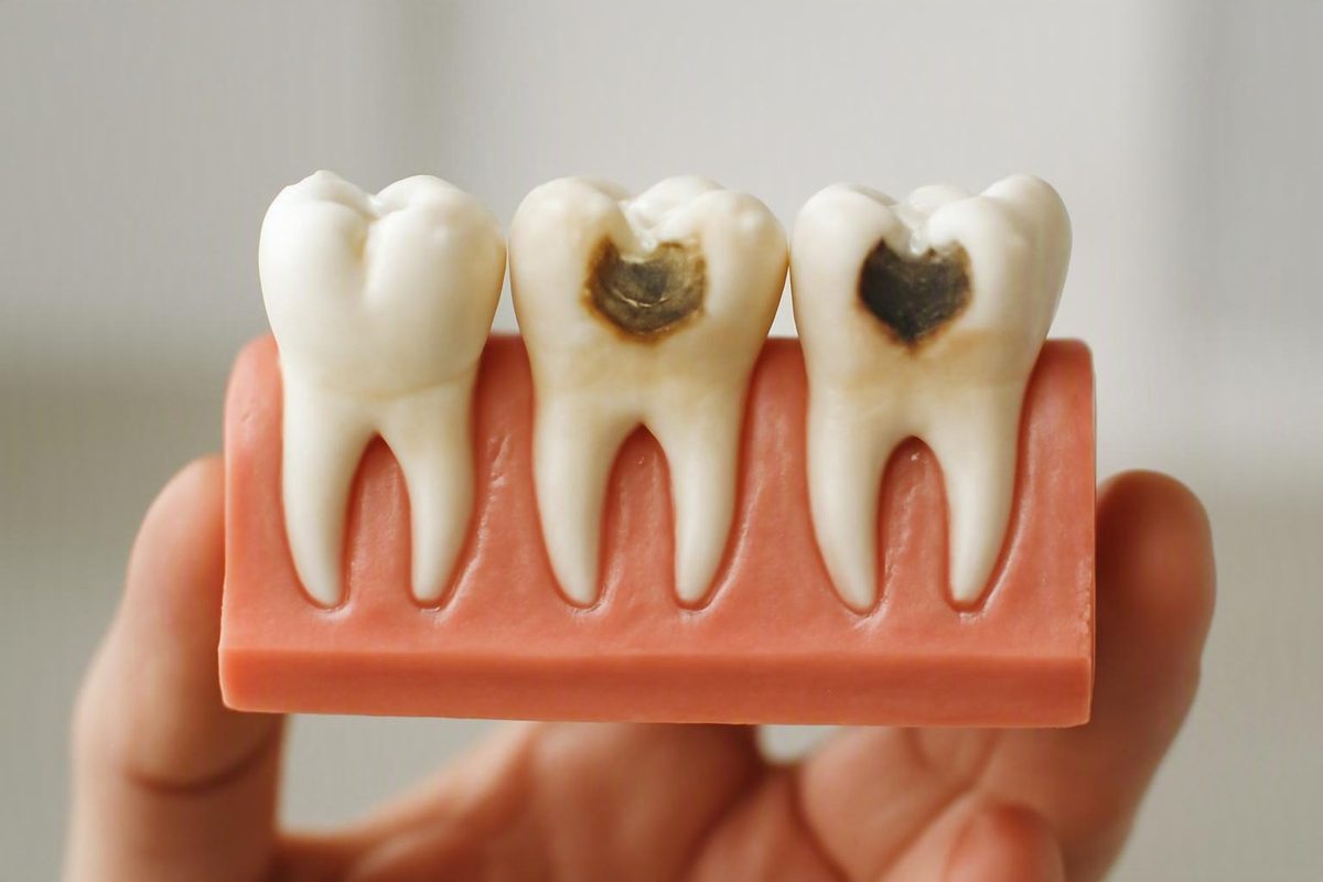
The five-stage model at a glance
A common explanation of the stages of cavities follows decay as it moves from the outside of a tooth toward its center and, in advanced disease, beyond the root:
- Demineralization
- Enamel decay
- Dentin decay
- Pulp involvement or damage
- Abscess
This is an educational model, not a universally standardized diagnostic classification. Tooth decay is continuous, and an individual lesion may not fit neatly into a numbered stage. Cleveland Clinic uses this five-stage sequence while noting that early damage may be painless and that deeper decay generally requires professional treatment in its medically reviewed overview of cavities.
For consumers, the most useful dividing line is often whether the enamel surface remains intact:
- Intact, noncavitated mineral loss may sometimes be stabilized or reversed through remineralization.
- A collapsed surface or formed hole represents permanent structural loss. The missing tooth structure does not naturally grow back.
- Spread into dentin, pulp, or tissues around the root increases the possibility of more extensive treatment and greater urgency.
| Stage | Affected tissue | Possible appearance | Possible symptoms | Reversibility | Typical care | Urgency |
|---|---|---|---|---|---|---|
| 1. Demineralization | Minerals within enamel; surface remains intact | Chalky, dull, or opaque white area; sometimes no visible change | Often none | Potentially reversible or stabilizable while the surface remains intact | Fluoride, plaque control, fewer sugar and starch exposures, saliva support, and professional monitoring (NIDCR) | Arrange a dental review; usually not an emergency |
| 2. Enamel decay | Enamel surface has broken down | Small pit, brown area, roughness, or visible hole | May remain painless | Not structurally reversible after a true hole forms | Commonly a filling; the approach depends on the defect and tooth (Sensodyne) | Dental assessment should not be postponed simply because there is no pain |
| 3. Dentin decay | Softer dentin beneath enamel | A larger or darker defect may appear, although some decay is hidden | Possible sensitivity to cold, sweets, biting, or chewing; sometimes no pain | No | A filling, inlay, onlay, or crown may be considered according to structural loss (Dental Solutions) | Prompt dental assessment |
| 4. Pulp involvement | Innermost tissue containing nerves and blood vessels | A deep cavity or damaged restoration may be present; appearance alone is unreliable | Lingering temperature pain, throbbing, nighttime pain, biting tenderness, redness, or swelling may occur | Lost structure is not reversible; pulp status requires clinical assessment | Possible root canal treatment followed by restoration; extraction if the tooth cannot be treated or restored predictably (Cleveland Clinic) | Prompt assessment, particularly with persistent pain or swelling |
| 5. Abscess | Infection has extended beyond the tooth, often near the root | Gum or facial swelling or pus may be visible; sometimes little is visible externally | Severe or radiating pain, bad taste, fever, swollen neck lymph nodes, or general illness may occur | No | Source control may involve drainage, root canal treatment, or extraction; antibiotics are not a substitute for treating the source (Smile Center Turkey) | Prompt care; facial swelling, fever, swallowing difficulty, or systemic illness increases urgency |
Sources count the stages differently because they place boundaries at different points. Some four-stage systems combine pulp infection and abscess formation in one advanced stage. Other systems divide enamel changes more finely or add tooth loss after extensive destruction. One six-stage guide, for example, counts tooth loss as the final stage. These frameworks describe broadly similar movement through enamel, dentin, pulp, roots, and surrounding tissues rather than fundamentally different diseases (Quarterpath Dental).
Plaque should not be classified as tooth damage itself. It is a sticky bacterial film involved in the process. Bacteria within plaque use sugars and starches and produce acids that promote mineral loss. Plaque can therefore be present before there is detectable tooth-tissue damage.
Progression stages must also be distinguished from cavity types. Pit-and-fissure cavities occur in grooves, smooth-surface cavities occur on broader surfaces, and root cavities occur on exposed root surfaces. These terms describe location, not depth. A lesion in any of these locations may be early, cavitated, or deep.
Editorial note: Decay Guide is written by a health writer, not a dentist, and its articles are not clinician-reviewed. This guide cannot diagnose or stage a particular tooth and does not replace a dental examination. Read more about how Decay Guide is written.
Stage 1: Demineralization before a hole forms
Demineralization is the earliest tissue change in the five-stage model. Oral bacteria use sugars and starches from foods and drinks. Acids produced during that process remove minerals from enamel. Saliva and fluoride act in the opposite direction by helping replace minerals.
This is an ongoing balance rather than a single event. Frequent acid exposure, plaque that remains on the teeth, limited fluoride exposure, or reduced saliva can shift the balance toward continued mineral loss.
Plaque matters because it keeps acid-producing bacteria against tooth surfaces. It is a bacterial film, not a cavity and not proof that permanent damage has already occurred. Removing plaque regularly helps reduce the conditions that allow repeated acid exposure.
One possible early sign is a chalky, dull, or opaque white area. This may reflect mineral loss beneath an enamel surface that has not collapsed. A white mark alone cannot confirm active decay, however, because enamel can look white for other reasons.
Early demineralization often causes no pain and may be difficult to see, particularly between teeth, along the gumline, around restorations, or in the grooves of back teeth. A person can therefore have early decay without noticing a visible or physical change.
What “remineralization” means
Remineralization is the return of minerals to weakened enamel. Saliva supplies minerals, while fluoride can reduce mineral loss and support mineral replacement. The National Institute of Dental and Craniofacial Research explains that early mineral loss may be reversed before a cavity forms, whereas continued loss can produce a permanent cavity requiring repair (NIDCR).
Measures that may help an intact early lesion include:
- Brushing consistently with fluoride toothpaste
- Removing plaque, including from between teeth
- Reducing how often teeth are exposed to sugary or starchy foods and drinks
- Seeking professional advice for persistent dry mouth
- Receiving professional fluoride care when considered appropriate
- Having the area monitored by a dentist
- Using a sealant in selected pits or grooves when appropriate
“Reversible” has a precise boundary. It does not mean every white mark will disappear, that missing enamel can grow back, or that home care can repair an established hole. It means that an early, noncavitated area may regain minerals or stop progressing while its outer surface remains intact.
A dentist may monitor an intact early lesion rather than restore it immediately. That is an individualized clinical decision; a photograph or mirror check cannot determine whether monitoring is appropriate.
Stage 2: Enamel breakdown and a formed cavity
If mineral loss continues, enamel becomes weaker until its surface can collapse, producing a pit or hole. This change is called cavitation. It marks the clearest transition from potentially reversible mineral loss to permanent structural damage.
Possible findings include:
- A small pit or rough area
- Brown discoloration
- A visible hole
- Food repeatedly catching in one place
- A defect next to an existing filling or other restoration
These findings do not establish depth or activity by themselves. A brown area may be stain, active decay, or a lesion that is no longer progressing. A cavity may also be difficult to see if it is between teeth or extends beneath a small surface opening.
An enamel cavity can remain painless. The absence of a toothache therefore does not prove that a visible defect is shallow or harmless.
Once a true hole has formed, the missing tooth structure does not naturally regrow. Fluoride can still protect surrounding enamel and reduce further mineral loss, but it cannot reconstruct a collapsed surface.
A dentist commonly treats an established cavity by controlling or removing the damaged tissue and restoring the defect with a filling. The appropriate approach depends on the lesion’s size and location, its activity, the remaining tooth structure, any existing restoration, and whether a durable repair can be placed.
This is why neither of the following statements is dependable:
- “Every enamel mark needs drilling.”
- “Every enamel lesion can be reversed.”
An intact early lesion may be managed without a filling, while a cavitated lesion represents permanent structural loss. Color alone cannot show which situation applies.
Stage 3: Decay reaches dentin
Dentin is the softer layer beneath enamel. Decay may advance more readily after reaching it, and a lesion that appears small at the surface can extend more broadly underneath the enamel.
Dentin contains microscopic tubules that communicate toward the pulp. This helps explain why sensitivity may become more noticeable as decay deepens.
Possible symptoms include:
- Sensitivity to cold food, drinks, or air
- A response to sweet foods or drinks
- Discomfort when biting
- Pain or sensitivity while chewing
- Food lodging in a defect
- An intermittent ache
Not every dentin cavity hurts. Symptoms vary according to the lesion, tooth, pulp response, and individual perception. Sensitivity can also result from exposed roots, enamel wear, cracks, gum recession, recent dental treatment, or other causes. It cannot establish lesion depth at home.
Treatment is not automatically “a crown” merely because dentin is involved. Depending on the amount and location of structural loss, a dentist may consider:
- A filling
- An inlay
- An onlay
- A crown
The choice depends on the depth and width of the defect, the surfaces involved, the sound structure that remains, the condition of the pulp, existing cracks or restorations, and the forces placed on the tooth.
Prompt assessment matters because dentin offers less resistance to continuing decay than enamel. Further progression may leave less sound structure available for a conservative repair.
Stage 4: Pulp involvement
The pulp is the soft tissue at the center of a tooth. It contains nerves, blood vessels, and connective tissue. When decay approaches or enters this space, bacterial irritation and inflammation may affect tissue confined within rigid surrounding walls.
Possible warning signs include:
- Pain that lingers after hot or cold exposure
- A deep or spontaneous ache
- Throbbing pain
- Pain that disrupts sleep
- Tenderness when biting
- Gum redness near the tooth
- Local swelling
These symptoms are not a home diagnostic checklist. Pain intensity cannot prove that decay has reached the pulp or determine whether irritated pulp tissue can recover. Conversely, a deeply damaged tooth may produce limited pain.
A dentist may combine several kinds of information when assessing suspected pulp involvement:
- The history and pattern of symptoms
- Examination of the tooth and surrounding gum
- Biting or percussion tests
- X-rays
- Pulp sensibility or vitality-related testing
- Assessment of cracks, restorations, and remaining tooth structure
No single response necessarily determines the diagnosis. Examination findings, imaging, symptoms, and pulp-test responses are interpreted together.
When the pulp is considered irreversibly affected, root canal treatment may provide a way to preserve the tooth. Treatment of the inside of the tooth must then be followed by an appropriate restoration to seal and protect the remaining structure.
Extraction may be considered if the tooth cannot be predictably treated, sealed, or restored. It is not inevitable merely because decay has approached or entered the pulp.
Persistent or worsening pain should be evaluated promptly. A reduction in pain does not necessarily mean that the underlying problem has resolved.
Stage 5: Abscess and spreading infection
An abscess can develop when infection extends through the tooth and into tissues near the root, creating a pocket of pus. It represents advanced disease, but not every toothache is an abscess and not every abscess produces the same symptoms.
Possible warning signs include:
- Severe, persistent, or radiating pain
- Swelling of the gum, jaw, or face
- Pus or drainage near the tooth
- A bad or unpleasant taste
- Fever
- Swollen lymph nodes in the neck
- Feeling generally unwell
If an abscess drains, pressure and pain may temporarily decrease. That does not mean the source of infection has been eliminated. Infection may also be present with limited pain.
Facial swelling, fever, difficulty swallowing, worsening pain, or systemic illness associated with a suspected dental infection warrants prompt assessment rather than waiting for a routine appointment. The supplied clinical guide identifies these as warning signs requiring prompt care (Smile Center Turkey).
Advanced dental infection can spread into nearby tissues or other parts of the body and can rarely cause life-threatening complications. This is not an inevitable outcome, but it is why facial swelling, fever, swallowing difficulty, and general illness should be taken seriously.
Treatment aims to control the source. Depending on the tooth and clinical findings, it may involve:
- Drainage
- Root canal treatment
- Extraction if the tooth cannot be retained
- An appropriate restoration if the tooth is preserved
- Antibiotics when a clinician determines that they are needed
Antibiotics do not rebuild a damaged tooth, remove decayed structure, reliably drain a collection of pus, or permanently seal an infected root-canal system. They may form part of treatment, but they do not replace treatment of the infected source.
An abscess does not automatically require extraction. Some teeth can be preserved through drainage and root canal treatment followed by a suitable restoration. Whether that is possible depends on the damage, infection, cracks, supporting tissues, and remaining tooth structure.
Why symptoms and tooth color cannot tell you the stage
The stages of cavities describe biological depth, but the clues available at home are indirect. Early decay is often symptomless, and some established cavities also produce little or no pain. Toothache may suggest deeper irritation, but pain does not map reliably to a stage number.
Color is similarly inconclusive:
- A white area may reflect demineralization, a developmental change, or another enamel difference.
- A brown area may be stain, active decay, or an inactive lesion.
- A black mark in a groove may be staining or decay.
- Decay between teeth may not be visible in a mirror.
Color cannot establish whether a lesion is active. Activity refers to whether mineral loss and breakdown are continuing; depth refers to how far the lesion extends. A dark lesion may be shallow and stable, while a less conspicuous lesion may extend beneath the surface.
Sensitivity is not a dependable staging tool either. Cold or sweet sensitivity may occur with dentin involvement but can also result from exposed roots, worn enamel, cracks, gum recession, or other conditions. A lack of sensitivity does not rule out a cavity.
What a clinical assessment adds
During an examination, a dentist can assess:
- Which tooth surface is affected
- Whether the surface is intact or cavitated
- How much structure appears to have been lost
- The relationship to fillings, crowns, or other restorations
- The condition of surrounding gum and supporting tissues
- Whether the tooth appears restorable
X-rays may reveal decay hidden between teeth or beneath areas that cannot be examined directly. They can also help estimate depth and identify changes around a root. Cleveland Clinic notes that dental examinations and X-rays can identify cavities, including decay that is not yet visible.
If pulp involvement is suspected, pulp testing may contribute further information. Responses to temperature or electrical stimuli can help assess how the pulp is responding, while biting and percussion tests may help identify inflammation around a tooth. These are professional diagnostic procedures, not do-it-yourself tests.
Treatment decisions depend on more than a stage number. Relevant factors include:
- Lesion depth and activity
- Remaining sound tooth structure
- Pulp status
- Location and accessibility
- Existing restorations or cracks
- Supporting gum and bone
- Whether the tooth can be predictably restored
Use the stage model to understand terminology and prepare questions—not to decide from a photograph that a tooth needs a filling, root canal, or extraction.
How to reduce the chance that decay progresses
Prevention aims to shift the balance away from repeated mineral loss and toward remineralization. It can reduce risk but cannot guarantee that every cavity will be prevented.
Use fluoride toothpaste consistently
Brush twice daily with fluoride toothpaste, as recommended in NIDCR’s public-health guidance. Fluoride can reduce mineral loss and support the replacement of minerals in early enamel lesions.
Brushing quality matters as well as frequency. Plaque may remain near the gumline, around crowded teeth, beside restorations, and in the grooves of back teeth. Brush thoroughly without using excessive force.
Clean between teeth
A toothbrush does not reliably clean every contact area between teeth. Interdental cleaning helps disrupt plaque in areas where decay may otherwise remain hidden until it is detected during an examination or on an X-ray.
The appropriate tool may be floss, an interdental brush, or another aid suited to the space and the person’s dexterity. A consistent routine is more useful than an elaborate one that cannot be maintained.
Reduce frequent sugar and starch exposure
Cavity risk is influenced not only by how much sugar or starch is consumed but also by how often teeth are exposed. Each exposure can support another period of acid production, leaving less time for saliva to neutralize acids and replace minerals.
Practical approaches include:
- Keeping sweet foods and drinks to mealtimes where possible
- Avoiding prolonged sipping of sugary drinks
- Reducing frequent sticky snacks
- Choosing water between meals
- Paying attention to repeated exposures, not only obvious sweets
Oral bacteria can also use fermentable starches. Prevention does not require treating all carbohydrates as forbidden; the useful focus is reducing frequent, prolonged exposure while maintaining plaque control and fluoride use.
Pay attention to dry mouth
Saliva helps clear food, buffer acids, and return minerals to enamel. Reduced saliva can therefore make the balance less favorable.
Persistent dry mouth should be discussed with a dental or medical professional rather than self-diagnosed. A professional can consider possible contributing factors and appropriate saliva-support measures. Do not stop prescribed medication without speaking to the prescriber.
Ask whether sealants are appropriate
Dental sealants form a barrier over pits and grooves, particularly on molars where plaque and food can become trapped. They may be appropriate for selected teeth in children, teenagers, or adults.
A sealant is not a substitute for assessing suspected established decay. Suitability depends on the tooth surface and clinical findings.
Attend examinations at a schedule based on individual need
There is no single recall interval that suits everyone. Monitoring needs may vary according to current decay, dry mouth, exposed roots, orthodontic appliances, existing restorations, and previous cavity experience.
Dental assessment matters because early lesions and cavities between teeth may be neither visible nor painful. Follow-up can also show whether a monitored area remains stable or is progressing.
Practical prevention checklist
- Use fluoride toothpaste twice daily.
- Clean between teeth consistently.
- Reduce frequent sugary and starchy food or drink exposures.
- Discuss persistent dry mouth with a dental or medical professional.
- Ask whether sealants are appropriate for vulnerable grooves.
- Attend dental examinations at the interval recommended for your needs.
Frequently asked questions
Which stage of a cavity can be reversed?
The stage with genuine potential for reversal is early demineralization before the enamel surface has collapsed. Saliva and fluoride may help return minerals to enamel, while plaque removal and fewer sugar and starch exposures can reduce continuing acid attacks.
Once a physical hole forms, the missing structure does not regrow. Treatment can repair the defect and help prevent further progression, but that is restoration rather than biological reversal.
Because it is difficult to determine at home whether a surface remains intact, a white mark should not automatically be assumed to be either reversible decay or harmless discoloration.
Can a cavity be advanced even if it does not hurt?
Yes. Early decay is often painless, and some deeper cavities also produce few or no recognizable symptoms. Pain depends on factors beyond depth, including how the pulp responds and whether inflammation or pressure is present.
A lack of toothache cannot rule out dentin involvement, pulp damage, or hidden decay between teeth. Visible defects, repeated food trapping, broken restorations, or unexplained discoloration should therefore be assessed even if the tooth feels normal.
How quickly does a cavity move through the stages?
There is no reliable universal timeline. Progression may be slow, remain stable for a period, or accelerate. Relevant influences include the frequency of fermentable-carbohydrate exposure, plaque control, fluoride exposure, saliva, tooth anatomy, lesion location, and ease of cleaning.
Some consumer dental sources describe cavities as developing over months or years, but such ranges cannot predict what will happen to an individual tooth. Only follow-up examinations can show whether a particular lesion is stable or advancing.
Why do some sources describe four stages while others describe five or six?
The numbered stages are educational frameworks rather than one universal diagnostic system. Four-stage explanations may combine pulp infection and abscess formation. Five-stage systems commonly separate abscess from pulp involvement. Six-stage versions may divide earlier changes more finely or add tooth loss after extensive destruction.
Despite the different numbering, these systems generally describe the same direction of progression: mineral loss in enamel, surface breakdown, extension into dentin, pulp involvement, and infection beyond the tooth. The tissues involved and whether the surface is intact are more useful than the exact number assigned.
Can antibiotics cure an abscessed tooth?
No. Antibiotics do not rebuild the tooth or eliminate the structural source of infection. Definitive care may require drainage, root canal treatment, or extraction, depending on whether the tooth can be preserved.
A clinician may decide that antibiotics should form part of care, but they are not a substitute for source control. Symptoms may temporarily improve while the underlying dental problem remains.
Do not use leftover antibiotics or rely on a wait-and-see approach for a suspected abscess. Facial swelling, fever, difficulty swallowing, worsening pain, or feeling systemically unwell warrants prompt assessment.
The central distinction to remember
Intact early mineral loss may sometimes remineralize, but a formed hole is permanent structural damage. As decay extends into dentin, pulp, or tissues beyond the tooth, the possibility of more extensive treatment increases.
Do not wait for pain or assign a stage from tooth color alone. A dentist can assess lesion depth, pulp health, and restorability through examination and, when appropriate, X-rays or pulp testing. Facial swelling, fever, difficulty swallowing, worsening pain, or systemic illness associated with suspected dental infection calls for prompt care.