
A Gum-Line Groove Is Not Always Enamel Erosion
Smooth, shiny grooves may reflect erosion, decay, recession or abrasion; intact weakened surfaces may strengthen, but lost structure cannot regrow.
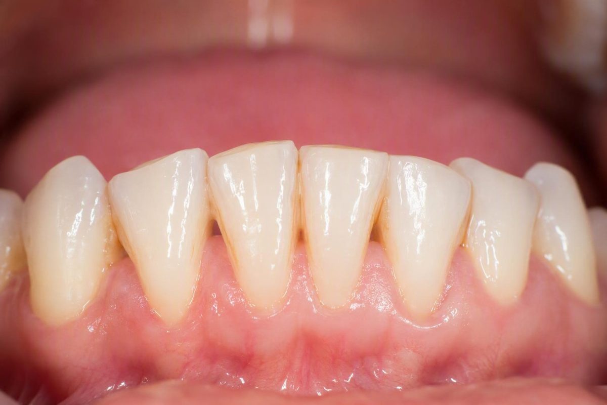
The short answer: early gum-line erosion has no single diagnostic appearance
Early-stage tooth erosion at the gum line does not have one reliable appearance or sensation. Possible clues include:
- A smooth or unusually shiny surface
- A shallow indentation or groove
- Roughness or small pits
- A change in color
These features can accompany erosive tooth wear, but none confirms it. The same findings can also occur with decay, gum recession, an exposed root, brushing-related wear, or another defect at the neck of the tooth (Better Health Channel’s dental erosion fact sheet).
A changed area may cause no pain at all. A tooth that feels normal is therefore not necessarily unaffected.
Color is especially easy to misinterpret. Yellow may become visible when enough enamel has been lost to reveal the dentine beneath it. Near the gum, however, a yellowish area may instead be an exposed root following gum recession. That is not the same as early enamel erosion.
Likewise, a wedge-shaped groove, dark mark, chalky patch, or sensitive spot is not a diagnosis. Each can have several explanations, and more than one process may be affecting the same tooth.
The practical boundary is simple: you can identify a change worth examining, but you cannot reliably name its cause from appearance, a photograph, or sensation alone.
What erosion is—and why the word is often confused with decay
Dental erosion is tooth-surface loss associated with direct exposure to acids from food and drink or acids that reach the mouth from the stomach. It is also called acid wear or erosive tooth wear. Acid may soften the surface before brushing, grinding, or other mechanical contact removes more tissue.
Tooth decay begins differently. Bacteria in dental plaque use sugars and starches and produce acids that remove minerals from the tooth. Continued mineral loss can eventually cause the surface to break down into a cavity, as explained by the National Institute of Dental and Craniofacial Research.
The two processes can overlap without becoming interchangeable:
- An acidic sweet drink can expose teeth directly to acid.
- Its sugar can also support bacterial acid production.
- A surface softened by direct acid exposure may be more vulnerable to mechanical wear.
- A recessed or worn gum-line area may collect plaque and later develop decay.
That overlap is one reason a gum-line mark can be difficult to interpret. Calling every groove “erosion” may overlook decay; calling every dark area a cavity may overlook staining, recession, or non-carious wear.
“Gum line” describes a location rather than a single kind of tooth tissue. When the gum margin is in its usual position, the affected area may be enamel on the crown. If the gum has receded, the visible area may be root tissue instead. Exposed roots do not have the same enamel covering as the crown and can become sensitive or vulnerable to decay.
This tissue distinction matters. Two areas that both look yellow and respond to cold may represent different conditions:
- Thinned enamel with dentine becoming more visible
- Gum recession with an exposed root
- Wear affecting an already exposed root
- Bacterial decay on the crown or root surface
- More than one of these processes at the same time
The correct response depends on which tissue is involved, whether the surface remains intact, and whether the change is stable or progressing.
Erosion, decay, abrasion, recession, or a cervical notch?
Several conditions can produce a mark where the tooth meets the gum. Their signs overlap, so this comparison is useful for orientation—not for self-diagnosis.
| Condition | Underlying process | Possible clues | Why an examination is still needed |
|---|---|---|---|
| Dental erosion | Repeated contact with dietary or stomach acid softens the surface and contributes to tooth-surface loss. Mechanical contact may then remove more softened tissue. | Smoothness, shine, shallow wear, pitting, rounded contours, sensitivity, or discoloration. | These findings are not unique to erosion. The distribution of wear, tissue involved, acid history, and evidence of progression all matter. |
| Gum-line or root decay | Plaque bacteria produce acids from sugars and starches, causing mineral loss that may progress to a cavity. | A white, brown, or black change; a pit or rough area; sensitivity to sweet, hot, or cold; pain when biting; or no early symptoms. | Color and symptoms cannot establish whether decay is present or how deep it is. A beginning cavity may cause no symptoms, while later decay may cause sensitivity, visible pits, staining, or pain (Mayo Clinic’s overview of cavities). |
| Brushing-related abrasion | Repeated mechanical friction may contribute to wear, particularly with vigorous scrubbing or a hard-bristled brush. An acid-softened surface may be more susceptible. | A smooth groove or notch near the gum, sometimes accompanied by sensitivity. | Brushing technique may be one contributor rather than the sole cause. A similar groove can occur in multifactorial wear or beside an exposed root. |
| Gum recession with an exposed root | Gum tissue moves away from its former position, exposing root tissue that lacks the crown’s enamel covering. | A tooth that looks longer, a yellowish area, a visible boundary near the gum, or sensitivity to temperature, touch, or brushing. | Recession does not establish whether erosion, abrasion, decay, gum disease, or other influences are also present. Exposed roots may be more vulnerable to sensitivity and decay (guidance on gum-line cavities and recession). |
| Wedge-shaped or cervical defect | Tooth structure is missing at the neck of the tooth, potentially through interacting chemical and mechanical influences. | A V-shaped or wedge-like notch, a sharp-feeling edge, sensitivity, or a stable symptom-free groove. | Shape alone cannot establish whether brushing, acid wear, decay, clenching, grinding, anatomy, or several factors produced it. |
The table does not provide a set of visual rules for identifying the cause. A dark area can be stain or decay. A shiny area can reflect wear but does not prove acid exposure. A wedge can be associated with mechanical wear but does not identify one particular force.
A useful rule is: the more the signs overlap, the less dependable visual self-diagnosis becomes. A dental assessment can place the color, texture, location, symptoms, root exposure, plaque retention, and wider pattern in context. Even after examination, some cervical defects may be considered multifactorial rather than assigned one simple cause.
Why damage may develop where the tooth meets the gum
There is rarely one universal cause of a gum-line lesion. Possible contributors are better grouped into acid exposure, reduced salivary protection, mechanical wear, and exposed-root or decay risks.
Acid exposure
Dietary acids may come from:
- Soft drinks
- Sports and energy drinks
- Citrus fruit
- Fruit juice
- Kombucha
- Vinegar-containing products
- Some supplements
Stomach acid can reach the teeth through reflux or recurrent vomiting. These exposures should be discussed without assuming why they are happening. Recurrent stomach-acid exposure may have medical or other personal causes that require individualized assessment.
Frequency and contact time matter. Having an acidic drink once with a meal creates a different exposure pattern from sipping it repeatedly over several hours. Prolonged or repeated sipping brings acid back into contact with the teeth and leaves less time for saliva to dilute and neutralize it.
The overall distribution may be more informative than one isolated groove. A dentist may consider which teeth and surfaces are involved alongside the history of dietary or stomach-acid exposure. Government guidance identifies both dietary acids and reflux or vomiting as potential sources of erosive tooth wear and explains that acid-softened surfaces can become more vulnerable to abrasion or grinding (Better Health Channel).
Reduced salivary protection
Saliva helps wash away and neutralize acids. It also supplies minerals that can support repair while a weakened tooth surface remains intact.
Dry mouth reduces those protective functions and may increase vulnerability to both erosive wear and bacterial decay. Some medications can contribute to dry mouth, but a gum-line mark cannot reveal whether medication, a health condition, breathing pattern, or another factor is responsible.
Persistent dry mouth is worth mentioning to a dentist or appropriate healthcare professional. Do not stop or change prescribed medication based on a tooth symptom without consulting the prescribing professional.
A clinician may need to consider when the dryness began, whether it is persistent, and whether it coincided with a health or medication change.
Mechanical wear
Forceful side-to-side scrubbing or use of a hard-bristled brush may contribute to gum-line wear. The interaction with acid is important: if a tooth surface has recently been softened, immediate vigorous brushing may add mechanical abrasion.
This does not mean brushing is harmful or should be stopped. It means that technique, pressure, brush type, and timing after acid exposure may matter. Brushing harder is not a useful response to a groove or stain.
Grinding or clenching may add mechanical stress or wear in some people, but a notch does not prove either behavior caused it. Claims about bite-related cervical defects remain uncertain enough that shape alone should not be used to assign a cause. Chemical exposure, brushing, anatomy, recession, plaque, and other mechanical forces may interact.
Exposed-root and decay risks
Gum recession can expose root tissue that differs from enamel-covered crown tissue.
For decay, repeated exposure to sugar or starch is particularly relevant because it supplies oral bacteria with fermentable material. Plaque accumulation, inadequate interdental cleaning, dry mouth, and frequent snacking or sipping may also increase risk.
An individual product may contribute to more than one pathway. A sweet acidic drink, for example, can expose the teeth directly to acid while also supplying sugar that plaque bacteria can use.
This is why “I brush well” does not settle the diagnosis. Brushing disrupts plaque, but it cannot eliminate reflux, restore normal saliva, reverse gum recession, regrow missing tooth structure, or prevent every direct acid exposure.
What can be remineralized and what cannot grow back
The word reversible needs careful definition. Mineral loss may sometimes be arrested or partly repaired while a surface remains intact. That does not mean a missing part of the tooth grows back.
A three-part framework makes the distinction clearer.
1. Intact and weakened surface
An intact tooth surface can lose minerals without yet developing a physical hole or groove. Saliva and fluoride may help return minerals to that weakened surface or make it more resistant to further acid attack.
This principle also applies to early bacterial decay. Before a cavity forms, improved mineral balance and fluoride exposure may sometimes stop or reverse the earliest stage. Once the surface has broken down into a hole, however, it is no longer merely an intact demineralized area (NIDCR’s explanation of early decay and cavities).
Remineralization is microscopic repair within remaining tissue. It does not create a new layer of enamel over a missing contour.
2. Visible wear or exposed tissue
Once erosion or abrasion has physically changed the tooth’s shape—producing a pit, depression, thinned edge, or groove—the lost structure does not biologically regrow.
Fluoride may help protect the surface that remains, and controlling the cause may slow or halt progression. Neither action restores the original contour.
Cleveland Clinic states that enamel cannot be replaced after it is gone, although further progression may be limited and the remaining tooth can be protected (Cleveland Clinic’s medically reviewed guide to tooth erosion).
3. Formed cavity or substantial structural loss
If decay has broken through the surface to form a cavity, fluoride alone cannot close the hole. A dentist may need to manage the compromised tissue and restore the defect.
More extensive structural loss may require a larger restoration. The appropriate response depends on the cause, depth, location, symptoms, cleanability, and amount of sound tooth remaining.
So, can early erosion at the gum line be reversed? An intact, weakened surface may be strengthened, but a visible loss of enamel or root structure cannot be biologically rebuilt.
The difficulty is that a person looking in a mirror cannot reliably determine whether an area is:
- Intact but demineralized
- Physically worn
- Cavitated
- Stained but sound
- An exposed root
- A combination of wear and decay
That distinction is central to deciding whether prevention and monitoring are enough or whether restorative treatment should be considered.
Low-risk steps that may limit further wear
While waiting for a dental assessment, the aim is to reduce additional chemical and mechanical stress without neglecting plaque control.
Keep cleaning the area
Do not stop brushing solely because the area is sensitive. Plaque left at the gum margin may increase the risk of decay and gum inflammation, including around a pre-existing wear defect.
Use a soft-bristled toothbrush and fluoride toothpaste. Apply light pressure instead of forcefully scrubbing the changed area. Continue careful cleaning along the gum line and between the teeth.
If ordinary cleaning remains painful, arrange an examination rather than repeatedly avoiding the tooth. Sensitivity can have several causes, including exposed root tissue, decay, wear, or another dental problem.
Change the timing after acid exposure
After acidic food or drink, reflux, or vomiting:
- Rinse the mouth gently with water.
- Do not scrub the teeth immediately.
- Wait at least 30 minutes before brushing.
This delay allows time for saliva to dilute and neutralize acid and reduces brushing against a recently softened surface. It is not a reason to skip brushing for the rest of the day. The waiting period, gentle pressure, soft bristles, and fluoride toothpaste are supported protective measures in the supplied dental guidance (tooth-erosion prevention guidance).
Reduce repeated acid contact
Focus on exposure frequency and contact time rather than assuming one food must always be eliminated. Practical changes may include:
- Avoiding prolonged sipping of acidic drinks
- Having acidic drinks less often
- Choosing water between exposures
- Keeping acidic drinks with meals rather than consuming them repeatedly throughout the day
- Rinsing gently with water afterward
If reflux or recurrent vomiting is exposing the teeth to stomach acid, protecting the teeth addresses only part of the problem. Persistent symptoms may warrant discussion with an appropriate healthcare professional, without making assumptions about their cause.
Support saliva and routine prevention
Stay adequately hydrated. If dry mouth persists, mention it during the dental visit, particularly if it began after a medication or health change. Do not stop prescribed medication without guidance.
Continue interdental cleaning and regular dental care. A mark that appears to be erosion may instead be plaque-related gum-line or root decay, so acid-focused changes should not replace decay prevention.
These measures may reduce future exposure and help strengthen an intact surface. They cannot:
- Fill a groove
- Replace missing enamel
- Rebuild lost root tissue
- Cover an exposed root
- Close a formed cavity
Avoid selecting prescription-strength fluoride, desensitizers, supplements, home remedies, or restorative materials for yourself based only on appearance. Those choices depend on the tissue involved and the diagnosis.
How a dentist may investigate a gum-line defect
A dental examination can gather information that a mirror or photograph cannot. Depending on the concern, the assessment may consider:
- The exact location of the change
- Whether it involves the crown or an exposed root
- Its shape, depth, texture, color, and margins
-
Whether the surface remains intact or has broken down
-
Gum recession or inflammation
- Whether food, plaque, or floss catches
- Similar changes elsewhere in the mouth
- Whether the area appears to be stable or progressing
No single feature necessarily settles the diagnosis. Texture, staining, sensitivity, and shape must be interpreted together rather than used as stand-alone tests.
The history can also matter. A dentist may ask about:
- Acidic foods and drinks
- Prolonged sipping
- Reflux or vomiting
- Dry mouth
- Brushing pressure and brush type
- Gum recession
- Sugar and starch frequency
- Grinding or clenching
- Recent health or medication changes
An X-ray may be used when decay, deeper damage, or the extent of a lesion is a concern. Clinical appearance, tissue type, history, symptoms, and the wider pattern remain important. Dental-practice guidance on gum-line lesions likewise describes examination and, when appropriate, X-rays as parts of assessment rather than visual appearance alone (gum-line decay assessment overview).
This is clinical context, not a universal requirement or a fixed monitoring schedule.
Treatment selection follows two main questions:
- What process or combination of processes is driving the lesion?
- How much tooth structure has been affected?
A shallow, intact, stable area with controlled risk factors may be managed differently from active root decay, rapidly progressing wear, or a deep defect that traps food.
Decay Guide describes its content as educational and states that it is not a dental practice. This article cannot diagnose or treat a particular gum-line change.
When protection may be enough—and when restoration may be considered
There is no universal sequence in which every lesion progresses from fluoride to bonding, then to a filling and finally to a crown. Management depends on the diagnosis, symptoms, progression, location, cleanability, and amount of missing structure.
When protection and monitoring may be enough
For an intact early surface, a dentist may consider:
- Monitoring for change
- Reducing dietary or stomach-acid exposure
- Improving plaque control
- Correcting forceful brushing technique
- Fluoride-based preventive care
- Managing sensitivity
- Addressing dry mouth or recession where relevant
The objective is to protect the remaining tissue and control the cause. Fluoride-based care may strengthen an intact surface, but it does not rebuild a notch.
A selected non-cavitated but sensitive defect may sometimes be managed with a professional desensitizing or sealing approach. That decision should follow examination because sensitivity alone cannot establish whether the area is worn, decayed, cracked, or root-exposed. Practice-based guidance describes such approaches for selected non-cavitated wear defects, while emphasizing that treatment depends on the lesion’s depth, location, and surrounding conditions (gum-line lesion treatment overview).
When a restoration may be considered
The words bonding and filling overlap in everyday use. What matters is whether the selected material and technique suit the defect’s location, moisture conditions, tissue type, and cause.
Crowns and more complex restorative care are generally considerations for greater structural loss, not automatic treatment for a small early gum-line mark. Medically reviewed erosion guidance describes management as severity-dependent, ranging from preventive fluoride care in earlier disease to bonding or crowns when damage is more advanced (Cleveland Clinic).
When to arrange an examination
Arrange a dental assessment for a gum-line area that is:
- New or enlarging
- Persistently sensitive
- Rough, pitted, chipped, or notched
- Newly white, brown, black, yellow, or otherwise discolored
- Trapping food or catching floss
- Painful during brushing
- Associated with visible root exposure
- Difficult to distinguish from a cavity
Contact a dentist more promptly for toothache, persistent mouth pain, pain when biting, or a broken tooth. These symptoms do not identify the cause, but they make timely assessment more important. Mayo Clinic specifically advises dental care for toothache or mouth pain and lists pain on biting among possible symptoms of advancing decay (Mayo Clinic).
Frequently asked questions
Can early tooth erosion at the gum line happen without pain?
Yes. Early erosive wear may be painless, and early tooth decay also commonly causes no symptoms.
Sensitivity may develop when tooth structure becomes thinner or a root is exposed, but its absence does not rule out damage. Pain is therefore a poor screening test. A visible or tactile change can still warrant assessment even when the tooth feels normal.
Does a wedge-shaped notch at the gum line prove that I brush too hard?
No. Vigorous brushing or a hard brush may contribute to gum-line wear, particularly if the surface has recently been softened by acid. A wedge shape does not, however, identify one cause.
Acid exposure, recession, mechanical wear, decay, anatomy, grinding or clenching, and several interacting factors may be relevant. The notch should be interpreted alongside its location, tissue type, symptoms, progression, and the overall pattern in the mouth.
Can fluoride regrow enamel that has already worn away?
It also cannot fill a visible groove, replace missing root structure, or close a formed cavity. When structure is missing, a dentist may monitor, protect, or restore the area depending on the diagnosis, extent, symptoms, and progression.
Why should I wait 30 minutes to brush after acidic food, drinks, reflux, or vomiting?
Acid can temporarily soften a tooth surface. Brushing immediately may add mechanical abrasion while that surface is more vulnerable.
Rinse gently with water, allow saliva time to dilute and neutralize the acid, and wait at least 30 minutes before brushing with fluoride toothpaste and a soft-bristled brush (tooth-erosion prevention guidance). This applies after dietary acid as well as stomach-acid exposure.
Can a dentist distinguish erosion from a gum-line cavity?
A dentist can make a more informed assessment than appearance alone allows, but some lesions remain multifactorial or uncertain.
The evaluation may include the area’s location, shape, texture, surface integrity, staining, root exposure, plaque retention, sensitivity, surrounding wear pattern, and relevant dietary or acid history. X-rays may help when decay or deeper damage is suspected, while comparison with previous records may show whether the defect is changing.
The conclusion may not always be that one label explains everything. Erosion, abrasion, recession, and decay risk can interact in the same area.
The central distinction is not whether a mark merely “looks like erosion.” It is whether the surface remains intact, has physically worn away, exposes root tissue, or has developed decay. A gum-line change can be noticed at home, but it cannot reliably be named there.
Gentle brushing with fluoride toothpaste, less frequent acid exposure, water rinsing, hydration, interdental cleaning, and delayed brushing after acid exposure are reasonable protective measures. They cannot replace lost tooth structure. A new, changing, painful, sensitive, rough, chipped, discolored, or food-trapping area should be examined so the response matches the actual cause.