
Is That Dark Mark a Stain, Tartar or a Cavity?
Some dark lesions are arrested decay that a dentist may monitor rather than drill. A new, persistent or changing mark still warrants an examination.
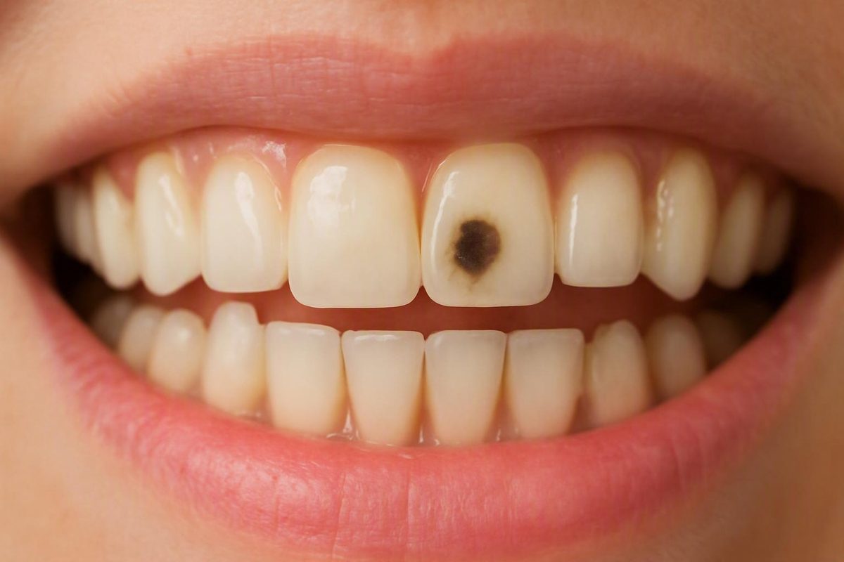
What a black spot can—and cannot—tell you
No—a black spot on a tooth is not automatically a cavity. It is a visible sign with several possible explanations, including:
- External staining from food, drinks or tobacco
- Tartar that has collected dark pigment
- Pigment trapped in a natural groove or small crack
- Active tooth decay
- Potentially inactive, or arrested, decay
- Discoloration associated with a filling, crown or other dental work
- An enamel defect
- Internal discoloration following an injury
These conditions can overlap in appearance. A stain and an area of decay may both look brown or black, especially in a photograph or the limited view available through a bathroom mirror. Color alone cannot establish the cause.
Lack of pain does not rule out a cavity. Some decay produces few or no early symptoms. Conversely, pain near a dark mark does not prove that the mark itself is responsible. Penn Dental Family Practice advises having even a painless black spot assessed because staining, decay and dental trauma can look similar from the outside (guidance on black tooth spots).
Arrange a dental examination if a mark is new, persistent, changing, rough, sensitive or painful. Do not try to diagnose it by scraping, pressing or probing it, and do not assume that whitening is appropriate before the cause is known.
Important editorial limitation: Decay Guide is written by a health writer, not a dentist, and its articles have not been reviewed by a clinician. The available evidence for this article consists largely of consumer dental information and commercially published dental-practice material rather than independent clinical guidelines. This guide is therefore informational and cannot diagnose a tooth or replace a dental examination and any imaging a dentist considers appropriate. Read more about how Decay Guide is written.
The main causes of a dark dot, line or patch
A dark mark may sit on the tooth’s surface, within a deposit attached to it, inside damaged tooth structure or in or around a restoration. The comparison below provides context, not a home diagnostic test.
| Possible cause | Typical context | Cautious clues | Will brushing help? | Possible professional management |
|---|---|---|---|---|
| External stain | Exposure to coffee, tea, red wine, berries, dark foods or tobacco | Flat discoloration; similar marks may appear on several teeth | It may reduce fresh surface pigment, but established stain can remain | Examination, cleaning or polishing; selected whitening after other causes are excluded |
| Stained tartar | Often near the gumline or between teeth | A deposit that remains after normal brushing | No, once plaque has hardened into tartar | Professional scaling or cleaning |
| Pigment in a natural groove | Often on a molar’s chewing surface | A dark dot or line following a pit or fissure | Cleaning may have little visible effect | Examination followed by reassurance, monitoring or treatment according to the findings |
| Active decay | May occur in grooves, between teeth, near the gums or beside dental work | A growing mark, pit, hole, food trapping, sensitivity or pain can increase concern | No | Restorative treatment based on the location and extent of damage |
| Arrested decay | A lesion professionally assessed as inactive | It may appear dark, hard and stable during a clinical assessment | No | Monitoring may be selected in some cases |
| Crack or enamel defect | Pigment collects in an irregularity | A narrow line, notch or localized surface defect | Usually not if pigment is embedded | Cause-specific monitoring or restorative care |
| Filling- or crown-related darkness | Beside or beneath existing dental work | A dark edge, shadow or change in a restoration | Usually not | Observation, polishing, repair or replacement depending on the diagnosis |
| Internal discoloration after trauma | Often follows a blow, fall or other injury | Much or all of one tooth turns gray or dark rather than developing a tiny surface dot | No | Assessment of the tooth and its inner tissues; treatment depends on the findings |
| Developmental or medication-related discoloration | Color developed while teeth formed or is associated with medical history | A pattern across several teeth may be present | Usually not | Assessment, followed by monitoring or selected cosmetic care if appropriate |
External staining
Pigments from coffee, tea, red wine, berries, other dark foods and tobacco can adhere to the outer tooth surface. This is called extrinsic discoloration. Exposure history can make staining plausible, but it cannot prove that a particular spot is only a stain. Someone who drinks coffee can still have decay, and decay can sit beneath or beside visible pigment.
Surface stains may affect broad areas, collect in small surface irregularities or appear on several teeth. Some may respond to professional cleaning or polishing. Whitening may be considered only after a dentist has determined that the issue is suitable discoloration rather than decay, tartar, a crack or damaged dental work (overview of possible causes and treatments).
Plaque, tartar and dark deposits
Plaque is a soft film that accumulates on teeth. If it remains in place, it can harden into tartar, also called calculus. Tartar can collect pigment and appear yellow, brown or black, particularly near the gums or between teeth.
Once tartar is established, ordinary brushing generally will not remove it. Professional scaling or cleaning is used instead. A dark gumline deposit may therefore be stained tartar, but decay near the gum margin can look similar and requires a dental assessment to distinguish it (overview of plaque and dark tartar).
Tooth decay
Tooth decay develops when acids associated with oral bacteria damage the mineralized tooth surface. If the surface breaks down, the affected area may eventually appear as a brown or black pit, patch or hole.
Color does not reveal how active or extensive the process is. A tiny dark point may be pigment in an intact groove, while damage in a less dramatic-looking area may be difficult to see.
Natural pits, fissures and small cracks
Molars contain natural pits and fissures that form part of their chewing surfaces. These narrow areas can retain both plaque and pigment, making a stained groove difficult to distinguish visually from decay. Small cracks and enamel defects can also trap color.
Do not test a groove or crack with a pin, dental instrument, toothpick or fingernail.
Dental work and material-related darkness
Older metal fillings and some crowns can create a dark appearance in or around a tooth. Pigment may also develop along the edge of a restoration. Possible explanations include material-related shadowing, superficial marginal stain, a damaged edge or decay beside or beneath the restoration.
A dark edge does not automatically mean that a filling or crown needs replacement. Equally, whitening the surrounding tooth is not an appropriate substitute for checking the restoration. The dental work and adjacent tooth structure need to be assessed together.
Developmental discoloration, enamel defects and medication history
Developmental discoloration generally differs from a newly appearing isolated spot. Fluorosis, for example, develops while enamel is forming and may affect several teeth in a pattern. Enamel defects can alter the surface and allow pigment to collect.
Some medications have also been associated with tooth discoloration. The type of medication, the timing of exposure and whether the discoloration is external or internal all matter. A medication history may help a dentist interpret the pattern, but it cannot diagnose the mark by itself (discussion of fluorosis and inactive dark lesions).
Trauma-related internal discoloration
A tooth may darken internally after an impact damages its inner tissues.
A tooth that changes color after an injury should be assessed even if it is painless. The appropriate response depends on what has happened inside the tooth and may be very different from treatment for a surface stain (causes of surface and internal darkening).
Use location as context, not as a diagnosis
Where a mark appears can help a dentist decide what to examine. Location still cannot establish the cause on its own.
At the gumline
A dark line or ridge where the tooth meets the gum may be stained tartar, especially if it appears to be attached to the tooth and remains after normal brushing.
Decay can also occur near the gum margin. Exposed root surfaces, nearby dental work and difficulty cleaning the area may affect the assessment. Tell the dental practice if the gum is swollen, bleeds locally or the mark is changing.
On a molar’s chewing surface
Natural molar pits and fissures can retain pigment and plaque. A dark line may therefore be a stained but intact groove, active decay or a previously affected area that is no longer active.
Good lighting or a close-up photograph may show the location clearly, but neither can confirm what is happening beneath the surface. A dentist may examine whether the groove is intact, whether the surrounding enamel has changed and whether the appearance has remained stable.
Between teeth or on the back of a tooth
Marks between teeth or on the back of a tooth can be difficult to inspect directly. Food trapping or floss catching may raise concern, but these signs are not specific to decay. A rough filling edge or another structural irregularity may cause similar experiences.
A dentist may consider an X-ray when the location and examination findings suggest that imaging would provide useful information, particularly where direct vision is limited. Not every visible stain requires imaging (location-based assessment of dark tooth marks).
Beside a filling or crown
Darkness at the edge of dental work may be superficial stain or material-related shadowing. It may also reflect a damaged restoration, a gap or decay in adjacent tooth structure.
Appearance alone is not a sound basis for replacing a filling or crown. The dentist will consider its condition, symptoms, changes over time and what can be seen during the examination or on an appropriate image.
When the whole tooth darkens
If most or all of one tooth turns gray or black after a blow, fall or other injury, the concern differs from an external stain. Internal tissues may have been affected even if the tooth did not chip and there was no immediate pain.
Arrange a dental assessment and explain when and how the injury occurred. Do not treat whole-tooth darkening as a cosmetic stain without first having the tooth evaluated.
A note for caregivers
There is no single visual rule for diagnosing a child’s black tooth spot. Age, tooth type, symptoms, development and dental history all require individual consideration.
Clues that raise or lower concern—without diagnosing the spot
The following is a non-diagnostic checklist. It can help you describe the mark and decide whether to contact a dentist, but it cannot confirm staining, tartar, active decay or inactive decay.
Change
Concern may be higher when the mark:
- Is new or appeared suddenly
- Is clearly enlarging
- Has recently become darker
- Has changed from a line into a visible pit
- Appeared with a new chip or fracture
A stable mark may be less suggestive of ongoing change, but stability does not prove that the tooth is healthy. Slow changes can also be difficult to notice without a reliable comparison.
Surface and nearby findings
Features worth reporting include:
- A visible pit or hole
- An area that looks rough or broken
- Food repeatedly lodging in the same place
- Floss catching or shredding
- A nearby filling or crown
- A gumline deposit that remains after brushing
- Localized gum redness, swelling or bleeding
Do not press, scratch or pick at the area to determine whether it is hard, soft or sticky.
Symptoms
Cold or sweet sensitivity, pain while biting and spontaneous toothache can make decay, a crack or a restoration problem more concerning. They do not identify the cause on their own.
Pain may come from another surface of the same tooth, a neighboring tooth or the surrounding gum. It is neither required for a cavity nor proof that the visible mark is responsible.
Features that may fit staining
A smooth-looking, flat, stable and symptom-free mark—particularly when similar discoloration affects several teeth—may fit surface staining better than a single enlarging pit. A history of exposure to coffee, tea, red wine or tobacco adds context.
These clues do not rule out decay beneath a stained groove or around dental work.
Features compatible with tartar
A deposit near the gumline that appears raised or attached and does not brush away may be tartar. Do not attempt to chip it off. Home inspection cannot confirm whether the mark is calculus, stained tooth structure or decay near the gum.
Active versus arrested decay
Decay is not always a choice between “harmless stain” and “rapidly progressing cavity.” A dentist may sometimes assess a dark lesion as arrested, meaning it appears inactive. A professionally evaluated lesion that is hard and stable may be monitored in selected cases, while surface breakdown, change or symptoms may raise concern about activity.
Do not apply these classifications at home. They depend on professional assessment of the lesion and the person’s wider dental history and risk.
Before the appointment, note:
- When you first saw the mark
- Whether its size, shape or color has changed
- Any cold, heat, sweet or pressure triggers
- Whether food or floss catches there
- Any recent or past injury
- Coffee, tea, wine, tobacco or other staining exposures
- Nearby fillings, crowns or previous repairs
- Relevant medication history
When to book a dental visit and which signs need faster attention
Arrange a dental examination for a black spot that is:
- New or suddenly noticeable
- Still present after normal brushing
- Enlarging or darkening
- Rough, pitted or associated with a visible hole
- Sensitive to temperature or sweets
- Painful at rest or while chewing
- Beside dental work that has changed
- Difficult to inspect because it lies between teeth
- Part of a tooth that darkened after trauma
A painless but persistent mark still deserves assessment. Early or hidden decay may not hurt, and appearance alone cannot establish that a stable-looking mark is harmless. Assessment does not mean that every spot will require drilling, an X-ray or another specific treatment.
Contact a dental practice promptly for individualized advice if you have persistent or sharp pain, pain while chewing, new temperature sensitivity, a visible hole, gum swelling, localized bleeding, a foul taste or odor, or a mark that is clearly growing. These findings can accompany several dental problems and require professional triage (warning signs associated with dark tooth marks).
The supplied evidence does not establish a complete emergency threshold or a universal deadline for every black spot. If swelling is substantial, symptoms are severe or escalating, or you are concerned that an infection may be spreading, contact an appropriate dental or medical service for urgent, individualized guidance rather than relying on this article.
A small, unchanged, symptom-free mark and a painful enlarging hole are not equivalent. Describe the location, appearance, symptoms and recent changes when contacting the dental practice so its team can advise you on timing.
A painless mark beside an old filling, a spot between teeth or a whole tooth that darkened after injury should be assessed before cosmetic whitening is considered.
How a dentist investigates a black spot
A dental visit usually combines several sources of information rather than relying on one conclusive test.
The dentist may ask:
- When did you first notice the mark?
- Has it changed in size, shape or color?
- Does cold, heat, sweetness or biting cause discomfort?
- Has the tooth been hit or injured?
- Is there a filling or crown in or beside it?
- Have you noticed food trapping, gum bleeding or a bad taste?
- Do you use tobacco or regularly consume strongly pigmented drinks?
The clinical examination may cover the dark area, surrounding enamel, neighboring teeth, gums and existing restorations. The dentist may clean or dry the surface to improve visibility and assess it with professional instruments. Surface findings contribute information, but they do not guarantee a complete diagnosis by themselves.
An X-ray may be considered if the mark’s location, symptoms or examination findings suggest that imaging could add useful information. This may be particularly relevant for areas between teeth or around restorations, where direct inspection is limited. Not every visible line or superficial stain requires an X-ray.
The possible outcome is not limited to “cavity” or “no cavity.” Depending on the findings, the dentist may recommend:
- Reassurance with no treatment
- Monitoring for change
- Professional cleaning or scaling
- Discussion of cosmetic stain removal
- Repair or replacement of a restoration
- A filling or another restoration
- Further assessment of an injured or internally discolored tooth
Assessment of the history, surface, restorations and selective imaging is used to distinguish among causes that may look similar externally (overview of dental assessment for a black spot).
To prepare, bring or record:
- The approximate date the mark appeared
- Dated photographs if they show a genuine change
- Symptom triggers and how long discomfort lasts
- Details of any blow or injury
- Tobacco and staining-food or drink exposure
- Current and relevant past medications
- The history of fillings, crowns or other work on that tooth
Treatment depends on the cause—not the color
The underlying cause should be established before removal, whitening or restorative treatment is recommended.
| Diagnosed cause | Main goal | Possible professional management |
|---|---|---|
| Surface stain | Remove or lighten external pigment | Cleaning, polishing or a selected whitening approach |
| Established tartar | Remove hardened deposits | Professional scaling or cleaning |
| Stained but sound groove | Confirm that the tooth surface is intact | Reassurance, cleaning or monitoring |
| Cavitated decay | Remove damaged tissue and restore the tooth | Filling; a larger restoration if damage is more extensive |
| Arrested decay | Confirm inactivity and watch for change | Professionally planned monitoring in selected cases |
| Crack or enamel defect | Protect the tooth and address symptoms | Monitoring or cause-specific restorative care |
| Darkness beside a restoration | Determine whether the issue is cosmetic or structural | Observation, polishing, repair or replacement |
| Trauma-related internal discoloration | Assess the condition of the tooth’s inner tissues | Cause-specific restorative or root-canal care |
| Intrinsic or developmental discoloration | Consider appearance after disease is excluded | Selected cosmetic treatment where appropriate |
These are possible pathways rather than recommendations for a particular tooth. Treatment may range from no intervention to cleaning, a filling, repair of dental work or more extensive care, depending on the diagnosis (diagnosis-dependent treatment overview).
Surface stains
Professional cleaning or polishing may remove some external stains. Whitening may be suitable for selected discoloration after the teeth, gums and existing dental work have been assessed.
Deeper or intrinsic discoloration may respond differently from pigment on the enamel surface.
Tartar
Established tartar is treated with professional scaling or cleaning because normal brushing does not remove the hardened deposit.
Cavitated decay
When decay has produced a cavity, treatment generally involves removing damaged tissue and restoring the area with a filling. More extensive loss of tooth structure may require a larger restoration or crown. If the inner pulp is affected, root-canal treatment may be considered.
These treatments are selected according to the actual location and extent of damage. A black color alone does not show that a crown or root canal is necessary.
Arrested decay
A lesion assessed professionally as inactive may sometimes be monitored. This does not mean that every dark or hard-looking spot should be left untreated.
The decision depends on the clinical findings and the person’s broader dental circumstances. There is no universal home test or follow-up schedule for an unexplained dark lesion.
Darkness around a filling or crown
Management may range from observation to repair or replacement. Superficial marginal stain or material-related shadowing may not indicate structural damage. A defective restoration or decay beside it requires a different response.
Replacement should not be recommended solely because a restoration looks dark.
Trauma-related internal discoloration
An injured tooth may need testing and follow-up to assess its internal tissues. Treatment is cause-specific and may include restorative or root-canal care after examination. Whitening intended for ordinary surface stains does not address internal pulp damage.
Bonding, veneers and other cosmetic options
Bonding, veneers or other cosmetic procedures may conceal selected discoloration after its cause has been identified. They are not substitutes for removing tartar, treating active decay, repairing defective dental work or addressing internal tooth damage.
What to do—and avoid—while waiting for an assessment
Continue gentle oral hygiene unless a dental professional has instructed you otherwise.
Brush twice daily with fluoride toothpaste and clean between the teeth using floss or another suitable interdental method. Brushing may reduce fresh surface pigment, but it cannot repair a cavity, remove established tartar or correct internal discoloration.
If the spot remains after ordinary brushing, do not escalate to forceful scrubbing.
Whitening products are intended for selected forms of discoloration. They do not treat:
- Tooth decay
- Tartar
- A cracked tooth
- A defective filling or crown
- Internal tooth damage after trauma
Avoid:
- Scraping the mark with a dental tool or metal object
- Picking it with a toothpick
- Pressing it with a fingernail
- Trying to widen or clean out a groove
- Aggressive abrasive powders
- Unspecified peroxide mixtures
- Baking-soda experiments
- Oil pulling or other unverified bleaching methods
The supplied evidence does not establish these experiments as safe or effective treatments for an unexplained black mark. Self-probing also cannot provide a reliable diagnosis.
Monitor rather than manipulate the area. Record whether the spot changes in size or color and whether you develop sensitivity, pain, food trapping, floss catching, gum swelling or bleeding. A dated photograph may document an obvious change, but it cannot determine the cause.
Measures that may support oral health and reduce staining, plaque accumulation or decay risk include:
- Brushing twice daily with fluoride toothpaste
- Cleaning between teeth regularly
- Limiting frequent sugary exposures
- Reducing strongly staining drinks
- Rinsing with water after dark beverages
- Avoiding tobacco
- Obtaining dental care appropriate to your individual risk and history
These measures cannot guarantee that every dark mark will be prevented, and they cannot determine the cause of a spot already present (prevention and professional-care guidance).
Frequently asked questions
Can a painless black spot still be a cavity?
Yes. Some cavities produce few or no early symptoms, and marks in molar grooves, between teeth or near the gumline can be difficult to assess at home.
A painless spot could also be stain, tartar, a pigmented groove or an inactive lesion. Neither color nor absence of pain can distinguish these possibilities. Arrange an examination if the mark is new, persistent or changing.
Can brushing or whitening remove a black spot from a tooth?
Sometimes, if the cause is suitable. Gentle brushing may reduce fresh surface pigment, while professional cleaning, polishing or selected whitening may improve some external stains.
Brushing will not remove established tartar or repair a cavity. Whitening will not treat decay, a failing restoration, a crack or trauma-related internal damage.
Can a dentist identify the cause without an X-ray?
Sometimes. Obvious surface stain, tartar or material-related discoloration may be identifiable from the history and clinical examination.
In other cases, an X-ray may add useful information, particularly for a mark between teeth or around existing dental work. Not every black spot requires imaging, and neither visual inspection nor an X-ray guarantees a complete answer in every case.
What does a black spot next to a filling or crown mean?
It may represent material-related darkness, marginal staining, a rough or damaged edge, or decay beside or beneath the restoration. These possibilities can look similar externally.
Do not assume the restoration needs replacement, but have a new or changing mark assessed before trying to whiten it.
Can a dark cavity be inactive and monitored instead of filled?
Sometimes. A dentist may assess a dark lesion as arrested or inactive and select monitoring rather than drilling.
That decision requires professional evaluation. Color, apparent hardness, stability or lack of symptoms is not enough to classify a lesion at home.
What to remember
A black tooth spot may be staining, tartar, pigment in a natural groove, active or inactive decay, restoration-related discoloration, an enamel defect or internal darkening after injury. These conditions can look alike, and some may be painless.
Avoid scraping, probing or indiscriminate whitening. Continue gentle fluoride brushing and interdental cleaning, document meaningful changes, and arrange a dental examination for any new, persistent, changing, sensitive, painful or otherwise concerning mark.