
What Decay Near the Gums Means—and What to Do Next
An area that is only beginning to lose minerals may sometimes be managed without a filling, while a permanent hole generally requires dental repair.
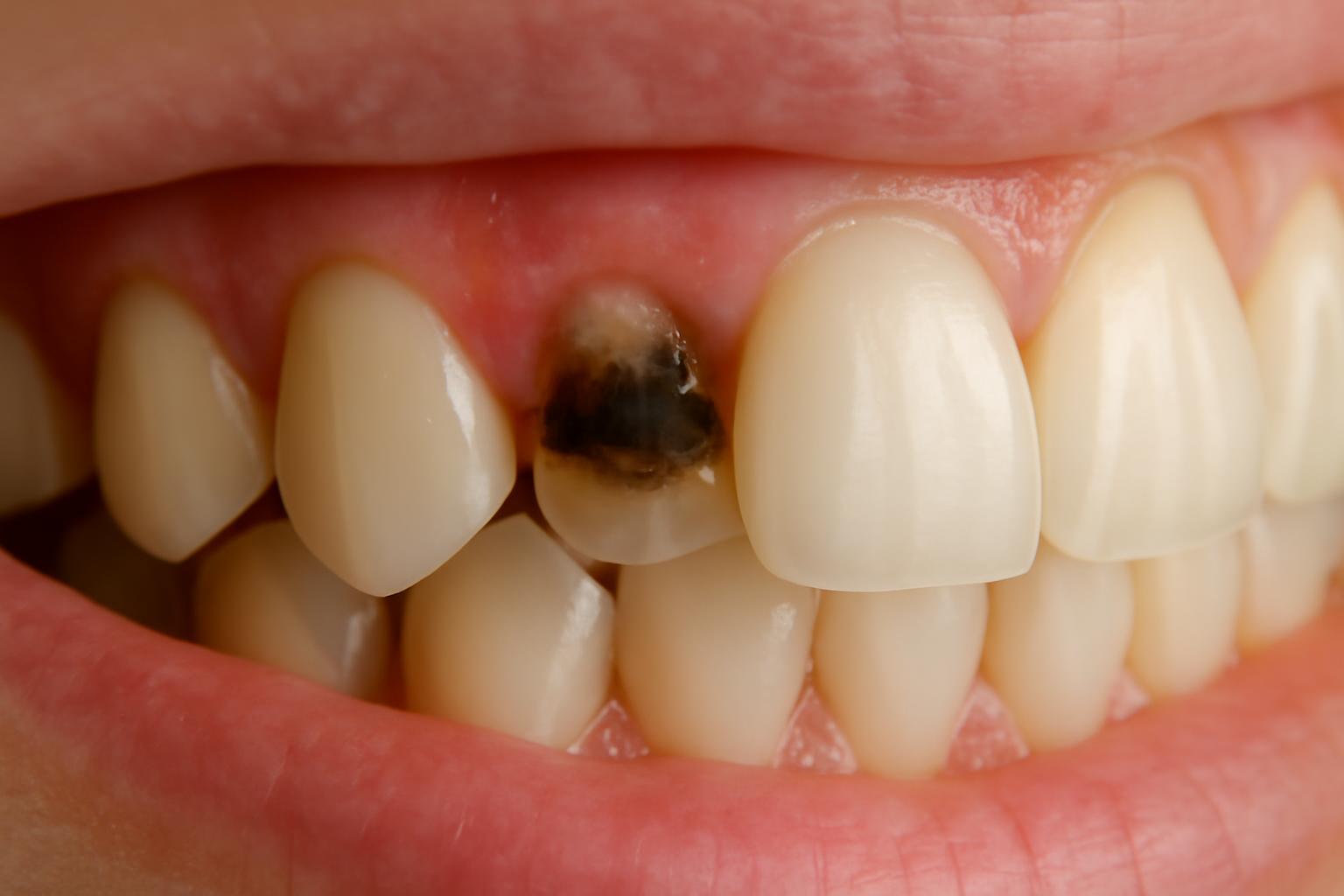
A dark mark, rough patch, notch, or sensitive spot where a tooth meets the gum can be concerning. It may be a gum line cavity, but stain, tartar, an exposed root, gum inflammation, or noncarious tooth wear can create a similar appearance. Looking in a mirror—or deciding that “smooth means stain” and “rough means cavity”—cannot reliably settle the question.
“Gum line cavity” describes a location, not one specific diagnosis. Decay may affect enamel near the gum margin or an exposed root, and that distinction can influence risk and treatment. An area that is only beginning to lose minerals may sometimes be managed without a filling, while a permanent hole generally requires dental repair.
Arrange an examination for a persistent mark, rough area, notch, floss-catching spot, recurrent sensitivity, or pain with chewing. Until then, gentle cleaning with fluoride toothpaste can reduce ongoing plaque exposure, but home care cannot confirm what the spot is or rebuild missing tooth structure.
What is a gum line cavity?
A gum line cavity is an informal name for tooth decay at the junction where a tooth meets the gum. It is not necessarily a separate formal category of cavity. Dentists assess the tooth surface involved, the extent of the lesion, and whether a permanent defect has formed.
Decay on the smooth, enamel-covered surface near or just above the gum margin may be classified as smooth-surface decay. If gum recession has exposed part of the root and decay develops there, it is called root caries or a root cavity. The cervical region is the neck of the tooth near the crown–root junction, but “cervical cavity,” “gumline cavity,” and “root cavity” do not always describe identical anatomy. Healthline distinguishes smooth-surface decay near the gum from decay on an exposed root.
The relevant anatomy includes:
- Crown: The portion of the tooth ordinarily visible above the gum
- Enamel: The hard outer covering of the crown
- Root: The portion that normally extends beneath the gum
- Cementum: The outer covering of the root
- Dentin: Tooth structure beneath enamel and cementum
- Pulp: The innermost tissue containing nerves and blood vessels
When gums recede, cementum and the dentin beneath it can become exposed to the mouth. These root tissues are less resistant to decay than the enamel covering the crown. Even so, a mark near the gum does not establish that the root is involved—or that the mark is decay. The visible location alone does not reveal which tooth surface is affected, whether the surface is intact, or how far any damage extends.
An illustration for this topic should show a tooth in cross-section and label the crown, enamel, gum margin, exposed root, cementum, dentin, and pulp. It should distinguish decay on enamel above the gum from decay on an exposed root and from a lesion extending beneath gum tissue. The central point is that two marks can appear to occupy the same “gumline” area while involving different surfaces.
How decay develops at the tooth–gum junction
A cavity near the gum develops through the same basic process as decay elsewhere. Dental plaque is a sticky bacterial film. When plaque bacteria metabolize sugars and starches, they produce acids that remove minerals from tooth structure. Saliva and fluoride can support mineral replacement, but repeated acid exposure may eventually outweigh that repair process. If enough structure is lost, an initially intact area becomes a permanent cavity.
Sugar does not directly drill a hole into a tooth. The process involves repeated carbohydrate exposure, bacterial acid production, mineral loss, and incomplete recovery. Frequent sipping or snacking can create repeated acid challenges instead of allowing longer recovery periods between exposures. The National Institute of Dental and Craniofacial Research explains this balance between mineral loss and replacement in its guide to the tooth-decay process.
The tooth–gum junction can retain plaque when brushing passes across the visible crowns but does not clean the margin effectively. Plaque can also remain between teeth, where a toothbrush does not reach every surface.
Plaque and tartar are related but not identical:
- Plaque is the bacterial film in which acid production occurs.
- Tartar, or calculus, is plaque that has hardened through mineral accumulation.
- Tartar may make effective cleaning more difficult and provide a surface on which plaque can remain.
- Tartar should not be described as the substance that directly produces decay-causing acid.
- Once a deposit has hardened into tartar, professional removal is generally needed; harder scrubbing at home is not the answer.
Gum recession changes the local environment by exposing a root surface that was previously covered by gum tissue. That surface lacks the crown’s enamel covering and is therefore more vulnerable to mineral loss.
For a broader explanation of demineralization and cavitation, see Decay Guide’s article on how cavities form in stages.
Risk factors: exposed roots, dry mouth, plaque, and frequent sugar exposure
A gum line cavity rarely has one isolated cause. Risk is better understood as an interaction among local exposure, saliva, diet, and cleaning. One person may have recession but effective plaque control and adequate saliva, while another may have modest recession combined with persistent dryness and frequent carbohydrate exposure.
Local exposure and exposed roots
Gum recession exposes a root surface that lacks the crown’s enamel covering. More exposed root area means more vulnerable surface is available for plaque to contact. Recession does not prove that decay is present, and not every cavity near the gum involves recession, but it changes the anatomy a dentist must assess.
Older adults may be more likely to have recession, exposed roots, dry mouth, or previous restorative dental work. Age itself does not prove that a cavity is present. The relevant issue is the individual combination of exposed surfaces, saliva, diet, previous decay, and cleaning ability.
Saliva and dry mouth
Saliva helps clear debris, neutralize acids, and provide minerals involved in tooth-surface repair. Reduced saliva removes some of that protection. Dry mouth may be associated with medications or health circumstances, but a person should not stop, replace, or reduce a medication independently. Persistent dryness should be discussed with dental and medical professionals so its possible causes and practical management can be considered safely. A dental-practice overview describes reduced saliva, recession, plaque accumulation, and frequent carbohydrate exposure as relevant factors in gumline decay (LandMark Dental).
Report persistent dryness and any recurring need to sip water for comfort. These observations do not measure saliva flow or confirm cavity risk by themselves, but they can help a clinician understand the circumstances surrounding the tooth concern.
Plaque and cleaning limitations
Plaque that remains at the gum margin gives bacteria a stable environment. Risk may rise when brushing repeatedly misses that edge or when interdental surfaces are not cleaned.
Brushing force should not be confused with cleaning quality. Scrubbing harder is not a treatment for decay or tartar. Forceful technique may coexist with recession, sensitivity, or noncarious wear, but it should not be assumed to have caused a cavity. A dentist or hygienist can help distinguish recession and wear from decay and demonstrate a technique suited to the person’s mouth.
Frequency of sugar and starch exposure
Plaque bacteria can use fermentable carbohydrates from sugary and starchy foods and drinks. The relevant pattern is not simply whether a person ever eats one particular food. Repeated snacks, sweetened drinks, or prolonged sipping repeatedly supply bacteria with carbohydrate and create additional acid challenges.
A practical reduction strategy is to examine frequency:
- Are sweetened drinks sipped over several hours?
- Are snacks spread throughout the day?
- Are sweetened beverages or lozenges used repeatedly because of dry mouth?
- Could some exposures be consolidated into more defined occasions?
This approach is more useful than treating one food as the sole cause of decay.
Higher-risk profile
Consider asking for an individualized cavity-risk discussion if you have one or more of the following:
- Visible gum recession or multiple exposed roots
- Persistent dry mouth
- Repeated cavities near the gums
- Difficulty cleaning because of sensitivity, limited dexterity, tooth position, or dental appliances
- Plaque or tartar that repeatedly accumulates at the gum margin
- Frequent sipping or snacking on foods and drinks containing fermentable carbohydrates
These factors do not confirm that a particular mark is decay. They indicate that individualized prevention and follow-up may be useful.
Possible signs—and why appearance alone is unreliable
A gum line cavity may cause symptoms, but early decay can be painless. Absence of pain therefore does not rule out early mineral loss. Conversely, sensitivity or discomfort does not prove that decay is responsible.
Possible clues include:
- Sensitivity to cold drinks, cold air, or sweet foods
- White, yellow, brown, or dark discoloration near the gum
- A rough area, pit, notch, or visible defect
- Floss repeatedly catching or fraying at one location
- Localized discomfort while brushing
- Pain or sensitivity when biting or chewing
Cold, sweets, biting, or pressure may raise suspicion of a tooth problem. Red, swollen, tender, or bleeding tissue may point more toward gum inflammation. These patterns overlap: decay may sit beside inflamed gum tissue, and both conditions may be present in the same area. Symptom patterns are tendencies rather than diagnostic rules, as explained in Minnetonka Dental’s comparison of tooth and gum pain.
A dark line is particularly nonspecific. It could represent external stain, tartar, the color of an exposed root, decay, or a noncarious defect. A notch may likewise have more than one possible cause.
A home “smooth versus rough” check is not reliable enough to diagnose or exclude decay. A dental-practice discussion of stains and cavities also labels such self-checks as non-diagnostic and recommends professional assessment for persistent roughness, sensitivity, notching, or floss catching (Matthews Family Dentistry).
The table below summarizes why common observations are inconclusive rather than offering rules for self-diagnosis:
| Clue only—not a diagnosis | Possible decay | External stain | Tartar | Recession | Gum inflammation | Abrasion or erosion |
|---|---|---|---|---|---|---|
| Change near the gum | May involve discoloration, a pit, notch, or sensitivity | May change color without proving structural loss | May form a hardened deposit on the surface | May expose a root that differs in color from enamel | May alter the appearance of the surrounding tissue | May produce a noncarious change in tooth contour |
| Why the clue is inconclusive | Symptoms and appearance cannot show lesion depth or confirm cavitation | Color alone cannot establish what lies beneath | A deposit may obscure the tooth surface | An exposed root may be sound, sensitive, worn, or decayed | Inflammation may coexist with a tooth defect | Tooth wear and decay may resemble one another near the gum |
| What assessment must establish | Whether tooth structure is demineralized or missing | Whether discoloration is external | Whether a removable deposit is present | Which root surface is exposed and whether it is intact | Whether the concern primarily involves soft tissue | Whether structure was lost through decay or another process |
Arrange an examination if a mark, rough area, or notch persists; if sensitivity repeatedly returns; if floss catches in the same place; or if biting and chewing cause discomfort. Professional cleaning may clarify whether a removable deposit is involved, but examination is still needed to assess the underlying tooth.
How a dentist evaluates suspected gumline decay
Diagnosis is a process, not a verdict based on one symptom. The clinician must determine whether the finding involves tooth structure, gum tissue, or both; whether the surface is enamel or exposed root; and whether there is mineral loss, a permanent defect, a deposit, stain, or noncarious wear.
An assessment may include:
- A symptom history. The dentist may ask what triggers discomfort, how long it lasts, and whether it occurs without an obvious trigger.
- Visual and surface examination. The clinician examines the location, contour, and condition of the area.
- Gum assessment. Recession, swelling, bleeding, tenderness, and the position of the gum margin may affect interpretation.
- Cavity-risk assessment. Dry mouth, exposed roots, previous decay, dietary frequency, fluoride exposure, and cleaning limitations may be considered.
- Targeted imaging when appropriate. Dental images may be used when an area is difficult to assess directly or more information is needed.
Imaging supports rather than replaces the clinical examination. Dentists combine the available findings to judge which surface is affected, whether a permanent defect is present, and how the area should be managed. A general overview from LandMark Dental describes visual and surface examination, risk assessment, and selective imaging.
Professional cleaning may help when tartar or external stain covers the area. It can remove a deposit and expose the surface for assessment, but it cannot replace tooth structure already lost through decay or wear.
The position of the gum also matters because a lesion extending beneath the tissue may be harder to reach. That does not mean surgery is automatically necessary; access and treatment must be decided from the individual examination.
How to prepare for the appointment
Before the visit, note:
- What triggers the symptom: cold, sweets, brushing, flossing, biting, or chewing
- How long sensitivity lasts after the trigger stops
- Whether discomfort occurs without a trigger
- When you first noticed the mark, notch, roughness, or sensitivity
- Whether its appearance or symptoms seem to have changed
- Whether you have persistent dry-mouth symptoms
- Whether floss catches, shreds, or breaks at that location
- Your current medications and supplements
Bring an up-to-date medication list, but do not stop or change medication because you suspect it contributes to dry mouth. Medication decisions should be discussed with the relevant prescriber and dental professional.
Treatment by stage: from early mineral loss to deeper decay
Treatment follows a stage-and-access pathway. Not every concern near the gum needs drilling, and not every formed cavity can be managed through prevention alone. The dentist considers whether the surface is intact, whether a permanent defect exists, how far the damage extends, whether the pulp is involved, and whether the area can be reached for treatment.
Stage 1: Early mineral loss without a permanent hole
If the surface remains intact and the lesion is at an early mineral-loss stage, professional preventive management may sometimes arrest or remineralize it. A plan may include fluoride, better plaque control, reduced frequency of sugar exposure, attention to dry mouth, and monitoring.
This is not a promise that every early lesion—particularly every root-surface lesion—will reverse. Suitability for preventive management depends on the surface involved, whether the lesion is active, and the person’s ongoing risk factors. NIDCR explains that early decay may sometimes be stopped or reversed before a permanent cavity forms.
Stage 2: A formed cavity
Once mineral loss has created a permanent hole or structural defect, home remineralization does not rebuild the missing anatomy. Brushing can reduce plaque around the area, but it cannot refill the defect. Whitening may change tooth color in suitable circumstances, but it does not remove decay or restore lost structure.
The important boundary is therefore not simply “small versus large.” It is whether the surface remains noncavitated and potentially manageable through preventive care or has become permanently damaged and requires repair.
Stage 3: Accessible, limited decay
A filling is a common treatment for accessible decay with limited structural damage. The dentist removes damaged tissue as appropriate and restores the defect so the surface can function and be cleaned.
The restorative approach is selected for the individual tooth.
Stage 4: Decay that is difficult to access
Decay extending beneath gum tissue may be more difficult to reach and restore. If gum tissue obstructs access, a dentist may sometimes consider a minor procedure to expose enough of the surface for treatment.
“Below the gum” does not automatically mean surgery. Some lesions can be treated without changing the gum, while others require an individualized access plan. A commercial oral-health overview notes that treatment can become more complicated when decay extends beneath the gum.
Stage 5: Extensive damage or pulp involvement
When substantial tooth structure has been lost, a dentist may consider a more extensive restoration. That decision depends on how much sound structure remains and what is required to protect and clean the tooth—not merely on the cavity’s proximity to the gum.
Root-canal treatment may be considered when decay reaches, infects, or seriously threatens the pulp. It is not routine treatment for every root cavity. A crown is also not automatic; it may be considered when the amount of structural loss and the tooth’s restorative needs justify it.
Treating the cavity is not the same as treating its contributors
Repairing the defect in the tooth does not, by itself, resolve:
- Gum recession
- Active gum inflammation or periodontal disease
- Persistent dry mouth
- Repeated plaque accumulation
- Frequent sugar exposure
- Cleaning limitations
- Noncarious abrasion or erosion
These concerns may require a parallel plan. A restoration can repair a cavity on an exposed root while separate assessment addresses recession or gum disease. Dry-mouth management may help reduce future risk but cannot replace an existing filling. Gum treatment does not remove decay from the tooth.
A useful visual decision pathway would have four branches:
- Noncavitated: Consider professional prevention, fluoride, risk control, and monitoring.
- Formed cavity: Assess for restoration.
- Difficult access: Determine whether direct restoration is possible or another access plan is needed.
- Deep or pulp involvement: Evaluate the need for more extensive restoration or pulp treatment.
What cleaning, whitening, and home care can—and cannot—do
Effective home care reduces ongoing plaque and acid exposure. It may help protect an intact surface, support management of eligible early mineral loss, and reduce risk around an existing restoration. It cannot determine whether a dark or rough area is decay.
Professional cleaning may remove tartar and some external stains. That may make the underlying surface easier to assess, but cleaning does not restore a true cavity or replace missing tooth structure.
Whitening changes the color of suitable tooth structure. It does not disinfect a cavity, remove decayed tissue, restore a notch, or rebuild an exposed root. A dental-practice comparison similarly notes that professional cleaning may remove external deposits, whereas whitening does not treat decay.
Brushing harder is not a treatment. Extra force will not remove a cavity and may make sensitivity, recession, or wear concerns harder to interpret. Use gentle, deliberate technique rather than trying to scrape the spot away.
Fluoride and improved plaque control may help manage an eligible early lesion before cavitation. That is different from regrowing a permanent hole. A dentist must determine whether preventive management, monitoring, or repair is appropriate.
Do not delay an examination solely because sensitivity improves or the area does not hurt. Symptoms can fluctuate, and early decay may be painless.
There is no basis in the supplied evidence for treating a branded mouthwash, water flosser, hydroxyapatite product, or do-it-yourself repair kit as a universally superior solution. Such products should not substitute for determining whether tooth structure has been permanently damaged.
Preventing new or recurring decay near the gums
Prevention should address the same factors that make the area vulnerable: plaque retention, exposed roots, reduced saliva, repeated carbohydrate exposure, and difficulty cleaning. The aim is a routine that is gentle, consistent, and sustainable.
Brush gently with fluoride toothpaste
Brush twice daily with fluoride toothpaste and a soft-bristled brush. Position the bristles so they clean the tooth–gum junction rather than passing only across the middle of the crowns. One commonly taught approach angles the bristles toward the gums and uses gentle pressure, but no exact angle or motion guarantees cavity prevention. The University of Illinois Chicago describes soft-bristled brushing with gentle pressure toward the gum margin, interdental cleaning, and risk-based prevention.
If recession, sensitivity, limited dexterity, or dental work makes cleaning difficult, ask a dentist or hygienist for a demonstration adapted to your mouth. Decay Guide also offers a detailed article on proper toothbrushing technique.
Clean between teeth daily
A toothbrush does not reach every surface between teeth. Use an interdental method you can perform consistently and comfortably. The appropriate tool may depend on the size of the spaces, the position of restorations, dexterity, and professional advice.
If floss repeatedly catches, frays, or breaks at one location, do not keep forcing it through. Arrange an examination to determine whether a deposit, irregular surface, restoration, or contact problem is involved.
Reduce repeated sugar exposure
Focus on how often teeth are exposed rather than trying to identify one forbidden food. Practical changes may include:
- Avoiding prolonged sipping of sweetened drinks
- Reducing grazing on sugary or starchy snacks
- Keeping sweetened foods and drinks to more defined occasions
- Choosing unsweetened hydration between meals where practical
- Checking whether products repeatedly used for dry-mouth comfort contain sugar
These steps reduce repeated opportunities for plaque bacteria to produce acid. They do not compensate for an established cavity.
Address persistent dry mouth
Drink water for hydration and discuss persistent dry mouth with dental and medical professionals. A clinician can consider the symptoms, possible contributing circumstances, cavity history, and appropriate preventive support.
Do not stop or alter medication independently. If medication may be contributing, the prescriber is best placed to decide whether any safe adjustment is appropriate.
Have tartar removed professionally
Once plaque has hardened into tartar, additional scrubbing at home is not the solution. Professional removal exposes the underlying surface and may make daily plaque control easier. Cleaning can remove the deposit but cannot repair a cavity or noncarious notch beneath it.
Use a risk-based follow-up schedule
There is no single examination or cleaning interval that is right for everyone. Follow-up should reflect cavity history, exposed roots, dry mouth, gum health, cleaning ability, current lesions, and the clinician’s findings.
People with recurring cavities, multiple exposed roots, persistent dry mouth, gum disease, or substantial cleaning limitations may need a different preventive plan from people at lower risk. If bleeding, swelling, or recession is also present, Decay Guide’s overview of the stages of gum disease explains why the surrounding tissues require their own assessment.
Ultimately, “gum line cavity” describes where a concern has appeared, not a diagnosis that can be confirmed in a mirror. A persistent dark mark, rough spot, notch, sensitive area, chewing discomfort, or floss-catching point may be decay, but it may also have another cause. Early mineral loss and a permanent cavity require different care, and treatment becomes more involved only when depth, structural damage, pulp involvement, or difficult access warrants it.
Arrange a professional assessment, then focus on sustainable risk reduction: fluoride toothpaste, gentle cleaning at the gum margin, daily interdental care, fewer frequent sugar exposures, hydration, dry-mouth management, and individualized dental follow-up.
Decay Guide is an informational publisher, not a dental practice. Its content is written by a health writer, has not been reviewed by a clinician, and does not provide diagnosis or replace a dental examination, as stated in the site’s health-content disclosure.
Frequently asked questions
Is every gum line cavity a root cavity?
No. A gum line cavity may involve the enamel-covered smooth surface near or above the gum margin, or it may involve an exposed root. Only the latter is root caries. The visible position of a mark near the gum does not reliably reveal which surface is affected. A dental-practice overview similarly distinguishes smooth-surface cavities near the gum from root cavities on exposed roots.
Can a gum line cavity be present without pain?
Yes. Early decay may produce no obvious symptoms. Pain is therefore not a reliable threshold for arranging an examination. Persistent discoloration, roughness, a notch, recurrent sensitivity, or floss catching can justify assessment even when the area is painless. A gumline-cavity overview from Rowlett Dental also notes that early lesions may lack noticeable pain.
Can fluoride reverse a gum line cavity?
Fluoride may help strengthen or remineralize an eligible area of very early mineral loss before a permanent hole has formed. It cannot rebuild missing tooth structure after cavitation. Root-surface lesions vary, so a dentist must determine whether preventive management, monitoring, or restoration is appropriate. This early-versus-established distinction is also described by Mercier Dental.
Does a dark line near the gum mean I have a cavity?
Not necessarily. A dark line may be decay, external stain, tartar, an exposed root, or a noncarious defect. Color and texture may offer context, but neither is reliable enough for self-diagnosis. Professional cleaning and examination can help distinguish a removable deposit from a defect in the tooth. A periodontist-authored overview likewise notes that a dark gumline spot may be tartar or staining rather than decay.
Does a cavity below the gumline always require gum surgery or a root canal?
No. Treatment depends on depth, access, structural damage, and pulp involvement. A below-gum lesion may be restorable without a gum procedure. A minor access procedure may be considered if tissue obstructs treatment, while root-canal treatment is tied to pulp involvement or serious risk to the pulp—not simply to the cavity being near or beneath the gum. These procedures are described as conditional rather than automatic in Channo DDS’s treatment overview.