
The Line Between Reversible Tooth Decay and a Permanent Hole
A white or chalky spot can occur with early mineral loss, but its appearance cannot establish the cause or confirm that the enamel is still intact.
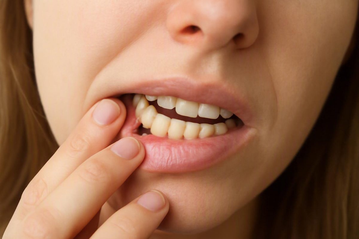
The short answer: early mineral loss may reverse, but a hole cannot heal
A true cavity—a physical hole caused by the loss of enamel or dentin—cannot close or disappear on its own. Saliva, fluoride, brushing, and dietary changes cannot regrow the missing tooth structure. Once a hole exists, the tooth needs professional assessment and case-appropriate management.
Very early tooth decay is different. Before the enamel surface breaks down, acids can remove minerals while leaving the underlying framework intact. This non-cavitated enamel demineralization may sometimes be stopped or reversed as minerals return to the weakened area. The National Institute of Dental and Craniofacial Research distinguishes this early mineral-loss stage from a formed cavity, which represents permanent damage that must be repaired by a dentist (NIDCR’s explanation of the tooth-decay process).
Much of the confusion is about terminology. People—and some educational sources—use cavity to describe both early decay and an actual hole. Clinically, however, these are importantly different conditions:
- An intact area of weakened enamel may be capable of remineralization.
- A broken surface or physical hole has already lost tooth structure and cannot rebuild itself.
A useful way to remember the distinction is remineralize versus restore. An intact enamel framework may regain minerals. Missing enamel or dentin must be professionally managed or restored.
Professional care does not automatically mean a conventional filling. A dentist’s recommendation may depend on whether the surface is intact, how deep and active the lesion appears to be, where it is located, whether it can be cleaned and monitored, and what other risk factors are present. Selected early lesions may be monitored or treated conservatively, while cavitated or deeper lesions may require restorative care.
Pain is not the dividing line. Early decay can be painless, but a more advanced cavity may also remain painless for some time. Nor can a white spot, dark mark, or episode of sensitivity establish whether the enamel surface is intact.
This article provides general education, not a diagnosis. It cannot determine whether a particular mark or symptom is reversible mineral loss, a formed cavity, a stain, erosion, a crack, or another dental condition.
How tooth decay moves from mineral loss to structural damage
Tooth decay is a process rather than a single event. It begins when bacteria in dental plaque use sugars and starches left in the mouth. The bacteria produce acids, and those acids remove minerals from enamel.
This mineral loss is called demineralization. Enamel is highly mineralized, so losing calcium and phosphate weakens its structure even before a visible hole appears. During the earliest stage, the surface may remain intact despite changes beneath it.
The mouth also has a counter-process: remineralization. Saliva helps neutralize acids and supplies calcium and phosphate that can return to weakened enamel. Fluoride can reduce further mineral loss and support mineral replacement. When conditions favor repair often enough, an early lesion may harden or become inactive rather than continuing to progress.
Remineralization is not tissue regeneration. It works within an existing enamel framework. It can reinforce enamel that has lost minerals, but it cannot recreate enamel or dentin that has physically collapsed or disappeared. Think of it as strengthening a weakened wall, not rebuilding a section that is missing.
The balance changes repeatedly throughout the day:
- Sugary or starchy food or drink reaches the mouth.
- Plaque bacteria metabolize those carbohydrates.
- Acids develop around the tooth.
- Enamel loses minerals.
- Saliva gradually helps neutralize the acids.
- Minerals and fluoride may be redeposited if the surface remains intact.
This is why the frequency of exposure matters, not only the total amount consumed. Repeated snacks, sips, or sweetened drinks create repeated acid attacks. Longer gaps between exposures give saliva more opportunity to neutralize acid and support mineral replacement. NIDCR describes this continuing balance between mineral loss and replacement and explains how frequent sugar and starch exposure can allow decay to advance.
If acid attacks repeatedly outweigh repair, the weakened area becomes more porous. Eventually, the enamel surface can lose its integrity and collapse. This is cavitation: an actual pit or hole has formed.
Once the surface is open, plaque and food may collect in an area that is difficult to clean. Decay can then progress through enamel into dentin, the softer layer beneath it. If it continues toward the center of the tooth, it may affect the pulp, which contains nerves, blood vessels, and connective tissue. Saliva and fluoride can support the surrounding enamel, but they cannot replace structure that has already been lost (overview of demineralization, saliva, and cavitation).
The point of no natural return is therefore not simply “the tooth looks different” or “the tooth hurts.” It is the loss of structural integrity. Determining whether the surface is truly intact—and how far a lesion extends—often requires a dental examination.
The stages of decay: what may reverse and what usually needs treatment
The following table is a simplified educational model based on the general progression from enamel mineral loss to cavitation, dentin involvement, and possible pulp disease. Real lesions do not always fit neatly into one stage, and treatment cannot be selected from symptoms alone (clinical overview of cavity progression and treatment).
| Stage | Surface condition | Possible signs | Reversibility | Likely management |
|---|---|---|---|---|
| 1. Early enamel demineralization | The enamel surface remains intact, although minerals have been lost | A white or chalky area may appear; often no pain | May sometimes be stopped or remineralized | Fluoride exposure, improved plaque control, less frequent sugar exposure, management of risk factors, and professional monitoring |
| 2. Enamel breakdown or cavitation | The surface has broken down, creating a pit or hole | A visible or detectable defect, discoloration, food trapping, or no obvious symptom | Missing structure cannot remineralize or close itself | Professional assessment; restorative or another case-appropriate treatment may be needed |
| 3. Dentin involvement | Decay has extended beneath enamel into dentin | Sensitivity, toothache, pain with eating, or no clear symptom | Lost enamel or dentin does not regrow | Often restorative care; treatment depends on depth, location, activity, and remaining tooth structure |
| 4. Pulp involvement or infection | Decay has approached or reached the inner tooth tissues | Significant or spontaneous pain, prolonged sensitivity, pain while chewing, swelling, pus, or changing symptoms | Not reversible through enamel remineralization | Depending on severity and whether the tooth can be retained, treatment may include root-canal treatment, a crown, drainage, or extraction |
At stage 1, minerals have been lost, but the enamel framework is still available for mineral redeposition. The area may look whiter or chalkier than the surrounding enamel, although it can also be difficult to see. Because early enamel decay is often painless, it may be discovered during a routine examination rather than after symptoms develop.
At stage 2, the problem has changed from chemical mineral loss to physical structural damage. A pit or hole means part of the surface is missing. Fluoride may still help protect nearby enamel and reduce future risk, but it cannot make the defect close.
At stage 3, decay has entered dentin. Sensitivity or pain may occur, but those symptoms cannot reliably reveal the lesion’s depth. A cavity may be deeper than it feels, while sensitivity can also result from conditions other than decay.
At stage 4, irritation or infection may affect the pulp and tissues around the tooth. Possible consequences include significant pain, an abscess, and eventual loss of the tooth. Treatment may become more extensive, but no single procedure applies to every case.
The table is not a self-diagnostic checklist. In particular:
- A painless tooth is not necessarily healthy.
- A painful tooth does not automatically have deep decay.
- A white spot is not automatically an active, reversible lesion.
- A dark mark is not automatically a cavity.
- A visible groove or pit may represent normal anatomy, staining, or a structural defect.
- Similar-looking lesions may need different management because their location, depth, activity, and cleanability differ.
Why a white spot, dark mark, or toothache cannot diagnose the stage
A mirror can reveal a change, but it cannot reliably show what is happening inside a tooth.
A white or chalky area is one possible sign of enamel mineral loss. It is not proof of tooth decay, however, and it does not prove that the lesion remains reversible. Even when a white area represents early decay, a dentist still needs to evaluate whether its surface is intact and whether the lesion appears active.
Other findings that deserve assessment include:
- A visible pit or hole
- White, brown, or black discoloration
- Persistent sensitivity to temperature or sweets
- Tooth pain
- Pain when biting or chewing
- A rough area or a location where food repeatedly catches
These findings can occur with decay, but none is specific enough to establish the stage or select treatment.
Early decay can be painless. Some people do not feel a cavity until it reaches dentin or pulp, and the timing and intensity of symptoms vary. The absence of pain therefore cannot rule out decay or show that a lesion has stopped progressing.
Pain also does not function as a depth gauge. Brief sensitivity, lingering pain, spontaneous aching, and pain while chewing can provide useful information, but they cannot show exactly how far a lesion extends. Similar symptoms may arise from several dental conditions.
Location makes home assessment harder. Decay may develop:
- Between neighboring teeth
- In the deep pits and grooves of back teeth
- Around the edge of an existing filling or crown
- Near or below the gumline
- On an exposed root surface
A lesion between teeth may not be visible in a bathroom mirror. A deep groove can look dark without being cavitated, while an apparently minor mark may conceal changes beneath the surface. Professional assessment may include examination of surface integrity, location, depth, and activity, with dental imaging when it is clinically appropriate (why professional examination and imaging may be needed).
A dentist may also consider:
- Whether the area can be cleaned effectively
- The condition of nearby enamel and restorations
- Plaque control and previous decay
- Saliva-related risks, including dry mouth
- Eating and drinking patterns
- Changes documented at earlier visits
Monitoring means more than waiting to see whether the tooth starts hurting. A reduction or change in symptoms does not by itself prove that a lesion has remineralized or become inactive.
What can support remineralization—and what cannot fill a cavity
Home care has an important role, but that role depends on whether the tooth surface remains intact.
For an early, non-cavitated lesion, the goal is to shift the balance away from repeated mineral loss and toward remineralization. For an existing hole, home care can improve the surrounding environment and reduce future risk, but it cannot replace missing tooth structure.
Practical measures include:
- Brush twice daily with fluoride toothpaste. Fluoride helps enamel resist acid and supports mineral replacement in early damage.
- Clean between the teeth. A toothbrush does not effectively clean every contact area, so an appropriate interdental method helps disrupt plaque where teeth meet.
- Reduce the frequency of sugary and starchy snacks and drinks. Fewer between-meal exposures mean fewer repeated acid attacks.
- Drink water and remain hydrated. Hydration supports normal saliva production.
- Keep professional monitoring appointments. Early lesions need reassessment to determine whether they are stable or progressing.
Twice-daily brushing with fluoride toothpaste, reduced between-meal sugar exposure, and regular dental care are established measures for prevention and early-lesion management. They support the mouth’s repair environment; they do not patch a formed hole (NIDCR guidance on fluoride, brushing, and limiting frequent snacks).
The benefit comes from regular plaque disruption and fluoride exposure—not from scrubbing hard enough to remove a mark or defect. A stain that does not brush away should not be treated as something that needs more pressure.
Eating patterns also matter. Constant sipping or snacking can repeatedly restart acid production. Combining exposures into meals rather than spreading them throughout the day may give saliva more time to neutralize acids between episodes. This does not make a particular meal schedule a treatment for a formed cavity; it reduces one contributor to continuing mineral loss.
Saliva is an important part of the protective environment. It helps clear substances from the mouth, neutralize acids, and deliver calcium and phosphate to enamel. Reduced saliva or persistent dry mouth can make remineralization more difficult. Because dry mouth has different possible causes, recurrent or persistent symptoms deserve professional attention rather than being ignored (saliva, dry mouth, and early mineral repair).
What home care cannot do is equally important. None of the following can close a physical hole or regrow missing enamel or dentin:
- Brushing more often or more forcefully
- Flossing
- Mouthwash
- Oil pulling
- Baking soda
- Vitamin or mineral supplements
- Eliminating a particular food
- Following a special “cavity-healing” diet
Some of these practices may have other purposes, but they do not provide a replacement framework for tooth structure that is already gone. Claims that a supplement, special diet, or home remedy can rebuild a formed cavity confuse mineral replacement within intact enamel with regeneration of missing tissue.
Their existence does not change the central boundary: early intact enamel may be supported, but an actual hole cannot be rebuilt at home.
There is also no reliable universal timeline for remineralization. Whether an early lesion stabilizes depends on its depth, activity, location, surface integrity, fluoride exposure, saliva, plaque control, eating pattern, and follow-up. A change in color or symptoms is not enough to confirm success.
The practical division is straightforward:
- Intact early lesion: home care can support a professionally supervised remineralization plan.
- Physical hole: home care remains useful for prevention and risk control but cannot perform the needed structural repair.
Professional treatment does not always begin with drilling
Dental assessment is not synonymous with receiving a filling. The first task is to determine what type of lesion is present.
A dentist may evaluate surface integrity, depth, location, activity, access for cleaning, oral hygiene, saliva-related risk, previous decay, symptoms, and whether reliable follow-up is possible. Imaging may be used when it can provide information that visual and tactile examination alone cannot.
For a selected non-cavitated lesion, management may include:
- Monitoring at planned intervals
- More focused plaque control
- Reducing the frequency of sugar exposure
- Professionally applied or prescription-strength fluoride
- Addressing dry mouth or other risk factors
- Protective measures appropriate to the lesion’s location
This is active management, not neglect. The purpose is to reduce mineral loss, support remineralization where possible, and identify progression before more tooth structure is lost.
Resin infiltration is a minimally invasive option that may be considered for certain non-cavitated lesions. It does not regrow missing tooth structure and is not appropriate for every lesion.
Silver diamine fluoride, commonly shortened to SDF, may be used to arrest decay in selected circumstances. It does not rebuild the missing part of a tooth, and it can permanently darken treated decayed tissue. That tradeoff may be acceptable in some situations and undesirable in others, particularly where appearance matters. Resin infiltration and SDF are therefore case-specific professional options, not universal alternatives to fillings (overview of conservative treatment options and selection factors).
When a formed cavity requires restoration, a filling is a common treatment. The dentist manages decayed tissue as appropriate and restores the defect with a dental material. The restoration replaces form and function; it does not cause the original enamel to regenerate.
More extensive damage may require different treatment:
- A crown may be considered when the tooth needs broader coverage or structural support.
- Root-canal treatment may be needed when the pulp is irreversibly damaged or infected but the tooth can be retained.
- Extraction may be considered when a tooth cannot be predictably restored or retained.
These are broad possibilities, not predictions for an individual tooth. Nor does every formed cavity necessarily receive a conventional filling. Tooth type, lesion location and activity, symptoms, restorability, medical circumstances, cooperation, access to follow-up, and the goals of care may all influence the plan.
The key distinction is that conservative professional care and self-healing are not the same thing. A dentist may use a non-drilling technique to arrest or manage selected decay, but that does not mean a structural hole has rebuilt itself.
Why waiting for pain can allow decay to become a larger problem
Waiting for a tooth to hurt can seem intuitive: if there is no pain, perhaps the problem is minor. Tooth decay does not reliably follow that rule.
Early decay often causes no symptoms. A lesion may progress through enamel before a person notices anything, and some cavities remain quiet after reaching dentin. Pain can signal that a problem deserves attention, but its absence cannot establish that a tooth is healthy, stable, or improving.
Once a lesion is cavitated, it may enlarge as additional enamel and dentin are lost. Possible consequences of progression include:
- Sensitivity
- Toothache
- Pain while biting or chewing
- Pulp inflammation
- Infection
- An abscess
- Loss of supporting tooth structure
- Tooth loss
Untreated decay can lead to pain, infection, abscess formation, tooth loss, and a need for more extensive care. Depending on the condition of the tooth, treatment may progress from preventive or restorative management to a crown, root-canal treatment, or extraction (possible complications and treatment escalation).
The concern is escalation, not a guaranteed sequence for every tooth. A lesion that initially qualifies for preventive management or a conservative procedure may later require a restoration. A small restoration can become a larger one if more structure is lost. Pulp involvement may introduce the possibility of root-canal treatment, while a tooth that cannot be retained may need extraction.
There is no universal timetable for this progression. The rate can vary with the lesion’s location, tooth structure, plaque control, saliva, fluoride exposure, eating frequency, age, and other factors. Claims that every cavity becomes severe within a fixed number of weeks or months are not reliable.
Symptoms may also fluctuate. Feeling better does not prove that decay has stopped, reversed, or become harmless. Only a professional reassessment can determine whether the clinical findings are stable.
For that reason, “watch it until it hurts” is not the same as professional monitoring. Monitoring involves a defined plan and comparison of examination or imaging findings. Passive delay provides no dependable information about whether the lesion is progressing.
A practical guide to routine, prompt, and urgent dental assessment
The appropriate level of attention depends on what has already been established and which symptoms are present.
Routine ongoing monitoring may be appropriate when a dentist has already identified an intact early lesion, assessed its risk, and established a preventive plan. That plan may include fluoride, changes to oral hygiene or eating frequency, and comparison of the lesion at later examinations. Keep the recommended follow-up rather than assuming that a lack of pain means monitoring is no longer necessary.
Prompt dental assessment is appropriate for a suspected cavity or an unexplained change such as:
- A visible pit or hole
- A new or concerning white, brown, or black area
- Persistent sensitivity
- Tooth pain
- Pain while chewing
- An area that repeatedly traps food
- A suspicious white or chalky spot
A white spot deserves assessment because appearance alone cannot determine its cause, activity, depth, or reversibility. A dark groove may be staining rather than decay, but that distinction cannot always be made at home.
There is no single appointment deadline that fits every suspicious tooth. Symptoms, access to care, medical circumstances, and the nature of the change differ. The useful decision rule is simpler:
- Do not wait for pain before seeking assessment.
- Do not assume a white or dark spot is reversible decay.
- Do not expect home care to close a physical hole.
- Seek urgent professional care for severe pain, pus, fever, or facial swelling.
Decay Guide publishes general educational information and cannot examine a tooth, diagnose its condition, or select individualized treatment. As its publisher description explains, it is not a dental practice and does not diagnose or treat patients.
Frequently asked questions
Can brushing with fluoride toothpaste make a cavity disappear?
Fluoride toothpaste can support remineralization when decay is at the early, non-cavitated stage and the enamel surface remains intact. Brushing also disrupts plaque and helps reduce continued acid production.
It cannot make an existing physical hole disappear. Once enamel or dentin has been lost, brushing cannot recreate it. Continue brushing because it protects the surrounding tooth and helps control future risk, but arrange a dental assessment rather than treating extra brushing as a repair.
Does a white spot on a tooth mean the decay is still reversible?
No. A white or chalky area can be associated with early mineral loss, but appearance alone does not establish its cause or confirm that the surface remains intact. White areas can have other explanations, and early decay lesions can differ in activity and depth.
A dentist may examine the surface, consider its location and risk factors, and monitor it over time. Treat a new or changing white spot as something to assess—not as proof that a cavity can be reversed at home.
Can I have a cavity even if the tooth does not hurt?
Yes. Early tooth decay is often painless, and some people do not feel a cavity until it has reached deeper tooth layers. Lack of pain does not rule out decay, prove that a lesion is inactive, or show that it has healed.
Routine examinations are useful partly because they can identify suspicious changes before symptoms become obvious. A visible hole, persistent discoloration, or another unexplained change deserves assessment even when the tooth feels normal.
Can a dentist treat early decay without a conventional filling?
Sometimes. Selected non-cavitated lesions may be managed with monitoring, professional fluoride, improved plaque control, and changes to risk factors. Resin infiltration may be considered for certain intact lesions, while silver diamine fluoride may be used to arrest decay in selected circumstances.
Suitability depends on the surface condition, depth, location, activity, oral hygiene, and ability to monitor the lesion. These approaches are not appropriate for every tooth, and they do not mean missing enamel or dentin has regrown.
What symptoms can indicate that a suspected cavity needs urgent attention?
Severe or escalating pain, pus, fever, and facial swelling warrant urgent professional evaluation because they may accompany dental infection.
Less dramatic findings—including persistent sensitivity, tooth pain, pain while chewing, a visible hole, or a concerning color change—still justify prompt dental assessment. Do not wait for them to become severe.
The distinction to remember is simple: intact, early enamel damage may sometimes regain minerals, but a physical hole cannot rebuild itself. Fluoride, saliva support, effective oral hygiene, and less frequent sugar exposure can support early-lesion management, but they cannot replace missing tooth structure. Because early decay may be painless and suspicious spots cannot be staged reliably at home, a concerning change should be assessed rather than watched until it hurts. Severe pain, pus, fever, or facial swelling warrants urgent professional evaluation. Decay Guide offers general education, not diagnosis or individualized dental advice.