
What Hidden Decay Between Teeth Means—and What May Happen Next
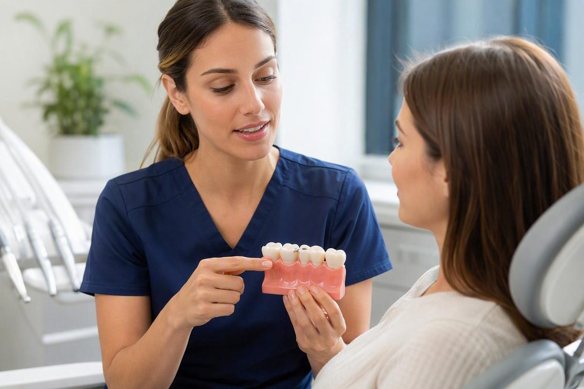
Interproximal cavities are areas of tooth decay on the adjoining side surfaces of neighboring teeth. In everyday language, they are cavities between teeth. Because these surfaces are difficult to see and may not be reached adequately by toothbrush bristles, decay can be present without an obvious hole, dark spot, or pain. Early interproximal decay may be difficult to see and may cause no symptoms.
That leads to two common misunderstandings. First, brushing regularly does not necessarily clean every surface where neighboring teeth meet. Second, sensitivity, trapped food, or floss that shreds may justify an examination, but none of those clues proves that decay is present.
The most useful distinction is between early, noncavitated mineral loss, which may sometimes be stopped or reversed, and an established physical cavity, where tooth structure has been permanently lost and professional repair is generally needed. The National Institute of Dental and Craniofacial Research explains that early decay can be reversed before a permanent cavity forms, but established structural damage requires repair.
What is an interproximal cavity?
An interproximal cavity is decay affecting an adjoining side surface between two neighboring teeth. It may affect one tooth at the contact area, or both facing surfaces may be involved. The term does not mean that both teeth are automatically decayed.
“Interproximal” is the clinical term a dentist may use in records or while discussing an X-ray. “Interdental cavity,” “cavity between teeth,” and “between-tooth decay” are plain-language descriptions of the same general location.
Understanding the basic tooth layers helps explain how decay progresses:
- Enamel is the hard outer covering.
- Dentin is the middle layer beneath the enamel.
- Pulp is the innermost soft-tissue area.
Decay begins in the tooth’s hard outer surface and may extend into deeper layers if the process continues. A dental overview of cavities between teeth and tooth anatomy describes enamel, dentin, and pulp in this order.
The contact between two teeth is less visible than a front surface or a molar’s chewing surface. One tooth can obstruct the view of the next, and toothbrush bristles may not reach the adjoining enamel adequately. A tooth can therefore appear normal in a mirror even when decay is developing on a hidden side surface.
The word cavity is also used loosely in everyday conversation. Sometimes it refers broadly to any finding associated with decay; at other times, it means a physical opening where tooth structure has been lost. That distinction matters because early mineral loss and an established hole do not have the same management options.
A dark line, food trap, sensitive contact, or rough area should not be labeled an interproximal cavity without professional assessment.
How decay forms between neighboring teeth
Tooth decay develops through repeated periods of mineral loss and repair rather than from one food or one missed brushing session. Bacteria in dental plaque use sugars and starches from foods and drinks and produce acids. Those acids remove minerals from enamel, a process called demineralization.
The mouth also has protective processes. Saliva supplies calcium and phosphate that can help replace minerals, while fluoride can reduce mineral loss and support mineral replacement. A cavity forms when repeated mineral loss eventually outpaces the opportunities for repair, according to the National Institute of Dental and Craniofacial Research’s explanation of the decay process.
This cycle helps explain why exposure frequency matters. Repeated sugary or starchy snacks and drinks provide recurring opportunities for plaque bacteria to produce acid. Sipping a sweetened drink over an extended period creates repeated exposures even if no single serving seems unusually large.
Why brushing may not be enough
A toothbrush cleans exposed outer, inner, and chewing surfaces, but its bristles may not pass through a tight contact between neighboring teeth. Brushing carefully remains important, but it does not guarantee that adjoining side surfaces have been cleaned.
This is why someone can brush consistently and still develop interproximal decay. Interdental cleaning has a different physical purpose from brushing: it is intended to reach areas between teeth that bristles may miss. A University of Illinois Chicago dental-school overview notes that brushing does not clean between-tooth surfaces and that prevention should reflect individual cavity risk.
Inadequate interdental cleaning and frequent sugar or starch exposure are modifiable contributors. They do not, by themselves, establish the cause of one person’s lesion.
Bacterial decay versus direct acid erosion
Tooth decay and dental erosion both involve mineral loss, but they are not the same process:
- Decay involves acids produced when plaque bacteria metabolize sugars and starches.
- Erosion is direct chemical damage from nonbacterial acids.
An acidic drink can affect dental health without explaining every cavity between teeth. A sweetened acidic drink may expose enamel directly to acid while also supplying sugar to plaque bacteria, but those are distinct mechanisms.
Possible signs—and why symptoms cannot confirm the diagnosis
Early interproximal decay may cause no pain, sensitivity, or visible change. Its location makes it difficult to inspect directly, and early disease may be discovered only during a professional assessment. Dental sources describing cavities between teeth consistently caution that symptoms may be absent when decay is beginning.
Possible symptoms as decay becomes more extensive include:
- Sensitivity to cold, heat, or sweet foods and drinks
- Discomfort when biting or chewing
- A persistent toothache
- A dark area or visible breakdown when disease is sufficiently advanced
These symptoms deserve attention, but they do not identify the cause by themselves. Sensitivity can come from several tooth or gum conditions. Chewing pain can arise from decay or another dental problem. A dark area may be decay, staining, a shadow, or existing dental work.
Other changes that may draw attention to one contact include:
- Food repeatedly lodging between the same two teeth
- Floss repeatedly catching, fraying, or tearing
- A persistent unpleasant taste
- Bad breath
- Bleeding or irritated gum tissue near the contact
These are reasons to investigate, not diagnostic signs. Food trapping may reflect a change in the contact between teeth.
| Clue | What it may mean |
|---|---|
| No pain or visible change | Possible but nonspecific: early decay can be symptom-free, but an absence of symptoms does not establish whether decay is present. |
| Cold, heat, or sweet sensitivity | Possible but nonspecific: decay is one potential explanation among several tooth and gum conditions. |
| Pain when biting or chewing | Possible but nonspecific: the source may be decay or another dental problem. |
| Persistent toothache | Possible but nonspecific: ongoing pain needs assessment, but pain alone does not identify the affected surface or treatment required. |
| Food repeatedly trapped at one contact | Possible but nonspecific: contact shape, tooth position, dental work, gum conditions, or decay may contribute. |
| Floss catches or shreds | Possible but nonspecific: a rough or irregular surface deserves investigation but does not prove a cavity. |
| Bad breath or an unpleasant taste | Possible but nonspecific: many oral and non-oral factors can cause these changes. |
| Nearby bleeding | Possible but nonspecific: irritated gum tissue is another possible explanation. |
| Dark line, shadow, or apparent hole | Possible but nonspecific: visible change raises concern, but staining or existing dental work can look similar. |
Treatment should not be selected from pain alone.
Do not repeatedly test a suspicious tooth with sweets, ice, hot liquids, hard biting, sharp objects, or home dental instruments. Instead, note what happens naturally: the location, trigger, duration, frequency, and whether the pattern is changing.
How dentists look for hidden decay between teeth
A dentist may assess suspected interproximal decay using several sources of information:
- Symptoms and history: where discomfort occurs, what triggers it, previous cavities, and existing restorations.
- Clinical examination: inspection of the teeth, contacts, dental work, and surrounding gums.
- Cavity-risk assessment: the person’s previous disease pattern, preventive routine, fluoride exposure, and relevant circumstances.
- Radiographs when appropriate: images that can reveal changes in areas hidden by neighboring teeth.
They can provide information about areas that are difficult to inspect directly. Dental-practice guidance on professional evaluation of cavities between teeth describes examination and bitewing radiographs as complementary methods.
Radiographs are important in many assessments, but they should not be presented as the only way to identify a problem or as something everyone needs on a fixed annual schedule. A dentist interprets imaging, when indicated, alongside the examination, symptoms, history, and overall cavity risk.
Treatment likewise should not be selected from one symptom or image in isolation. The supported general principle is that management depends on the stage and extent of decay, the condition of the tooth, symptoms, previous dental work, and the person’s broader risk.
How findings affect the treatment discussion
Useful questions include:
- Which tooth and surface are involved?
- Is the finding being described as early decay or an established cavity?
- How extensive does it appear to be?
- Are symptoms suggesting that deeper structures need assessment?
- What is the condition of the remaining tooth?
- How does the person’s previous cavity history affect the recommendation?
- Why is monitoring, preventive care, or restoration being proposed?
The University of Illinois Chicago describes a risk-based approach to cavity prevention and visit planning.
What the evidence does not establish
There is no supported single numerical accuracy rate here that applies to every bitewing image, tooth, lesion stage, and clinical setting. Nor is there a supported universal rule that everyone needs dental X-rays annually or dental examinations every six months. The appropriate schedule depends on professional assessment of the individual’s findings and risk.
Can an interproximal cavity be reversed?
Sometimes—but only when cavity is being used broadly to describe early mineral loss before a permanent hole has formed. An established physical cavity is different.
During early decay, enamel has lost mineral but has not necessarily suffered permanent structural breakdown. Fluoride, improved plaque control, fewer repeated sugar or starch exposures, and professional monitoring may help shift the balance toward mineral replacement.
Once enough tooth structure has been destroyed to form an established cavity, the missing structure does not grow back. Professional repair is generally required.
| Stage | What it generally means | General management goal |
|---|---|---|
| Early mineral loss | Enamel has lost mineral, but a permanent opening has not necessarily formed. The area may be symptom-free. | Reduce continuing mineral loss, support remineralization, improve preventive care, and reassess the area. |
| Established cavity | Tooth structure has physically broken down, and home care cannot regenerate what is missing. | Professionally manage the decay and restore the tooth where appropriate. |
| Deeper decay approaching or involving the pulp | Disease has extended toward the tooth’s inner tissue and may affect pulp health or the amount of sound structure remaining. | Assess the pulp and the tooth’s restorability, then select an appropriate treatment. |
These are broad stages, not personal treatment instructions. The supplied evidence does not support a universal numerical cutoff—such as a particular fraction of enamel—that automatically determines whether a lesion should be monitored or filled.
A professional recommendation should explain what has been found, how extensive it appears to be, the condition of the tooth, and why preventive management or repair is favored. If the dentist uses terms such as active, stable, noncavitated, or cavitated, ask what findings support that description.
Treatment options as decay becomes more extensive
Treatment is stage-dependent and individualized. The following options describe a range, not an automatic sequence that every tooth follows.
Early noncavitated lesions
When decay is at an early stage and preventive management is considered appropriate, care may involve:
- Fluoride exposure
- More effective plaque removal, including interdental cleaning
- Fewer repeated sugar and starch exposures
- Professional follow-up to assess the area
The aim is to reduce continuing mineral loss and support repair before a permanent cavity forms.
Established cavities and fillings
A filling is a common treatment when a physical cavity has formed. The general process may include:
- Removing decayed or irreversibly damaged tissue as appropriate
- Cleaning and shaping the area
- Placing restorative material
- Finishing the restoration
A healthcare-provider overview of interproximal decay describes removing decay, cleaning and shaping the cavity, and placing a filling.
Larger restorations and crowns
A larger restoration or crown may be considered when more of the tooth requires protection. A crown is not an automatic consequence of discovering a cavity between teeth. Many cavities can be treated with fillings, while teeth with more extensive damage or previous dental work may require a different approach.
Decay affecting the pulp
If decay reaches or seriously affects the pulp, root-canal treatment may be considered when the tooth remains restorable. Additional restoration may then be needed to return the tooth to function and protect the remaining structure. A general account of interproximal cavity treatment describes fillings for typical cavities and root-canal treatment when decay has reached the pulp chamber.
Pulp treatment is not selected merely because a tooth is sensitive. Symptoms, examination findings, imaging when appropriate, pulp health, and restorability all require professional assessment.
Extraction
Extraction may be considered when extensive decay, infection, or structural damage means a tooth cannot be saved. It is not the expected result of an ordinary cavity between teeth, and pain alone does not establish that extraction is necessary.
Across these treatment categories, relevant decision factors include:
- The stage and extent of decay
- Symptoms
- Pulp health
- Remaining tooth structure
- Whether the tooth can be restored
- Previous fillings, crowns, or other dental work
- The person’s pattern of cavity risk
No article can determine which treatment is appropriate for an individual tooth. A useful explanation from a dentist should connect the recommendation to the actual findings rather than relying only on the generic label “cavity.”
Preventing decay on surfaces a toothbrush may miss
A practical prevention routine has three foundations:
- Brush twice daily with fluoride toothpaste.
- Clean between the teeth daily using a suitable method.
- Reduce repeated sugary or starchy exposures.
Brushing applies fluoride and disrupts plaque on exposed surfaces. Interdental cleaning is intended to reach adjoining surfaces that toothbrush bristles may not clean adequately. Diet timing affects how often plaque bacteria receive sugars or starches that they can convert into acid.
Brushing harder or longer does not replace interdental cleaning. Use a separate interdental method suited to the available space, dental work, technique, and dexterity.
String floss
Guide it through gently rather than repeatedly snapping it into the gum.
If floss consistently shreds or catches at one point, do not keep forcing it. Report the location to a dentist so the tooth, contact, and any existing restoration can be examined.
Interdental brushes
It should pass through without being forced.
Water flossers
A water flosser may be a practical alternative for someone who has difficulty using conventional floss. Dental-practice guidance identifies water flossers and interproximal brushes as possible alternatives when flossing is difficult.
That does not establish that all tools are equally effective for preventing interproximal cavities in every person. The practical goal is safe, consistent cleaning with a method suited to the relevant spaces.
Why exposure frequency matters
Repeated sugary or starchy snacks and drinks can produce recurring periods of mineral loss. Practical changes may include:
- Reducing between-meal grazing on sugary or refined-starch foods
- Avoiding prolonged sipping of sweetened drinks
- Keeping sweet foods and drinks to fewer occasions
- Choosing unsweetened options more often
- Continuing fluoride-toothpaste use rather than relying on dietary changes alone
The goal is not a perfect diet. It is to reduce repeated acid-producing opportunities while supporting plaque control and mineral replacement.
Professional prevention should be risk-based
People with recurring interproximal decay may need a different preventive and review plan from those with a long period of stability. Professional care may include reviewing cleaning technique, checking existing restorations, discussing fluoride and dietary exposures, and deciding when monitored areas should be reassessed.
Mouthwash should not be treated as a universal requirement. If one is recommended, ask what ingredient is relevant and how the product fits with—not replaces—fluoride toothpaste and interdental cleaning.
When a dental assessment is worth arranging
Professional assessment is reasonable if you have:
- Persistent tooth pain
- Recurring sensitivity in the same area
- Discomfort when biting or chewing
- Food repeatedly trapping at one contact
- Floss consistently catching, hurting, or shredding in the same place
- A visible change that does not disappear with ordinary cleaning
- A previously identified area that has not been reassessed as planned
These findings do not establish the cause. They provide a reason to have the tooth, contact, dental work, and surrounding gum examined.
Waiting for severe pain or a clearly visible hole may miss an earlier symptom-free stage. Conversely, a painless finding on an X-ray still needs professional interpretation in the context of the examination and the person’s cavity risk.
This general article cannot assess urgency or provide a complete emergency-triage checklist. If symptoms are severe, rapidly worsening, or causing concern about infection, contact a dentist or an appropriate local urgent-care service rather than relying on self-diagnosis.
Before an appointment, record the symptom’s location, trigger, duration, frequency, and whether the pattern is changing. Avoid repeatedly provoking the tooth.
Questions to ask at the appointment
- Which tooth and exact surface are involved?
- Is this early mineral loss or an established physical cavity?
- Is the lesion thought to be active, and what supports that conclusion?
- What findings favor preventive or fluoride-based care?
- What findings favor a filling?
- If a larger restoration is recommended, why is a smaller repair unsuitable?
- How will stability or progression be checked?
- Which interdental method is most suitable for this contact?
- How does my cavity risk affect the examination or imaging schedule?
The useful dividing lines are straightforward: decay between teeth may be hidden and painless; symptoms are clues rather than proof; and early mineral loss is not the same as an established cavity. A dentist can combine the history, clinical findings, individual risk, and radiographs when appropriate to explain whether preventive care, monitoring, or restoration is justified.
In the meantime, the broad preventive priorities are fluoride toothpaste, consistent interdental cleaning, and fewer repeated sugar or starch exposures. Professional care should be tailored to the individual rather than imposed on a universal timetable.
Decay Guide describes itself as an independent general-information publisher, not a dental practice. This article cannot diagnose a tooth, select treatment, assess an emergency, or replace individualized advice from a qualified dental professional.
Can I have an interproximal cavity without any pain?
Yes. Early interproximal decay may cause no pain, sensitivity, or visible change, and its position between adjoining teeth makes it difficult to see directly. Symptoms are not always present when an interproximal cavity is beginning.
Pain is not a reliable dividing line between decay and no decay. A painless finding still requires interpretation, while a painful contact may have another cause.
Can a dentist find a cavity between teeth without an X-ray?
Sometimes. Dentists may also use radiographs when appropriate because adjoining teeth can make the facing surfaces difficult to inspect directly. An overview of cavities between teeth describes examinations, dental instruments, and X-rays as possible detection methods.
Imaging decisions should reflect the examination, previous findings, symptoms, and cavity risk rather than a universal annual rule.
Can fluoride reverse a cavity between teeth?
Fluoride may help stop or reverse early noncavitated mineral loss, when a permanent hole has not yet formed. It cannot regrow tooth structure that has already been physically lost.
A dentist must determine which stage is present and whether fluoride-based care and follow-up are appropriate.
Does floss that catches, shreds, or hurts mean I have a cavity?
No. Repeated catching or shredding is a possible but nonspecific clue. It may be associated with decay, but a rough filling, tartar, an irregular contact, damaged tooth structure, gum irritation, or technique could also explain it. Practice guidance on possible signs notes that snagging floss and repeated food trapping warrant investigation but are not conclusive.
Do not repeatedly snap or force floss through the area. Note where the problem occurs and ask a dentist to inspect that contact.
Can I use an interdental brush or water flosser instead of string floss?
Possibly. String floss often suits tight contacts, an appropriately sized interdental brush may suit spaces where it fits without force, and a water flosser may be an option for someone who struggles with conventional floss.
These methods should not be assumed to be equally effective for every person or space. The goal is consistent, safe cleaning with a suitable tool; a dentist or dental hygienist can help match the method to the contact.