
How Tooth Decay Can Change From a White Spot to a Visible Hole
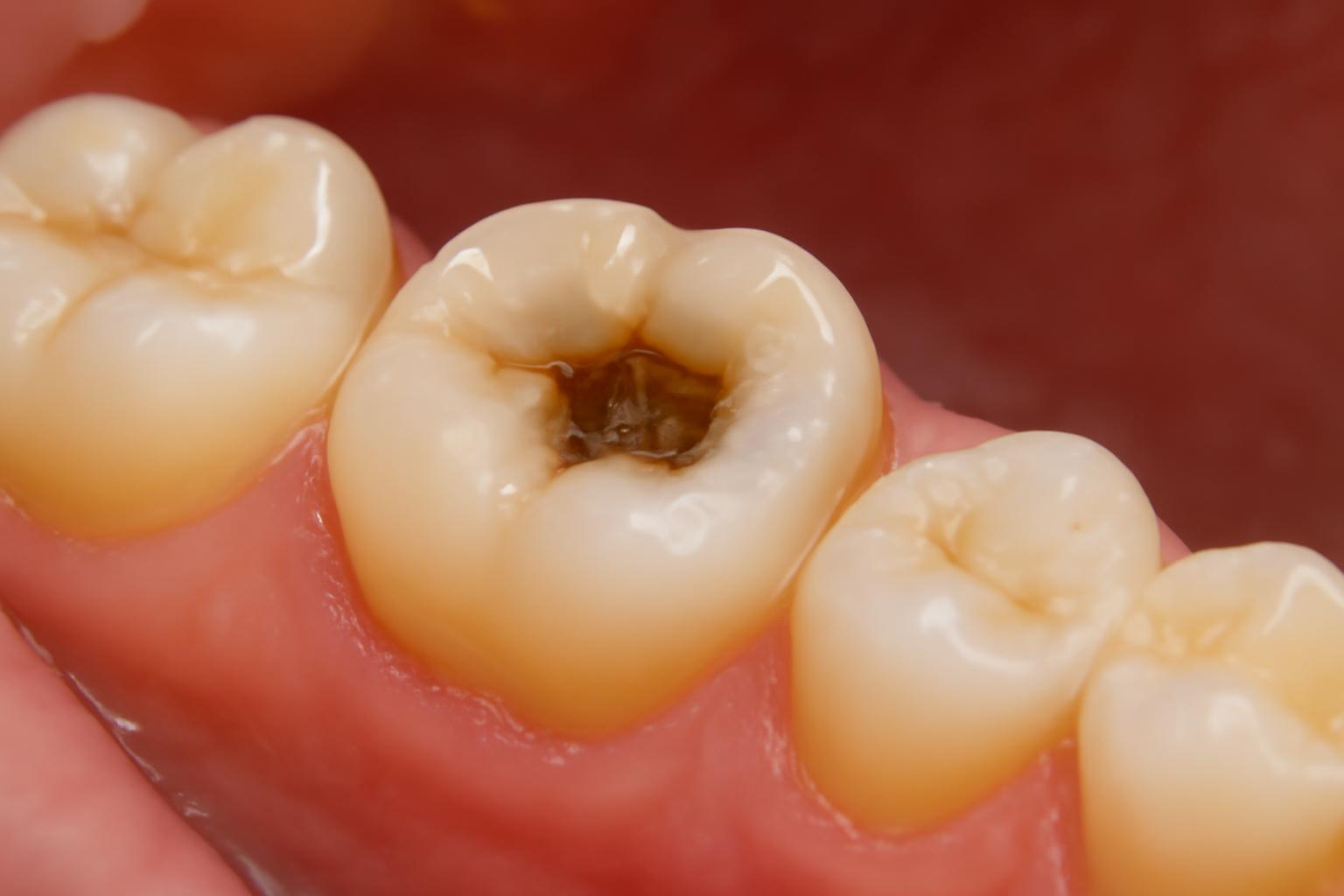
Possible decay may appear as a subtle white enamel change, a rough, discolored pit or a visible hole, but stay hidden between teeth or under dental work.
Important: This article is educational, not diagnostic. Decay Guide states that its articles are written by a health writer, not a dentist, and have not been reviewed by a clinician. A dentist must examine a suspicious tooth to determine whether decay is present.
The short answer: what a cavity may look like
Possible signs of tooth decay include:
- A dull, matte, milky, or chalky-white patch
- Localized yellow or brown discoloration
- A dark dot or line
- A rough or sunken patch
- A small pit or indentation
- A visible opening or hole
- A broken edge or missing tooth structure
Early decay may be white rather than dark. A chalky-white area can represent enamel losing minerals before an open defect has formed. As the process advances, discoloration, roughness, pitting, or visible structural damage may develop. Early decay can also be painless and difficult to see, according to the Cleveland Clinic’s overview of cavities.
A cavity is not always visible. Decay may develop between teeth, in deep chewing-surface grooves, near the gumline, on an exposed root, or beneath a filling or crown. A dental examination and, when appropriate, X-rays may be needed to find these areas.
Visual summary
What you notice What it could look or feel like Possible symptoms Can you assess it reliably at home? Color change Chalky white, yellow, brown, or black Often none; sometimes sensitivity No. Decay, stains, tartar, and enamel changes can overlap in appearance. Surface change Rough patch, pit, depression, broken edge, or hole Food trapping, biting discomfort, or sensitivity No. Structural change is concerning but does not reveal the cause or depth. Hidden change No visible mark Sensitivity, floss catching, or no symptoms No. Examination or imaging may be needed. Advanced change Larger opening, fractured tooth, or missing tooth material Persistent pain, swelling, bad taste, or drainage No. Professional care is needed, and infection warning signs may require urgent attention.
No color, mark, photograph, or texture can confirm a cavity by itself. The most useful observations are where the change is located, whether the surface looks damaged, whether symptoms are present, and whether the area is new or changing. Those clues still need professional interpretation.
How tooth decay can look at different stages
People often use “tooth decay” and “cavity” interchangeably, but there is a useful distinction:
- Tooth decay is the disease process in which tooth structure loses minerals and may progressively break down.
- An early white-spot lesion is an area of enamel mineral loss that may still have an intact surface.
- A cavity, in the structural sense, is an area where tooth material has broken down into an opening or hole.
The patterns below are educational examples, not stages that can be diagnosed from a mirror. Teeth do not necessarily progress through a visible white-to-yellow-to-brown-to-black sequence. A darker area is not automatically deeper, more severe, or more active than a lighter one.
| General pattern | Likely appearance | Surface or texture | Possible symptoms | How visible is it? | Appropriate next action |
|---|---|---|---|---|---|
| Early enamel mineral loss | Matte, dull, milky, or chalky-white patch; sometimes only a loss of normal shine | Often intact, without an open hole | Frequently painless | Easy to overlook and may be hidden by location | Arrange an assessment if the patch is new or persistent |
| Progressing enamel breakdown | White, yellow, tan, or light-brown area; possibly a small dot or line | May appear rough, pitted, depressed, or sunken | None or occasional sensitivity | Sometimes visible under good light | Book an assessment rather than waiting for pain or a larger defect |
| Possible deeper involvement—not visually diagnosable | Brown or dark area, sometimes with a clearer pit or opening | Loss of tooth structure may be more evident | Sensitivity to sweets, heat, or cold; possible biting discomfort | May be obvious but can remain hidden between teeth | Seek a prompt dental appointment |
| Advanced structural damage | Larger opening, fractured edge, or missing tooth material; discoloration may be present | Definite breakdown of the tooth | Persistent or severe pain, lingering sensitivity, swelling, bad taste, or drainage | Often visible, unless beneath dental work or between teeth | Obtain prompt care; use urgent dental services for infection warning signs |
These general appearance and symptom patterns are consistent with Harvard Health’s cavity guide, which also notes that early decay may cause no symptoms and hidden areas may require X-rays.
Early enamel mineral loss: A chalky-white area can occur when enamel has lost minerals but has not formed a hole. It may look less glossy than the surrounding tooth, particularly when dry. Because the surface can remain intact and painless, the change may be easy to overlook or confuse with a non-decay enamel difference.
Progressing enamel breakdown: The affected area may become yellow or light brown, or its surface may look less uniform. A small pit, indentation, or depression can appear.
Possible deeper involvement: A clearer pit or opening and sensitivity to sweet, hot, or cold items can occur as damage extends farther into a tooth. Brown or black discoloration may be present, but it cannot reveal how deeply decay extends.
Advanced structural damage: Greater loss of tooth material may produce a larger opening, weakened or fractured edges, persistent pain, or swelling.
There is therefore no dependable “cavity color chart.” Darkness can reflect staining, trapped pigment, inactive decay, or exposed underlying tooth material. Dentists consider the surface, location, symptoms, progression, surrounding tooth structure, and imaging findings rather than using color as a depth gauge.
Why cavities look different depending on where they form
On a molar’s chewing surface: Back teeth have natural pits and fissures. Decay may appear as a suspicious dot, discolored groove, rough patch, pit, or hole. Normal anatomy and harmless groove staining can look similar, so a black line in a molar groove is not automatically a cavity.
Between teeth: Decay can begin where neighboring teeth touch, leaving little or no visible change on the surfaces you can inspect. A person may notice food trapping, sensitivity, or floss repeatedly catching, but there may be no clue at all. X-rays can reveal some between-tooth decay that cannot be seen directly.
Near the gumline or on an exposed root: Decay may occur where a tooth meets the gum or where gum recession has exposed part of the root. The area may show discoloration, an indentation, roughness, or structural loss, but it does not have a single diagnostic color. Tartar, staining, wear, and non-decay root-surface changes can affect the same region.
Around a filling or crown: New decay can occur at a restoration’s margin or beneath existing dental work. At home, someone might notice a discolored line, rough edge, or small gap—or nothing unusual. A line around a filling or crown does not by itself prove decay, and hidden damage may require imaging or another form of clinical assessment.
These location-specific patterns, including cavities in molar grooves, between teeth, near exposed roots, and around restorations, are described in Grand Oaks Dentistry’s visual cavity guide.
On a front tooth: A suspicious change may appear as a white or discolored area near the gumline or between adjacent teeth. Front teeth are easier to inspect than back teeth, but greater visibility does not make home diagnosis reliable. Developmental enamel differences and other non-decay conditions can also produce white patches.
On a baby tooth: Decay in a primary tooth can follow the same broad visual pattern as decay in an adult tooth: a white area may be followed by darker discoloration, a pit, or a visible hole. Children may also show pain, irritability, or cheek swelling. Some childhood cavities remain hidden between teeth and are found through an examination or X-ray, as explained in this pediatric guide to early cavity signs.
Across these locations, ask four practical questions:
- Is the area new or changing?
- Does the surface appear intact or structurally damaged?
- Are sensitivity, pain, swelling, or food trapping present?
- Is the area in a location that can actually be inspected?
The answers can help you describe the concern to a dentist, but they cannot confirm decay.
Cavity or stain? Why appearance alone cannot tell you
A brown or black mark can be decay, but it can also be staining. Early mineral loss may look white, while non-decay enamel changes may also appear white. Cavities and other tooth changes can therefore overlap in color.
Structural change generally raises more concern than color alone. A new pit, sunken patch, rough or broken edge, or visible loss of tooth material may indicate that the surface has broken down. Uncomplicated discoloration may leave the tooth intact. Even so, a mirror or photograph cannot reliably determine whether the surface is intact or whether a lesion is active.
The distribution of discoloration is also only a weak clue. Widespread color change may suggest generalized staining, while a persistent localized mark deserves attention. But staining can be localized, and decay can affect more than one tooth. Neither pattern is a dependable home test.
Several findings can resemble a cavity:
- Pigment trapped in a normal molar groove
- Surface or internal tooth staining
- Tartar buildup
- Fluorosis or another developmental enamel change
- Worn, chipped, or eroded enamel
- An inactive or arrested area of previous decay
- Discoloration around an existing restoration
Visual overlap between stains and cavities can be substantial, particularly early in the process. A dentist may need to determine whether the tooth surface is damaged and whether an area is active rather than simply stained, as discussed in Putney Dental Care’s comparison of stains and cavities.
Dentists do not classify every dark groove as a cavity based on color alone. They consider the area’s location, surface condition, progression, symptoms, surrounding tooth structure, restoration history, and—when appropriate—imaging.
Arrange an evaluation if a mark is:
- New or unexplained
- Still present after normal brushing
- Enlarging or changing
- Rough, pitted, sunken, or visibly open
- Associated with repeated food trapping
- Sensitive or painful
- Near a filling or crown that looks or feels different
Do not assume that a stain is harmless, smooth, reversible, or removable with whitening. Tooth discoloration has several possible causes, and cosmetic treatment should not replace an assessment of whether tooth structure is damaged.
What a cavity may feel like—and why pain is an unreliable test
Early tooth decay often causes no pain. A comfortable tooth can still have enamel mineral loss, a hidden cavity, or damage that has not yet irritated deeper structures. Waiting for toothache may allow a concern to progress unnoticed.
Possible symptoms that can accompany decay include:
- Sensitivity to sweet foods or drinks
- Sensitivity to heat or cold
- Pain or discomfort when biting
- A spontaneous or persistent toothache
- Food repeatedly lodging in one place
- Persistent bad breath
- An unpleasant or foul taste
Symptoms can become more likely as damage reaches deeper tooth structures, but symptom severity cannot determine cavity depth at home. These symptoms can accompany cavities but may also have other causes, as summarized in this guide to cavity symptoms.
Texture-related clues are also nonspecific. You might notice a rough spot with your tongue, a small indentation, floss fraying in one location, or food repeatedly trapping between two teeth. These observations identify an area worth discussing with a dentist; they do not diagnose decay. A chipped edge, rough restoration, tartar, or another structural issue can produce similar experiences.
Sensitivity and pain can also result from gum recession, enamel wear, a crack, a damaged restoration, inflammation, or another dental condition. An examination is needed to identify the cause.
Myth versus fact
Myth: “No pain means no cavity.” Fact: Early and hidden decay may cause no pain. Comfort does not rule it out.
Myth: “Every cavity is black.” Fact: Early mineral loss may look chalky white, some cavities are not visible, and dark marks can be stains or inactive lesions.
Myth: “A visible hole must appear before decay needs attention.” Fact: Tooth decay can be assessed before an open defect develops. A persistent change may deserve evaluation even when no hole is visible.
The absence of pain shows only that the tooth is not currently painful. It does not establish that the tooth is healthy.
What you can check safely at home—and what requires a dentist
Home inspection can help you notice and document a change. It cannot diagnose it.
Under good light, use a mirror to look for:
- A new white, yellow, brown, or dark patch
- A dot or line that looks different from nearby grooves
- A pit, indentation, or sunken area
- A rough-looking or broken edge
- A visible opening
- Discoloration or a gap near a filling or crown
- Swelling or a bump on the gum
Record when you first noticed the area. If practical, take a clear photograph without forcing the mouth open or touching the tooth. Note whether the mark changes and whether you experience sensitivity, pain when biting, food trapping, swelling, bad taste, or drainage.
Do not probe, pick, scrape, or press into the area with a pin, needle, metal tool, toothpick, or another sharp object. Such testing cannot establish a diagnosis. The safe role of home inspection is to look, document, and arrange professional assessment rather than attempting to test the tooth yourself. At-home checks are not a substitute for the professional methods described in the Grand Oaks Dentistry cavity overview.
A dentist can assess discoloration, pits, surface breakdown, fractures, restoration problems, and other clinical findings. X-rays may be used when decay is suspected between teeth, beneath dental work, or in another area that cannot be inspected directly.
In plain language, dense enamel generally looks lighter on a dental X-ray because it blocks more of the X-ray beam. Tooth structure that has lost density through decay may appear relatively darker. That is why an area between two teeth can look normal in a bathroom mirror but appear as a darkened region on an X-ray. This radiographic distinction is described in Lakeshore Dental’s explanation of cavity detection.
An X-ray is not simply a photograph of a hole. Its interpretation depends on factors such as the image angle, tooth anatomy, location, restoration history, and clinical examination. Imaging is one part of an assessment, not a do-it-yourself visual test.
Neither a home photograph nor a symptom checklist can establish:
- Whether a mark is active decay
- Whether a dark area is inactive or arrested
- Whether the surface has broken down
- How far damage extends into the tooth
- Whether decay is present beneath a restoration
- Which treatment, if any, is appropriate
Self-inspection is useful when it helps you notice a change and describe it clearly while arranging care.
Can an early cavity heal, or will it need a filling?
Not every suspicious white patch or discolored area requires a filling. The central distinction is between early enamel mineral loss with an intact surface and established structural breakdown.
Some early enamel lesions without an open defect may remineralize under professional guidance. Fluoride and improved oral hygiene may help minerals return to weakened enamel in suitable cases. Not every white patch is decay, however, and not every early lesion is active or reversible.
Once tooth structure has broken down into an established hole, the missing material does not grow back or heal by itself. A dentist commonly removes damaged material and restores the tooth with a filling. Depending on the location and extent of damage, treatment may instead involve a crown, root-canal treatment, or extraction. These are general treatment categories, not recommendations for any particular tooth, as outlined in this cavity progression and treatment overview.
The practical distinction is:
- White or discolored but intact-looking area: It may represent early mineral loss, staining, fluorosis, or another enamel change. It needs assessment, not an automatic filling.
- Pitted, broken, or visibly open area: Structural breakdown is more concerning and should be examined promptly.
- No visible finding: Hidden decay remains possible when symptoms or examination findings raise concern.
Do not try to “remineralize” a visible hole at home while postponing an examination. Remineralization applies to selected early lesions, not missing tooth structure.
For more background on the biological progression, see Decay Guide’s guide to how cavities form in stages. That material is educational and cannot determine whether a particular white spot, dark groove, or hole requires treatment.
When to book a dental visit—and when symptoms are urgent
Arrange a dental assessment for a tooth change that is new, persistent, enlarging, rough, pitted, sensitive, painful, or visibly open—even if it does not currently hurt.
A prompt appointment is particularly appropriate for:
- A visible pit or hole
- A broken edge or missing tooth material
- Recurring pain when biting
- Lingering sensitivity to heat or cold
- Food repeatedly lodging in the same area
- A changing mark near a filling or crown
- A persistent change on a child’s tooth
Seek urgent dental care for possible infection or abscess signs, including:
- Severe or throbbing pain
- Facial, jaw, or gum swelling
- Fever
- Drainage or pus
- A foul or suddenly unpleasant taste
- A pimple-like bump on the gum
These symptoms can accompany an infection requiring timely professional attention. Oral-B’s reviewed patient guide identifies swelling, a raised gum bump, throbbing pain, fever, and a foul taste from drainage as warning signs requiring immediate dental help in its cavity and abscess guidance.
This article addresses only the dental warning signs supported by the cited material; it is not a complete emergency-medical triage tool. If you believe you are experiencing a medical emergency or cannot safely wait for dental advice, contact local emergency services or seek urgent medical help.
A three-level framework can help:
- Document and arrange routine assessment: You notice a new or persistent patch or dot but have no pain, swelling, or obvious structural loss. Monitor only long enough to record what you see and arrange a dental visit.
- Seek a prompt appointment: There is a pit, visible hole, broken area, repeated food trapping, biting pain, or lingering sensitivity.
- Seek urgent dental help: There is severe throbbing pain, fever, swelling, drainage, a foul taste, or a gum bump.
Lack of pain is not a reason to ignore a persistent change.
Frequently asked questions
Can a cavity be white instead of brown or black?
Yes. Early enamel demineralization may appear as a dull, matte, milky, or chalky-white patch before an open hole forms. The area may be painless and easy to overlook.
A white patch is not necessarily decay. Fluorosis, developmental enamel differences, and other changes may also look white. A dentist must determine whether the area represents active mineral loss, an inactive change, or something unrelated to decay. Harvard Health describes white, brown, or black spots as possible appearances while emphasizing that early decay may be symptom-free in its cavity appearance guide.
Does a black dot or dark line in a molar groove always mean a cavity?
No. Molar chewing surfaces contain natural grooves that can collect pigment. A dark dot or line may reflect staining, normal groove anatomy, inactive decay, or an active cavity.
Concern increases if the area is new, changing, pitted, rough, sensitive, painful, or associated with repeated food trapping. Those clues still cannot confirm decay. The overlap between dark staining and cavities is discussed in this stain-versus-cavity guide.
Can I have a cavity if my tooth does not hurt?
Yes. Early decay commonly causes no symptoms, and some cavities remain painless until damage reaches deeper tooth structures. Decay between teeth or beneath dental work may also progress without an obvious visible sign.
Do not wait for pain if you notice a persistent patch, pit, rough area, or other change. Pain does not prove a cavity either, because sensitivity and toothache can have several causes. Cleveland Clinic notes that a person may not feel a cavity until it reaches dentin or the tooth’s inner tissue in its cavity symptoms overview.
What does a cavity between teeth look like on an X-ray?
Decay between teeth may appear as a relatively dark area or shadow compared with the lighter, denser enamel around it. The contrast reflects reduced density in affected tooth structure.
Its exact appearance and visibility depend on the location, extent, anatomy, and angle of the image. An X-ray must be interpreted alongside a clinical examination; a dark region is not suitable for self-diagnosis. A dentist’s explanation of this light-versus-dark density difference is available in the Lakeshore Dental radiography guide.
Can an early cavity go away without a filling?
An early area of enamel demineralization may sometimes remineralize if the surface has not broken down. Professional guidance, fluoride, and improved oral hygiene may support that process in appropriate cases.
An established hole cannot regrow missing tooth structure or heal by itself and generally requires dental treatment. Because color and photographs cannot reliably show whether the surface is intact, a dentist should assess a suspected early lesion before deciding whether restoration is necessary. This distinction between potentially remineralizable early damage and an established cavity is explained in the Crest cavity-stage guide.
A possible cavity can range from a subtle white enamel change to a rough, discolored pit or visible hole—and it may be invisible between teeth or beneath dental work. Because color, pain, texture, and photographs are unreliable diagnostic tests, a persistent or changing mark deserves dental assessment. Seek urgent dental care for severe throbbing pain, fever, swelling, drainage, a foul taste, or a gum bump.
Decay Guide is not a dental practice and does not diagnose or treat dental conditions. Its articles are written by a health writer, are not clinician-reviewed, and cannot replace a dental examination, as stated in Decay Guide’s non-clinical disclosure.