
When Decay and a Crack Damage the Same Tooth
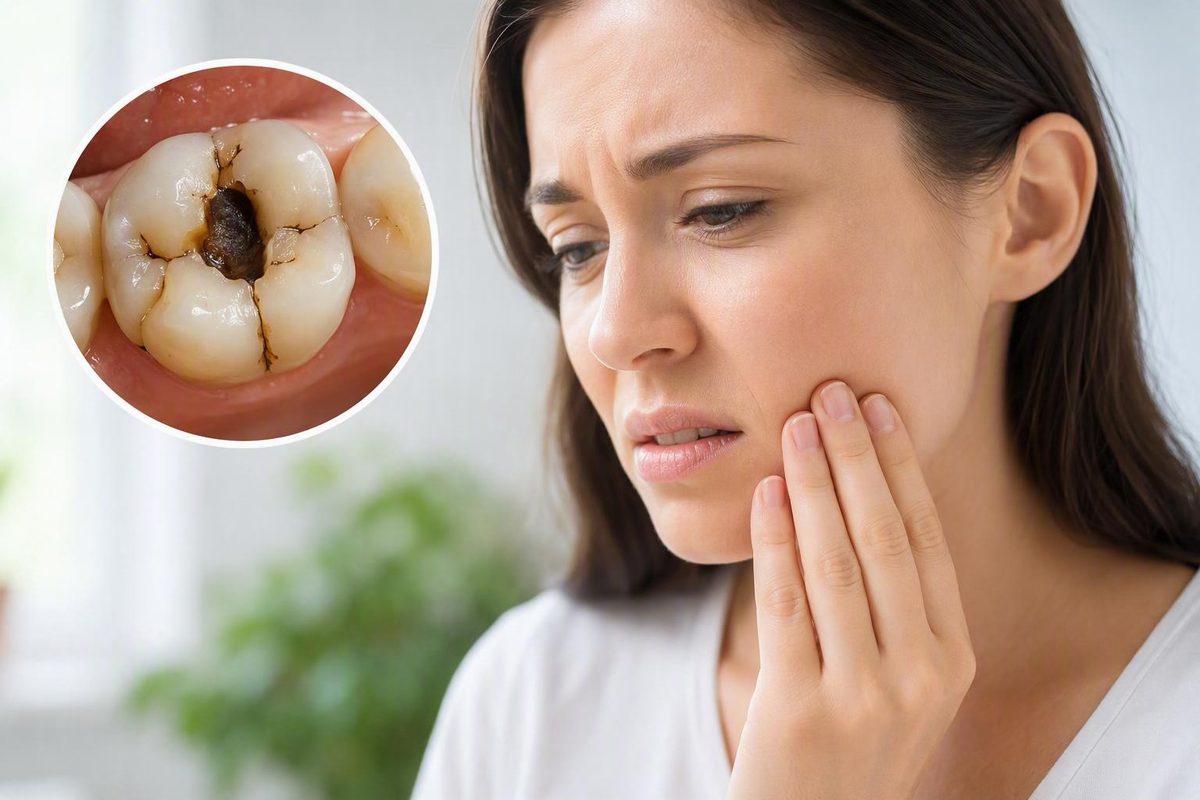
A cavity and a crack can affect the same tooth at the same time. Decay removes tooth structure and may leave the remaining enamel and dentin too weak to tolerate normal chewing forces. The tooth may then crack, lose a cusp, or crumble. The interaction can also work in the other direction: a fracture may create a pathway through which bacteria reach deeper tissue.
Visible damage does not necessarily reveal the full problem. A small opening may conceal more extensive decay, while a fine crack may travel farther than it appears to on the surface. Pain is also an unreliable measure of severity. Some structurally damaged teeth produce little discomfort; others hurt sharply even though the crack’s endpoint is not yet known.
Treatment for a cracked tooth with a cavity therefore depends on the depth of the decay, the crack’s direction and endpoint, the condition of the pulp inside the tooth, the amount of healthy supported structure left, and whether the tooth can be predictably rebuilt.
What it means when a tooth has both decay and a crack
A cavity can contribute directly to a tooth breaking. As decay progresses, it removes mineralized tissue and may hollow out or undermine parts of the tooth. The remaining walls and cusps must then carry chewing pressure with less structural support. A sufficiently weakened tooth may crack, fracture, or crumble when biting something hard or during ordinary use.
Decay and cracking are related but distinct processes:
- Tooth decay is progressive destruction and loss of tooth structure.
- A crack is a physical fracture. It may be a shallow line confined to enamel, a fractured cusp, an incomplete crack extending toward the root, or a split that separates parts of the tooth.
- A broken area may represent decay-related collapse, a fracture, or both.
- A dark line, stain, or hole cannot establish how deeply either problem extends.
Once a crack reaches dentin or the pulp, bacteria may gain access to tissue that was previously protected. A tooth can therefore have both structural damage and disease inside it; the crack and cavity do not simply exist side by side.
Other forces may contribute. Large or repeated restorations leave less original tooth structure, while grinding, clenching, trauma, and chewing hard objects can place concentrated or repeated stress on a weakened tooth. A peer-reviewed review identifies dental caries, restorative intervention, repeated restorations, clenching, grinding, and hard-object chewing as relevant factors when evaluating a suspected crack (cracked tooth syndrome review).
The part visible in a mirror may not be the most important part. Decay can extend beneath enamel or an old restoration. A crack can be too fine to see, travel under the gum, or continue toward the pulp or root. Conversely, a prominent surface line may prove to be limited to enamel. Appearance alone cannot show whether the tooth can be restored.
Neither active decay nor a structural crack can be definitively repaired at home. Toothpaste, rinses, pain medicine, and improvised repair materials cannot remove diseased tissue, restore missing support, assess the pulp, or establish where a crack ends. The tooth needs professional evaluation even if the discomfort temporarily settles.
Symptoms can offer clues, but they cannot identify the problem
Pain patterns can help a dentist decide what to investigate, but they cannot determine with certainty whether the cause is decay, a crack, pulp inflammation, or a combination of problems.
| Symptom or finding | What it may suggest | Why it is not conclusive |
|---|---|---|
| Sharp pain while biting | A cracked segment may move slightly under pressure | Decay, pulp inflammation, bite imbalance, or a damaged restoration can also make chewing painful |
| Pain when pressure is released | Often associated with a crack and sometimes described as release or rebound pain | It does not prove that a crack is present or reveal its depth |
| Sensitivity to sweets | Exposed or decayed tooth structure | A crack involving dentin can produce similar sensitivity |
| Cold or heat sensitivity | Decay, exposed dentin, or irritation of the pulp | Both cracks and cavities can affect the same tissues |
| Lingering or spontaneous toothache | Possible deeper pulp inflammation or infection | The symptom does not identify whether decay, a crack, or both caused the pulp problem |
| Discoloration or a dark area | Decay, staining, exposed inner tissue, or changes within the tooth | Surface color cannot establish structural depth |
| Swelling near the tooth | Possible inflammation or infection | Gum and periodontal conditions can cause similar swelling |
| No symptoms | A shallow defect or meaningful damage that has not yet irritated the pulp | Absence of pain does not prove that the tooth is sound |
Sharp, intermittent pain during chewing—particularly pain as pressure is released—can raise suspicion of a crack. The discomfort may occur only when food presses on a particular cusp or when the person bites in a certain direction. It can be difficult to reproduce consistently or even to identify which tooth hurts.
Sweet and temperature sensitivity are often associated with cavities, but they are not specific to decay. A crack that reaches dentin may cause similar sensitivity. Either condition can also affect the pulp, producing prolonged sensitivity, aching, spontaneous pain, discoloration, or swelling. Some cracks and small cavities cause no symptoms at all. These overlapping presentations are described in dental guidance on cracked-tooth symptoms and diagnosis.
Cracked-tooth symptoms can resemble cavities, pulp inflammation, exposed-dentin sensitivity, periodontal disease, sensitivity related to a filling or another restoration, bite trauma, and some non-dental causes of facial pain. Pain on biting or release is therefore suggestive rather than diagnostic.
Pain intensity also does not determine whether a tooth can be saved. A painless tooth may have lost substantial supporting structure.
Self-diagnosis can consequently point in the wrong direction. Sweet sensitivity does not establish that the problem is “just a cavity,” and biting pain does not prove that the tooth has split. Symptoms guide the investigation; an examination determines the diagnosis.
How quickly should the tooth be assessed?
Not every visible line or chipped edge requires same-day treatment. Cracks range from shallow enamel lines to fractures that divide a tooth. Nevertheless, contact a dentist promptly about a suspected cracked, broken, or crumbling tooth, even when pain is mild, intermittent, or absent.
When arranging care, explain:
- Whether a piece broke away suddenly
- Whether the tooth is continuing to crumble
- Whether pain is increasing
- Whether biting or releasing the bite triggers pain
- Whether heat, cold, or sweets cause lingering sensitivity
- Whether the gum, jaw, or face is swollen
- Whether you have fever, tender glands, or bad breath associated with the area
- Whether the damage followed a fall, impact, or other trauma
Swelling, fever, increasing pain, tender glands, and bad breath may accompany infection and are reasons to seek urgent dental assessment. A crack can allow bacteria to enter the pulp, potentially contributing to an abscess or spread of infection beyond the tooth. Crack severity and treatment vary, so the appropriate timing must be based on the individual findings (Cleveland Clinic guidance on cracked teeth).
A shallow enamel defect is not equivalent to an acutely broken, painful tooth with swelling. When you call, the dental office can use your symptoms, the extent of breakage, the trauma history, and the services available locally to determine how quickly you should be seen. Do not minimize major acute breakage simply because the pain is currently tolerable.
This article cannot provide complete emergency triage. If swelling or illness appears to be spreading beyond the immediate tooth area, do not wait for a routine appointment; seek urgent professional assessment through the appropriate dental or medical service in your area.
If part of the tooth has separated, retrieve it if you can do so safely. Keep the fragment in milk or saliva and take it to the dentist because reattachment is sometimes possible. This fragment-preservation advice and the possible treatments for a broken tooth are outlined in NHS guidance on chipped, broken, or cracked teeth.
A temporary reduction in pain does not mean the crack has closed or that the cavity has stopped progressing. Prompt assessment is intended to identify the damage before further structural loss or infection narrows the available treatment options; it cannot guarantee that every tooth will be saved.
Why diagnosis usually takes more than an X-ray
A routine dental X-ray may reveal decay, an existing restoration, bone changes around a root, or other useful findings. It may not display a fine crack. Whether a fracture is visible depends partly on its width, location, and orientation relative to the X-ray beam.
A normal routine X-ray therefore does not rule out a cracked tooth. Diagnosis is usually an uncertainty-reduction process in which the dentist combines the history, examination, tests, and imaging rather than relying on one result.
A typical evaluation may include:
- Reviewing the symptom history. The dentist may ask when the discomfort began, which foods or temperatures trigger it, whether pain occurs during biting or release, and whether symptoms are spontaneous or linger.
- Reviewing dental and injury history. Large or repeated restorations, grinding, clenching, recent dental work, hard-object biting, or trauma may provide useful context.
- Inspecting the tooth and restorations. The dentist looks for decay, missing structure, fracture lines, unsupported cusps, wear, damaged restorations, and changes in color.
- Trying to reproduce the biting pain. Controlled pressure may be applied to individual cusps or parts of the chewing surface to help localize the problem.
- Assessing the pulp. The tooth’s response may be compared with nearby teeth to investigate whether the pulp appears healthy, inflamed, infected, or nonresponsive.
- Examining the surrounding gum and support. Probing around the tooth may identify a narrow, localized change associated with a fracture, although gum findings can have other causes.
- Using imaging where appropriate. Routine X-rays may identify decay or related disease. Selected cases may warrant cone-beam computed tomography, but no imaging method makes every fine crack visible.
- Reassessing after removing decay or an old restoration. Some structural damage becomes apparent only after diseased or obscuring material has been removed.
Other potential methods include magnification, transillumination with a bright light, and dye. No single finding should automatically be treated as conclusive.
Cleveland Clinic lists examination, bite testing, transillumination, dye, X-rays, cone-beam CT, and gum assessment among the methods that may be used to investigate a cracked tooth (diagnosis of a tooth fracture). The exact combination depends on the tooth and clinical findings.
Diagnosis may remain difficult because symptoms can come and go, pain can be hard to localize, and a very fine crack may not be visible. The dentist combines the available evidence to answer four practical questions:
- How far does the crack appear to travel?
- Is the pulp healthy or diseased?
- Is the tooth adequately supported by the surrounding tissues?
- Is enough healthy, supported structure left for a predictable restoration?
The treatment decision: decay depth, pulp health, and restorability
A cracked tooth with a cavity does not automatically need a filling, crown, root canal, or extraction. Treatment should follow the damage found rather than a predetermined ladder through which every tooth progresses.
The decision centers on five questions.
1. How much decay is present?
A limited cavity removes less structure than decay extending beneath a cusp, around an old filling, or close to the pulp. The dentist must consider not only the visible opening but also the softened or undermined tissue that needs to be removed.
2. Where does the crack travel?
A shallow enamel line has different implications from a fractured cusp, a crack approaching the pulp, a split into separate segments, or a fracture extending deeply toward the root. The endpoint may matter more than the line visible on the chewing surface.
3. How much healthy, supported tooth remains?
Useful tooth structure must remain after decay, weakened enamel, and unsuitable restorative material are removed. A thin wall that looks intact may not provide meaningful support if decay has undermined it or a fracture has separated it from the rest of the tooth.
4. Is the pulp healthy?
The pulp is the soft tissue inside the tooth. If it remains healthy, the tooth may need removal of decay and structural restoration without internal treatment. If the pulp is exposed, irreversibly inflamed, infected, or necrotic, root-canal treatment may be needed if the tooth is otherwise restorable.
5. Can the tooth support a predictable restoration?
This is the question of restorability. In plain language, it means whether enough healthy, supported tooth and surrounding support remain to rebuild the tooth, protect it against chewing forces, and use it with a reasonable expectation of function. It does not mean that any procedure can guarantee indefinite survival.
Assessment sometimes requires removing decay or an existing restoration. That step clears diseased or obscuring material and reveals the structure underneath. A small-looking defect may prove deeper than expected, or the crack may turn out to be limited enough for a more conservative restoration. Guidance specifically addressing a cracked tooth with decay describes removing decay before evaluating crack depth and remaining healthy structure.
The initial plan may therefore change. A dentist might begin expecting to place a filling but discover that a cusp has too little support. In another case, the pulp may be healthier and the crack more limited than the symptoms suggested. A revised plan does not necessarily mean that the original assessment was careless; sometimes the complete condition cannot be seen until damaged material is removed.
A broad, qualified decision path is:
- Limited decay and a small, stable defect: a filling or bonding may be possible.
- A larger defect or loss of cusp support: an onlay or crown may be considered to cover vulnerable chewing surfaces.
- Exposed or diseased pulp: root-canal treatment may be required if the tooth remains restorable.
- Pulp disease plus major structural weakness: root-canal treatment and a protective restoration may serve different but complementary purposes.
- A split, deep root-level fracture, inadequate support, or insufficient remaining structure: extraction may be necessary.
Tooth location and chewing forces also affect planning. Back teeth generally carry substantial chewing loads, while crack direction, the number of supported walls or cusps, the person’s bite, and suspected grinding or clenching can influence the structural protection considered. These factors require individualized planning rather than a universal rule.
Filling, onlay, crown, root canal, or extraction?
These treatments are possibilities tied to different findings. They are not stages that every cracked and decayed tooth must pass through.
Filling or bonding
A filling replaces tooth structure after limited decay has been removed. Bonding may rebuild a small chipped or fractured area with restorative material. Either may be considered when:
- The defect is relatively small and stable
- Sufficient sound tooth surrounds it
- The crack does not undermine important supporting structure
- The pulp does not require internal treatment
- The restoration can tolerate the forces placed on that tooth
A filling cannot compensate for every crack. If a cusp moves independently or too little supported tooth remains, placing material in the cavity may not provide adequate protection.
Onlay
An onlay is a restoration that replaces a larger damaged area and covers one or more cusps or parts of the chewing surface. It may be considered for a broken cusp or larger defect when the tooth requires more reinforcement than a direct filling can provide.
The boundary between a large filling, an onlay, and a crown is not universal. Crack position, decay extent, remaining walls, bite forces, restorative material, and the ability to create a stable design all matter.
Crown
A crown covers and reinforces a substantially weakened but restorable tooth. It may be considered when a large cavity, fracture, extensive previous restoration, or major loss of structure leaves insufficient support for a filling alone.
A crown does not treat infection inside the tooth. It addresses the tooth’s external shape, coverage, and strength. It may be used while the pulp is healthy or after root-canal treatment when the remaining tooth needs structural protection.
Root-canal treatment
Root-canal treatment addresses tissue inside the tooth. It involves removing diseased or infected pulp, cleaning and shaping the internal canal space, and sealing it.
A crack alone does not make root-canal treatment automatic. Internal treatment may be needed when decay or a crack has exposed the pulp or when the pulp is irreversibly inflamed, infected, or necrotic. A tooth can also require a root canal because of pulp disease even if the visible crack seems small.
Root canal and crown together
A root canal and a crown are not interchangeable:
- Root-canal treatment addresses diseased tissue inside the tooth.
- A crown reinforces weakened structure outside the tooth.
Both may be used when the pulp requires treatment and the remaining tooth is structurally weak but restorable. Tooth position and chewing forces influence the final restoration, so root-treated teeth do not all receive an identical design. The distinction between internal treatment and external reinforcement is explained in this clinical comparison of crowns and root-canal treatment.
Extraction
Extraction removes a tooth that cannot be predictably restored or retained. It may be necessary when:
- The tooth is split into separate segments
- The fracture extends deeply toward or through the root
- The crack travels too far below the gum for a stable restoration
- Too little healthy, supported structure remains
- The surrounding support is inadequate
- Decay and fracture together make reconstruction impractical
The decision is not based on pain alone. A painless split tooth may be nonrestorable, while a painful tooth with treatable pulp inflammation and adequate remaining structure may still have restorative options.
No procedure can guarantee that a cracked tooth will last indefinitely. Treatment may remove decay, address pulp disease, cover weakened structure, and reduce movement, but the original fracture does not biologically heal like a broken bone. A treated crack may still enlarge, making follow-up and realistic expectations important.
What to do while waiting for a dental appointment
The immediate goals are to reduce additional stress, keep the area reasonably clean, and manage discomfort safely. These measures do not replace examination or treatment.
- Avoid chewing on the affected tooth. Use the other side if comfortable and choose foods that require little force.
- Avoid hard foods and objects. Ice, hard candy, unpopped kernels, tough foods, and non-food objects can concentrate pressure on weakened structure.
- Rinse gently with warm water. This may help clear loose debris without placing tools into the damaged area.
- Use an external cold pack if swelling is present. Apply it to the outside of the face rather than directly to the tooth.
- Protect a separated fragment. Keep it in milk or saliva and bring it to the appointment.
- Do not glue the tooth. Household adhesives are not dental restorative materials.
- Do not treat a home repair as definitive. Covering a rough edge or reducing discomfort does not show whether decay, pulp disease, or a deep fracture remains.
An over-the-counter pain reliever may be considered only according to its label and when compatible with your health conditions, allergies, pregnancy status, and other medicines. No pain reliever is suitable for everyone. Ask a pharmacist, dentist, or another qualified healthcare professional if you are uncertain. Do not exceed label instructions or place aspirin or numbing medicine directly on the tooth.
Avoiding chewing, using an external cold pack, taking medicine only as directed, and preserving a fragment in milk or saliva are temporary measures described in university-affiliated guidance for a broken tooth. They may reduce discomfort or further loading, but they do not repair the damage.
Temporary measures cannot:
- Remove infected or decayed tissue
- Seal the full depth of a crack
- Restore missing structural support
- Determine whether the pulp is healthy
- Treat an abscess
- Establish whether the tooth is restorable
If symptoms change while you are waiting—particularly if swelling, fever, worsening pain, or other signs of infection develop—contact the dental office again and explain the change. The appropriate timing may need to be reconsidered.
What delaying treatment can change
Mild, intermittent, or absent pain does not prove that the damage is superficial. Early decay can be painless, and some fractures do not irritate the pulp immediately. A fractured cusp may also have a different symptom pattern and outlook from a split tooth or vertical root fracture.
Waiting may allow:
- Additional loss of tooth structure
- An unsupported wall or cusp to break
- A fracture to extend
- The pulp to become inflamed or infected
- An abscess to develop
- The tooth to become less predictably restorable
- Eventual tooth loss
These outcomes are possible, not inevitable on a fixed timetable. Symptoms alone cannot predict whether a crack will remain stable or progress. Earlier assessment may preserve more diagnostic and restorative options, but it cannot guarantee that the tooth will be saved.
A crack does not heal like a fracture in living bone. Dental treatment may stabilize parts of the tooth, remove decay, treat diseased pulp, and restore function, but the fracture line may remain a long-term vulnerability. Educational guidance on cracked-tooth diagnosis and treatment similarly notes that treatment depends on the crack’s type, location, and severity.
After treatment, practical prevention focuses on reducing avoidable stress and identifying new problems before extensive structure is lost:
- Maintain regular dental care based on your needs and professional advice.
- Have damaged, loose, or failing restorations evaluated.
- Discuss suspected grinding or clenching with a dentist before choosing an appliance.
- Avoid chewing ice, hard candy, pens, and other hard objects.
- Follow the dentist’s cleaning and monitoring instructions.
- Report renewed biting pain, sensitivity, swelling, or movement promptly.
The central distinction is straightforward: symptoms may raise suspicion, but they do not settle the treatment decision. The decisive questions are how deeply the decay extends, where the crack ends, whether the pulp is healthy, and whether enough supported tooth remains to restore. Until those questions are answered in person, protect the tooth from chewing pressure and arrange prompt professional assessment.
Frequently asked questions
Can a dentist fill a cavity if the same tooth is cracked?
Sometimes. A filling or bonding may be possible when the decay and fracture are limited, the pulp is unaffected, the defect is stable, and enough healthy supported tooth remains. If the crack undermines a cusp or the cavity has removed too much support, a filling alone may not adequately protect the tooth.
The dentist may need to remove decay or an old restoration before deciding. A defect that initially appears suitable for a filling may require an onlay or crown if deeper structural loss is uncovered. Treatment selection depends on decay depth, structural damage, and pulp health (filling, crown, and root-canal treatment considerations).
Does every cracked tooth with decay need a root canal?
No. Root-canal treatment is used when the pulp is exposed or diseased, not merely because a crack exists. A tooth with healthy pulp may instead be treated with a filling, bonding, onlay, or crown, depending on the structural findings.
If the pulp does require treatment, the remaining tooth may also need structural protection. Root-canal treatment addresses internal tissue; the restoration addresses the external tooth. A cracked tooth does not always require a root canal, and cracks and cavities can produce overlapping symptoms (cracked tooth versus cavity).
Why does biting hurt if the crack did not appear on my X-ray?
A routine X-ray may still look normal because crack visibility depends on the fracture’s width and orientation relative to the X-ray beam.
Dentists may therefore combine imaging with bite testing, magnification, transillumination, dye, gum assessment, and pulp testing. Standard two-dimensional radiographs often fail to display fine cracks, so biting pain remains a clue rather than proof (crack detection and routine dental X-rays).
Can a cracked tooth with a cavity heal or be repaired at home?
No. A crack does not biologically knit together like a broken bone, and an established cavity represents tooth structure that has already been lost. Home measures cannot remove decay, assess the pulp, or rebuild the tooth.
Avoid chewing on it, rinse gently with warm water, and use an external cold pack if swelling is present. A suitable over-the-counter pain reliever may be considered according to its label and personal medical safety. Do not glue the tooth or assume that reduced pain means the condition has resolved. Cracked or broken teeth cannot be definitively treated at home (fractured and broken tooth guidance).
Can a painless cracked and decayed tooth still need treatment?
Yes. Small cavities and some cracks may cause no symptoms. A fractured cusp may also be less painful than expected even though meaningful structural weakness remains. Absence of pain does not establish that the cavity is shallow, the pulp is healthy, or the crack is harmless.
A dentist should assess a cracked or broken tooth even when it does not hurt. Depending on the damage, treatment may include fragment reattachment, a filling, a crown, root-canal treatment, or another plan after the tooth has been examined (NHS guidance on cracked and broken teeth).
Decay Guide publishes general educational information and does not diagnose dental conditions, treat patients, or provide individualized advice. Only an in-person dental examination can determine whether a particular tooth needs a limited restoration, cuspal coverage, root-canal treatment, extraction, or a revised plan after the full damage becomes visible. Learn more about Decay Guide.