
What a Spot or Hole on a Front Tooth Could Mean—and What Happens Next
Once a hole has formed, missing tooth structure cannot regrow, so professional restoration is generally needed instead.
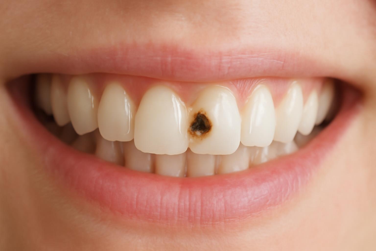
A front tooth can develop a cavity even though its broad surfaces are generally easier to clean than the grooves of a back tooth. Decay may occur on the visible face, near the gumline, between two teeth, or on the surface facing the tongue or palate. Early changes can be painless and difficult to interpret at home.
The key distinction is between an intact area of early mineral loss and an established hole where tooth structure has broken down. Selected early lesions may sometimes be stopped or remineralized. Once a hole has formed, the missing structure cannot regrow and generally needs professional restoration.
Color, sensitivity, and pain cannot establish which situation applies. A dental examination is necessary to distinguish decay from staining, erosion, a crack, previous injury, or another dental condition—and to determine how deeply any decay extends.
Quick guide: what to do about a suspicious front tooth
A white, brown, gray, or dark mark is not automatically a cavity. Neither is sensitivity. Conversely, early decay may cause no pain, and symptoms may not appear until the process reaches deeper tooth layers. Cavities can affect any tooth surface, including front teeth, according to the Cleveland Clinic’s medically reviewed overview of cavities.
Use this guide to decide when to arrange care rather than trying to diagnose the tooth in a mirror:
| What you notice | Practical next step |
|---|---|
| A new or unexplained white, brown, gray, or dark mark | Arrange a dental evaluation, especially if the mark persists or changes. |
| A rough area, sharp edge, or recurring sensitivity | Have the tooth examined rather than assuming it is decay or ordinary sensitivity. |
| Floss repeatedly catches, frays, or breaks at one location | Tell the dentist exactly where this happens. Decay is one possibility, but the finding is not diagnostic. |
| A visible pit or apparent hole | Arrange an appointment. An opening may indicate structural loss, but its nature and depth cannot be judged at home. |
| Toothache, significant pain when biting, or difficulty chewing | Contact a dentist promptly for assessment. These symptoms can have several causes and may indicate a problem beyond the outer enamel. |
| Pus, facial swelling, or another possible sign of infection | Seek prompt professional assessment rather than waiting for a routine visit. |
Earlier assessment may allow a dentist to preserve more sound tooth structure and reduce the likelihood of more extensive treatment. That does not mean anyone can predict how quickly a particular mark will progress. Progression varies, and appearance alone cannot reveal lesion depth, plaque exposure, fluoride exposure, saliva flow, dietary frequency, or the condition of the tooth.
Do not postpone an examination simply because the tooth does not hurt. At the same time, do not assume that discomfort proves you have a cavity. Sensitivity or pain can also occur with enamel erosion, a crack, an exposed root surface, an injury, or another dental problem.
Editorial disclosure: Decay Guide is an informational publisher, not a dental practice, and it does not diagnose or treat patients. Its articles are written by a health writer, have not been reviewed by a clinician, and cannot replace a dental examination. These limitations are described in the site’s privacy and writing disclosure.
How decay develops on a front tooth
Plaque is a film containing bacteria that accumulates on teeth. Certain bacteria use sugars and starches from food and drink, producing acids that remove minerals from enamel. Sugar does not directly drill a hole into a tooth. Instead, repeated periods of bacterial acid production can shift the balance toward mineral loss.
The mouth also has protective mechanisms. Saliva helps clear food and acids and supplies calcium and phosphate that can return to enamel. Fluoride can limit mineral loss and support mineral replacement. Decay develops when repeated acid attacks outweigh these protective processes long enough for enamel—and eventually the underlying tooth structure—to break down. The National Institute of Dental and Craniofacial Research explains this cycle of mineral loss and repair.
Factors that can create more opportunities for decay include:
- Frequent sugary or starchy foods and drinks. Each exposure can give plaque bacteria another opportunity to produce acid.
- Repeated snacking or sipping. Slowly consuming a sweetened drink over an extended period may result in recurring acid attacks.
- Inadequate plaque removal. Plaque can remain around the gumline, between teeth, or on surfaces that receive less attention.
- Insufficient fluoride exposure. Fluoride is one part of the tooth’s defense against mineral loss.
- Dry mouth. Reduced saliva weakens natural clearance and mineral replacement.
Front teeth are not immune merely because they lack the deep chewing grooves found on molars. Plaque can remain near the gums, between adjacent teeth, and on the back surfaces. Decay between two front teeth or on the palate-facing surface may not be visible in an ordinary mirror.
Acid erosion is related to decay but is not the same process. That overlap is another reason not to identify a cavity from color or sensation alone.
What a front tooth cavity can look and feel like
An early area of enamel mineral loss may appear chalky, opaque, or whiter than the surrounding tooth. At this stage, the surface may still be intact. A white area is not conclusive, however, because not every difference in enamel color is decay.
As decay advances, possible findings include:
- Brown, gray, or dark discoloration
- A visible pit, notch, or apparent hole
- A rough or sharp area
- A surface that feels different to the tongue
- Floss that repeatedly catches or frays at the same point
- A weakened or broken edge
Possible symptoms include sensitivity to cold, heat, or sweet foods; pain when biting; discomfort while chewing; or a persistent toothache. Early decay may be entirely painless. Pain or sensitivity becomes more likely when decay reaches the dentin beneath the enamel or affects the pulp inside the tooth. Even then, symptoms do not establish the cause by themselves, as the Cleveland Clinic cautions in its discussion of cavity symptoms and diagnosis.
A few distinctions are especially important:
- A dark mark may be a stain rather than a cavity.
- Sensitivity may result from erosion, an exposed root surface, a crack, or another dental condition.
- A sharp edge may come from a chip or previous injury.
- A tooth that changes color after an injury still requires assessment, even if no hole is visible.
- Pain when biting can have causes other than decay.
Location also affects what can be seen. Decay on the visible face of an incisor may be conspicuous, while a problem between teeth or on the back surface may remain out of view.
Do not probe a suspicious area with a pin, scraper, home dental tool, or another sharp object. Continue gentle brushing and interdental cleaning, note any changes or symptom triggers, and let a dental professional inspect the tooth.
Can an early front tooth cavity be reversed?
“Reversing a cavity” is often used too broadly. Whether mineral loss can be reversed depends on whether the tooth surface is still intact.
An early, non-cavitated enamel lesion is an area where minerals have been lost but the outer structure has not collapsed into a hole. Under professional guidance, selected lesions may sometimes be arrested or remineralized. Management may include fluoride, more effective plaque removal, and reducing the frequency of sugary or starchy exposures.
An established cavity involves structural breakdown. Once enamel or other tooth tissue is missing, the body cannot rebuild the tooth’s original shape. Toothpaste, supplements, dietary changes, and home remedies cannot regrow a formed hole. The missing area generally needs professional restoration.
The intact surface and lesion depth must be evaluated clinically. Color does not reliably separate the stages. A white area may represent early mineral loss, but it may have another cause. A dentist must assess the surface, location, extent, and surrounding tooth before deciding whether preventive management or restoration is appropriate. The NIDCR limits reversal to early mineral loss before a permanent cavity has formed.
Fluoride and brushing remain important after a hole develops. They help control plaque and protect the remaining surfaces of the affected tooth, as well as the rest of the mouth. What they cannot do is replace structure that is already gone.
The practical distinction is:
- Minerals lost from intact early enamel may sometimes be replaced.
- Missing tooth structure cannot grow back.
Be cautious with claims that a special paste, supplement, oil, restrictive diet, or mineral product can “heal” a visible hole. Better oral-care habits can reduce future risk, but they cannot restore a lost contour, manage damaged tissue, or determine whether deeper treatment is necessary.
How a dentist confirms the problem and judges its depth
A diagnostic visit begins with the history of the tooth. The dentist may ask when you first noticed the mark or symptom, whether it has changed, what triggers sensitivity, whether pain lingers, whether biting hurts, and whether the tooth has previously been injured or restored. Dietary patterns and dry-mouth symptoms may also help place the finding in context.
The examination may include:
- Visual inspection. The dentist examines the visible face, gumline, back surface, and between-tooth areas as access permits.
- Surface assessment. A mirror, lighting, and careful use of dental instruments can help assess the tooth’s contour and surface integrity.
- Evaluation of nearby tissues and teeth. This helps determine whether symptoms appear to originate from the suspected tooth.
- Assessment of structure and function. The dentist considers broken or weakened areas, previous restorations, and how the teeth meet.
- Dental X-rays when appropriate. Imaging may help identify decay that is not readily visible or clarify its extent.
The decision depends on the examination, symptoms, location, previous images, and individual risk. A cavity between teeth or on a less visible surface may be difficult to assess by direct inspection alone.
Mayo Clinic describes cavity diagnosis as combining symptom history, visual examination, careful use of dental instruments, and X-rays when needed. Treatment then depends on how serious the decay is and the condition of the tooth, rather than on color alone (Mayo Clinic).
Treatment selection depends on several related questions:
- Is the surface intact, or has it broken down?
- Is the change limited to enamel, or does it extend into dentin?
- How much sound tooth structure remains?
- Is an edge or a substantial part of the tooth weakened?
- Has the pulp been affected?
- Can the tooth be restored predictably?
- Where will the restoration sit, and what forces will act on it?
- How important will shade, contour, translucency, and texture be in that location?
No online photograph, symptom quiz, or article can answer all of these questions. Even a clear picture provides no reliable measurement of internal depth. A photograph may help a patient describe the concern when arranging an appointment, but it cannot select between fluoride management, a filling, a crown, root-canal treatment, or extraction.
Treatment by stage: from fluoride care to advanced restoration
Treatment is based on the clinical stage and the condition of the tooth—not simply on the fact that the tooth is visible. A small intact lesion and a deeply weakened front tooth are fundamentally different problems.
| Clinical situation | Treatment purpose | Major limitation |
|---|---|---|
| Selected early enamel lesion with an intact surface | Professional preventive management, including fluoride and control of plaque and sugar frequency, may help arrest or remineralize mineral loss. | Appropriate only when the surface and lesion depth meet clinical criteria; color alone cannot establish this. |
| Small or moderate formed cavity | Manage decayed tissue and replace missing structure, commonly with tooth-colored composite on a visible front tooth. | Composite is not suitable for every defect; location, cavity size, moisture control, bite, and remaining structure all matter. |
| Substantial structural loss or a significantly weakened tooth | A crown may cover and reinforce the tooth more extensively. | A crown covers more tooth than a small filling and is not justified merely because the tooth is visible. |
| Decay that reaches or seriously affects the pulp | Root-canal treatment may be required before the definitive outer restoration. | Root-canal treatment addresses the pulp and canal space but does not by itself replace all missing external structure. |
| Tooth that cannot be restored | Extraction removes the non-restorable tooth. | This is a last-stage option, not the expected outcome of every front tooth cavity; replacement planning may also be necessary. |
Very early decay does not automatically require drilling. Professional fluoride care and risk control may be appropriate when mineral loss is confined to an intact surface. Once a hole has formed, restoration is generally required because the original contour cannot remineralize back into place.
For a small or moderate cavity, a tooth-colored composite filling is a common conservative option. The dentist removes or otherwise clinically manages decayed tissue and rebuilds the missing area with resin. Composite can be selected and shaped to resemble adjacent enamel, which is useful on a tooth visible in a smile.
A filling is not always sufficient. If substantial structure has been lost or the remaining tooth is weakened, a crown may be considered because it covers the tooth more extensively. If decay reaches and seriously affects the pulp, root-canal treatment may be needed before the tooth receives its definitive restoration. A tooth that cannot be restored may require extraction. These stage-based options are outlined in the Mayo Clinic’s cavity treatment guide.
Several treatment terms are commonly confused:
- Composite filling: Primarily replaces tooth structure lost to decay or damage.
- Cosmetic bonding: Often uses a similar resin but primarily reshapes or enhances a tooth.
- Veneer: Covers the front surface mainly to change appearance.
- Crown: Covers most or all of the clinical tooth to restore and protect more extensive damage.
Materials and techniques can overlap, particularly between a composite filling and cosmetic bonding. The clearest distinction is purpose: a filling restores a decay-related defect, while cosmetic bonding primarily changes contour or appearance. A clinical comparison of white fillings, cosmetic bonding, and veneers also emphasizes that the options serve different roles and require individual assessment.
A veneer should not be used simply to conceal untreated decay. The underlying disease must first be assessed and clinically managed. Veneers, bonding, inlays, onlays, and fillings are not routine interchangeable treatments for an active front-tooth cavity. Suitability depends on the affected surface, the amount of sound tooth remaining, the health of the underlying tooth, the bite, and the intended function.
What to expect from a tooth-colored front filling
The exact appointment varies with the cavity’s location, size, depth, and proximity to the pulp. A typical direct composite procedure may include the following steps:
- Shade selection. The dentist chooses a resin shade intended to coordinate with the surrounding enamel. This may be done early because a tooth can appear different as it dries.
- Local anesthesia when needed. The tooth and nearby tissues may be numbed depending on the lesion and planned procedure.
- Management of decayed tissue. The dentist removes or otherwise manages damaged tissue while preserving sound structure where possible.
- Cleaning and moisture control. Suction, cotton rolls, a dental dam, or other methods may be used to control moisture.
- Surface preparation and bonding. The tooth is prepared so the composite can bond to the remaining structure.
- Layered resin placement. Composite may be placed in increments rather than as one large mass.
- Light curing. The material is hardened with a curing light as appropriate.
- Shaping and contouring. The dentist recreates the tooth’s outline, contact area, edge, and surface form.
- Bite adjustment. The restoration is checked to make sure it does not contact the opposing teeth too early or interfere with movement.
- Finishing and polishing. The surface is refined to improve smoothness and appearance.
This is a typical sequence rather than a universal protocol. Front-tooth work gives particular attention to shade, translucency, contour, texture, bite, and polish. A dental-practice description of the front-tooth composite procedure includes shade selection, decay removal, layered placement, light curing, sculpting, bite checking, and polishing, while noting that individual treatment varies.
Natural teeth do not have one flat color. Surface shape and polish also influence how a restoration looks and feels.
The goal is usually for the composite to blend with the tooth—not a guarantee that it will be invisible in every light, from every angle, or at every distance. The result depends on the size and position of the restoration, the underlying tooth color, the amount of available enamel, and the selected material.
Composite is maintainable rather than permanent. Over time, it may:
- Pick up surface stain
- Lose polish or begin to feel rough
- Wear at an edge or contact point
- Chip or fracture
- Develop a more visible margin
- Need refinishing, localized repair, or replacement
A later color mismatch is also possible. Natural enamel may change with age or whitening, while an existing composite does not whiten in the same way. If you are considering tooth whitening, discuss the sequence with the dentist before a visible restoration is placed.
Do not rely on universal promises about appointment length, how long numbness will last, how much sensitivity is expected, or how many years a filling will remain unchanged. These vary with the tooth, cavity, technique, restoration size, bite, habits, and individual response.
After treatment:
- Follow the treating dentist’s instructions about eating and drinking.
- If your mouth remains numb, avoid chewing until sensation returns if instructed, reducing the chance of biting your lip or cheek.
- Resume careful brushing and interdental cleaning as directed.
- Avoid repeatedly testing the new restoration with hard foods or objects.
- Notice whether your bite feels balanced after numbness has resolved.
Contact the dentist if pain is severe or worsening, substantial sensitivity persists, the tooth meets too early when you bite, an edge feels sharp, the restoration fractures, or the filling comes out. A high contact point or rough margin may be adjustable, but it should be assessed rather than tolerated or altered at home. These reasons for follow-up are consistent with published dental-practice guidance on post-filling warning signs.
Preventing another cavity—including decay in children
Prevention is not about eliminating one “bad” food or brushing more forcefully. It is about reducing repeated acid attacks while supporting the mouth’s protective factors.
Core habits include:
- Brush twice daily with fluoride toothpaste.
- Clean between the teeth every day using a method appropriate for the spaces.
- Pay particular attention to the gumline and the contacts between front teeth.
- Reduce the frequency of sugary or starchy snacks and drinks.
- Avoid slowly sipping sweetened beverages over long periods.
- Discuss persistent dry mouth with a dentist or appropriate healthcare professional.
- Attend dental follow-up at an interval based on personal risk and clinical findings.
Frequency matters because repeated intake creates repeated opportunities for plaque bacteria to produce acid. Having a sweet or starchy item with a meal is different from grazing or sipping it throughout the day. Water can avoid adding another sugar exposure, but it does not replace plaque removal or professional dental care.
Saliva is part of the tooth’s protective system, so dry mouth deserves attention. Reduced saliva may weaken food clearance and mineral replacement.
Dental follow-up should be individualized rather than fixed at one universal interval. Someone with active decay, dry mouth, or new restorations may need a different schedule from someone whose teeth and risk factors are stable. General preventive guidance for front-tooth decay includes fluoride brushing, daily interdental cleaning, limiting frequent sugary intake, and professional follow-up (Suncreek Dental).
Front-tooth decay in babies and young children
Young children can develop decay on their upper front teeth. Prolonged or bedtime exposure to milk, formula, juice, or sweetened drinks from bottles or sippy cups is associated with this pattern. During sleep, liquids may remain around the teeth while protective saliva flow is reduced.
Practical precautions include:
- Put only water in a bedtime bottle.
- Avoid allowing a child to sip milk, formula, juice, or sweetened drinks continuously throughout the day.
- Begin age-appropriate tooth cleaning when teeth appear.
- Use fluoride toothpaste according to advice from the child’s dentist or another qualified healthcare professional.
- Arrange dental assessment for new chalky white areas, brown changes, roughness, or visible breakdown on a child’s front teeth.
Colgate’s consumer oral-health guide describes the association between prolonged bottle or sippy-cup exposure and front-tooth decay in children, particularly when milk, formula, juice, or sweetened drinks remain around the teeth for extended periods.
This article does not provide a personalized fluoride amount, dose, or schedule for a child. Age, swallowing ability, fluoride exposure, decay risk, and local professional guidance may affect recommendations.
Parents should not wait for a child to report pain. A dental examination can determine whether a white area represents mineral loss or another enamel change.
Decay Guide also publishes related educational articles on:
- The stages of cavity formation
- How sugar contributes to tooth decay
- Proper toothbrushing technique
- Children’s cavity prevention
- What happens at a dental checkup
- Veneers compared with composite bonding
These articles remain non-clinical educational material and cannot determine what is happening in an individual tooth.
Frequently asked questions
Can a front tooth cavity go away without a filling?
Possibly, but only if “cavity” is being used for very early mineral loss before a hole has formed. A selected non-cavitated enamel lesion may sometimes be arrested or remineralized with professional fluoride care and better control of plaque and sugar frequency.
Once the surface has broken down into a hole, the missing structure cannot grow back and generally needs restoration. A dentist must determine whether the surface is intact. Color or lack of pain cannot answer that question reliably.
Will a composite filling on a front tooth be visible?
It may blend closely, but invisibility cannot be guaranteed. Dentists can select tooth-colored resin and reproduce the tooth’s contour, texture, and polish. The result still depends on the restoration’s size and position, the underlying tooth color, available enamel, material properties, and lighting.
Composite may later stain, chip, wear, or lose polish. Natural enamel can also change color with age or whitening while an existing composite does not whiten in the same way. The aesthetic goal is close integration, not a promise of an undetectable or permanent repair (Le Sueur Family Dental).
When does a front tooth cavity need a root canal instead of a filling?
Root-canal treatment may be required when decay reaches or seriously affects the pulp inside the tooth. Symptoms contribute to the assessment, but pain alone does not prove pulp involvement.
The dentist considers the examination, depth of decay, remaining tooth structure, pulp condition, and imaging when appropriate. Root-canal treatment manages the pulp and canal space; the tooth still needs a suitable definitive restoration afterward (Fioritto Family Dental).
What should I do if a front tooth filling feels high, sharp, or increasingly painful?
Contact the treating dentist. A filling that meets the opposing tooth too early may require bite adjustment, while a sharp or rough edge may need finishing or polishing. Severe or worsening pain, persistent substantial sensitivity, fracture, or a lost filling also warrants reassessment.
Do not file or reshape the restoration yourself. Until it is checked, avoid repeatedly biting on the area or testing it with hard objects. Published post-filling guidance likewise advises follow-up for an uneven bite, significant pain, or a broken or lost restoration (Eagle Falls Dentistry).
Why do babies and young children get cavities on their upper front teeth?
One important pattern is prolonged exposure of the upper front teeth to milk, formula, juice, or sweetened drinks in a bottle or sippy cup, particularly at bedtime. Plaque bacteria can use the available carbohydrates to produce acids, while protective saliva flow is reduced during sleep.
Bedtime bottles should contain only water. New white spots, discoloration, roughness, or visible breakdown on a child’s front teeth should be assessed rather than watched until pain begins.
A mark on a front tooth is not automatically a cavity, and an intact early lesion is different from a formed hole. Because appearance and symptoms cannot establish depth, the practical next step is a dental examination.
If restoration is needed, tooth-colored composite is commonly considered for a small or moderate defect on a visible tooth. Deeper decay or more extensive structural loss may require a crown, root-canal treatment, extraction, or another individualized approach. In the meantime, continue twice-daily fluoride brushing, clean between the teeth daily, reduce the frequency of sugar exposure, and seek prompt professional assessment for significant pain, difficulty chewing, pus, facial swelling, or another possible sign of infection.