
What Chalky White Spots Can—and Cannot—Tell You About Your Enamel
Explains why chalky white patches are not a diagnosis by themselves, how surface integrity changes the outlook, and which treatments fit each stage.
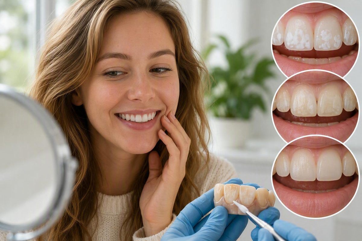
A chalky white patch can be an early sign that enamel has lost minerals, but it is not a diagnosis by itself. The important questions are whether the enamel surface remains intact, whether the lesion is active, and whether tooth structure has physically broken down.
That distinction determines what “reversal” can realistically mean. Early, noncavitated mineral loss may be arrested or partly remineralized. The enamel may become harder and less likely to progress even if the white mark remains. Once the surface has collapsed into a hole, remineralization cannot regrow the missing enamel, and dental repair is generally required (NIDCR’s explanation of the tooth-decay process).
What tooth decalcification means
Tooth decalcification commonly means that enamel has lost minerals, especially calcium and phosphate. Demineralization is the more precise term because the process involves more than calcium alone.
Enamel is an extremely mineral-rich outer layer. Its tightly packed crystals provide strength, but mature enamel does not contain living cells capable of rebuilding lost tissue through ordinary cellular growth. Remineralization reincorporates calcium and phosphate into enamel that remains structurally present; it does not grow a new piece of enamel where the original tissue is missing. A 2021 narrative review in Dimensions of Dental Hygiene explains this distinction between mineral replacement and cellular regeneration (review of enamel demineralization and remineralization).
Plaque-related decalcification can be an early stage of the tooth-decay process:
- Plaque bacteria metabolize fermentable carbohydrates and produce acids.
- Those acids remove minerals from enamel.
- The enamel becomes more porous and may look white or chalky.
- Continued mineral loss weakens the surface.
- The surface may eventually collapse and form a cavity.
The boundary that matters most is surface integrity:
- Demineralized but intact enamel: Mineral has been depleted, but the surface has not collapsed. Arrest or partial remineralization may be possible.
- Cavitated enamel: Tooth structure has physically broken down. It cannot regrow through remineralization and generally needs restorative treatment.
- A white mark from another cause: Remineralization may not address the appearance because active plaque-related mineral loss may not be the underlying problem.
This is also why decalcification should not be treated as a straightforward dietary calcium deficiency. Plaque-related enamel mineral loss reflects conditions at the tooth surface: acid exposure, plaque retention, saliva, fluoride, cleaning, and eating or drinking patterns.
How enamel loses—and regains—minerals
Enamel is not in a permanently fixed mineral state. Teeth undergo repeated periods of mineral loss and mineral replacement throughout the day.
Dental plaque contains bacteria that metabolize sugars and other fermentable carbohydrates. As those bacteria process carbohydrates, they produce acids that dissolve calcium and phosphate from enamel beneath the plaque.
The outcome is not determined by one snack. Each exposure to a sugary drink, sweet snack, or readily fermentable starch can provide another opportunity for bacterial acid production. Frequent sipping or grazing can therefore restart the acid challenge repeatedly, even when each serving seems small.
Between those challenges, saliva provides an important defense. It helps dilute and neutralize acids and carries calcium and phosphate to tooth surfaces. When conditions become favorable, some minerals can return to porous enamel. Fluoride supports this process and makes the resulting mineral structure more resistant to later acid exposure. NIDCR describes decay as an ongoing balance between mineral loss and saliva- and fluoride-supported replacement rather than a one-time event (NIDCR guidance on demineralization and remineralization).
Factors that influence this balance include:
- How effectively plaque is removed
- How often fermentable carbohydrates are consumed
- Whether fluoride is regularly available at the tooth surface
- The amount and properties of saliva
- Whether food and plaque are retained around orthodontic appliances
- Whether a lesion is intact or already cavitated
- Whether acidic drinks or stomach acid are directly eroding enamel
Reduced saliva flow weakens both acid neutralization and mineral delivery. Medication use, dehydration, and some health conditions may contribute to dry mouth, but persistent low saliva flow warrants professional assessment rather than condition-specific self-treatment. A dental-practice overview describes saliva as a source of calcium and phosphate and identifies reduced production as a cavity-risk factor (Cactus Point Dental’s remineralization overview).
Fluoride does not grow new enamel. It helps shift the demineralization–remineralization balance toward retaining or replacing minerals in enamel that is still present. It is therefore most useful before surface collapse, together with plaque control and fewer acid challenges.
What decalcification may look like—and what else can look similar
An early demineralized lesion may appear:
- Opaque rather than naturally translucent
- Dull, matte, or chalky white
- Flat but visibly different from nearby enamel
- Rough-looking or less glossy
- Near the gumline
- Around an orthodontic bracket, band, or attachment
These features are clues, not diagnostic rules. A mirror inspection or photograph cannot reliably establish whether a lesion is active, how deeply it extends, or whether the surface is microscopically intact.
White spots also have several possible causes.
Plaque-related decalcification is acquired after a tooth erupts. It develops where the local environment favors repeated mineral loss, often in a plaque-retentive area.
Dental fluorosis develops while teeth are forming, before they erupt. It is associated with excessive fluoride intake during tooth development, with the effect depending on factors such as timing and exposure. Developmental history and distribution may help a clinician distinguish fluorosis from acquired decalcification, but there is no dependable do-it-yourself visual test. A commercial orthodontic patient guide also distinguishes developmental fluorosis from bacteria-associated decalcification while recommending professional diagnosis when the cause is uncertain (Frigo Orthodontics’ decalcification guide).
Other possibilities include:
- Developmental enamel defects or hypomineralization
- Direct acid erosion
- An early or established cavity
- A residual mark from an inactive lesion
- Natural variations in enamel structure or color
Sensitivity and roughness are similarly nonspecific. Cold or sweet sensitivity can occur for several reasons, while some demineralized lesions produce no noticeable symptoms. Roughness may accompany porous enamel, deposits, erosion, or surface breakdown. Either symptom may justify evaluation, but neither confirms decalcification.
A dental examination addresses questions that a photograph cannot settle: Is the surface intact? Does the lesion appear active or inactive? Is there a hole or weakened margin? Is the pattern more consistent with plaque retention, erosion, or a developmental condition? Depending on the location and findings, a dentist may also use careful drying, lighting, magnification, radiographs, or other clinical tools to distinguish early mineral loss from established decay (clinical overview of examination and imaging for early decay).
Why white spots often appear around braces
Braces do not chemically extract calcium from teeth, and aligner attachments do not directly decalcify enamel. The risk comes from the way orthodontic appliances change the cleaning environment.
Brackets, wires, bands, and attachments create edges and sheltered areas where plaque and food can remain. They also make it harder for toothbrush bristles and interdental tools to reach every part of the tooth. If plaque repeatedly stays around a bracket, bacterial acid production becomes concentrated in that area.
This can create a recognizable pattern: chalky patches surrounding the former bracket position, beside bands and wires, or along the gumline. The shape reflects where plaque was retained rather than a bracket mechanically pulling minerals from enamel.
Removable aligners are not automatically protective. Both fixed braces and aligners can create cleaning challenges even though neither appliance directly removes enamel minerals (Coastline Orthodontics’ overview of orthodontic decalcification).
A practical, brand-neutral routine during orthodontic treatment includes:
- Brush with fluoride toothpaste at least twice daily.
- Angle the brush to clean above, below, and around every bracket.
- Clean carefully along the gumline and bracket edges.
- Clean between the teeth every day.
- Use floss threaders, interdental brushes, or other orthodontic aids as professionally instructed.
- Clean removable appliances according to professional instructions.
- Avoid replacing aligners over visible food debris or sugary residue.
- Continue routine dental cleanings and examinations during orthodontic treatment.
- Ask the dentist or orthodontist to monitor any developing white areas.
Technique and consistency matter more than trying to scrub a white spot away. Aggressive brushing does not replace access to plaque-retentive edges and can contribute to surface wear.
There is no responsible universal timetable for when a visible lesion will appear. Risk varies with plaque levels, eating and drinking frequency, saliva, fluoride exposure, appliance design, baseline enamel condition, and individual susceptibility. A spot noticed after braces may have developed during treatment, but its appearance alone cannot show exactly when it began or whether it remains active.
Can decalcified enamel be reversed?
The most accurate answer depends on the lesion’s stage.
If the enamel surface is intact
An early, noncavitated lesion may be arrested and may regain some mineral. Effective plaque control, fewer dietary acid challenges, fluoride, saliva, and professional monitoring can shift the local environment toward remineralization.
In this context, “reversal” does not necessarily mean that the tooth will look exactly as it did before. A meaningful result may be that the lesion:
- Becomes harder
- Becomes inactive
- Stops enlarging
- Becomes less porous
- Is less likely to progress into a cavity
Its color may improve, remain unchanged, or become less conspicuous without disappearing completely.
If the surface has collapsed
Once enamel has physically broken down into a hole, remineralization cannot recreate its original shape. A filling or another restoration is generally needed to replace missing structure and create a surface that can be maintained.
Visual appearance alone is therefore a poor basis for choosing treatment.
If the lesion has stabilized but remains white
Mineral replacement may improve physical stability without completely restoring the enamel’s original optical properties. A dental-practice article similarly notes that saliva-supported remineralization may help protect the surface while visible marks remain (Bhandal Dental Practice’s decalcification overview).
This separates two goals:
- Disease control: Stop active mineral loss and prevent cavitation.
- Appearance management: Decide whether a stable residual mark warrants cosmetic treatment.
Disease control comes first. Altering a spot’s appearance without controlling plaque, dietary frequency, or acid exposure does not address why it developed.
There is no universal recovery timeline. Outcomes vary with lesion depth and activity, surface integrity, location, plaque control, fluoride exposure, saliva, diet, and follow-through. Fluoride remains the established standard for conventional remineralization care, but not every intact lesion will completely remineralize or become invisible (narrative review of fluoride and adjunctive remineralization strategies).
Dental reassessment is the practical way to determine whether an area is stable, improving, progressing, or already cavitated. Informal comparisons in changing bathroom light are not a dependable substitute.
Prevention and home care: what has the strongest support
Home care should focus on changing the conditions that permit repeated mineral loss rather than trying to bleach, scrape, or supplement away a white spot.
The main priorities are:
- Brush twice daily with fluoride toothpaste. Clean every accessible surface, especially the gumline and areas around orthodontic attachments.
- Clean between the teeth daily. A toothbrush does not reliably reach every contact area.
- Reduce frequent sugary snacks and drinks. Each exposure can create another period of bacterial acid production.
- Choose water for routine sipping. Water does not supply sugar to plaque bacteria and can help clear residues.
- Maintain regular dental care. Monitoring can identify progression or cavitation that is not obvious at home.
- Have persistent dry mouth assessed. Hydration supports normal saliva function, but water alone may not resolve reduced salivary flow.
These measures align with government guidance on preventing early mineral loss from progressing to permanent decay.
Why grazing and sipping matter
Eating a sweet food once and nibbling it repeatedly over several hours do not create the same exposure pattern. Repeated intake supplies fermentable carbohydrates again and again, potentially restarting bacterial acid production before enamel has had much opportunity to recover.
The same principle applies to slowly sipping sweetened coffee, sports drinks, soda, juice, or other sugary beverages. Reducing the number of exposures may help even when the total amount consumed does not seem large. The purpose is not to impose a rigid eating schedule, but to avoid turning sugary intake into an all-day event.
Saliva is supportive, not a cure
Adequate hydration supports normal saliva function. Saliva helps neutralize acids, clear residues, and deliver minerals, but it cannot close a formed cavity.
Sugar-free gum may stimulate saliva in suitable people, but it does not replace fluoride toothpaste, brushing, interdental cleaning, monitoring, or necessary treatment. Persistent dry mouth—particularly when accompanied by rapid decay or difficulty eating or speaking—warrants dental or medical assessment.
Bacterial demineralization is not the same as erosion
Plaque-related demineralization occurs when bacteria metabolize fermentable carbohydrates and produce acid at the tooth surface. Erosion is direct chemical damage from acids that do not require bacterial metabolism. Acidic drinks can contribute, as can recurrent reflux or vomiting.
The distinction matters because plaque removal cannot resolve recurrent stomach-acid exposure. After a clearly acidic drink, reflux episode, or vomiting, rinse gently with water rather than scrubbing the teeth immediately. Use a soft brush and light pressure when brushing later. A dental-practice guide recommends rinsing with water and delaying brushing after acidic exposure while emphasizing that fully lost enamel does not regrow (Hilltop Dental Studio’s enamel-remineralization guide).
Recurrent reflux or vomiting requires appropriate professional care for the underlying problem as well as dental assessment. Toothpaste alone cannot address the source of repeated acid exposure. For a broader explanation of why brushing cannot repair erosion or missing tooth structure, see What Daily Brushing Cannot Fix.
Fluoride use should fit the person
Over-the-counter fluoride toothpaste is a foundation of prevention, but professional varnishes, gels, rinses, and prescription-strength toothpastes are not interchangeable or appropriate for everyone. Product selection depends on factors such as age, cavity risk, lesion location, and whether the surface remains intact.
Children require age-appropriate fluoride use and adult supervision to reduce swallowing. Parents should follow the product label and guidance from the child’s dentist or healthcare professional rather than applying an adult or prescription regimen by default.
Treatment options by lesion stage and goal
Treatment is easier to understand when divided into four goals: control the cause, arrest intact enamel, improve residual appearance, and restore missing structure. One product or procedure does not accomplish all four.
1. Control the cause
Before treating appearance, identify what is driving the change. Plaque around a bracket calls for better cleaning access. Repeated sugary sipping calls for fewer exposures. Reduced saliva flow requires assessment. Direct erosion from drinks or recurrent stomach acid requires a different response from bacterial demineralization.
Cause control may include:
- More effective plaque removal
- Daily interdental cleaning
- Fluoride toothpaste
- Fewer fermentable-carbohydrate exposures
- Water as the routine drink
- Professional cleaning where deposits cannot be removed at home
- Review of saliva, diet, and erosive exposure
- Monitoring during orthodontic treatment
Without that foundation, the tooth may remain vulnerable around a treated area or elsewhere in the mouth.
2. Arrest or remineralize intact enamel
For an early, intact lesion, management may include fluoride toothpaste, improved plaque control, fewer sugar exposures, and professional monitoring. A clinician may also select a topical fluoride treatment or prescription product when the lesion and overall risk justify it.
Prescription-strength fluoride should not be borrowed, improvised, or assumed necessary for every white spot. Professional concentrations and schedules are lesion- and tooth-specific rather than universal self-treatment instructions.
Calcium-phosphate products may be considered in selected cases. Suitability depends on the formulation and the person using it. These products should be treated as possible adjuncts rather than universal replacements for fluoride, plaque removal, and dietary control.
Fluoride has the most established role in conventional remineralization care. Hydroxyapatite, calcium-phosphate agents, xylitol, arginine, and probiotics have different proposed mechanisms and differing or incomplete evidence. The supplied evidence does not establish them as universally equivalent or superior to topical fluoride.
Oil pulling and baking soda should not be described as cavity cures. A properly formulated baking-soda product may affect acidity or assist cleaning, but it does not biologically regrow missing enamel. No home method can close a structural hole.
3. Improve the appearance of a stable white mark
If a lesion is inactive but still conspicuous, a dentist may discuss cosmetic options. The choice depends on the diagnosis, depth, location, surface condition, and surrounding enamel.
Possible approaches include:
- Microabrasion: Removes a small, superficial amount of enamel to reduce selected surface discoloration.
- Resin infiltration: Introduces resin into selected noncavitated porous enamel, altering how the area transmits light and potentially making it less visible.
- Composite bonding: Covers or replaces part of the visible surface with tooth-colored restorative material.
- Veneers: Cover a larger part of the front surface and represent a more extensive cosmetic intervention.
These procedures do not regrow enamel. Microabrasion removes material, infiltration changes lesion porosity and optics, and bonding or veneers cover or replace visible surfaces. Educational material from a registered dental hygienist lists these options for selected persistent or advanced white spots while emphasizing that early lesions may only partly reverse (overview of white-spot management after braces).
It changes tooth color rather than replacing minerals or repairing a cavity.
4. Restore missing tooth structure
When the enamel surface has physically cavitated, management generally changes from remineralization alone to restoration. A filling or another restorative approach replaces missing structure, re-establishes shape, and creates a maintainable surface.
The type of restoration depends on how much tooth structure is affected and whether decay extends beyond enamel. That decision requires clinical examination.
A useful sequence is:
- Confirm what the mark is.
- Determine whether the surface remains intact.
- Control plaque, sugar frequency, and other drivers.
- Arrest or remineralize intact enamel where possible.
- Reassess stability.
- Address residual appearance separately if desired.
- Restore areas that have physically broken down.
When a dentist should assess the tooth
Any uncertain or persistent white spot is worth showing to a dentist because plaque-related decalcification, fluorosis, developmental defects, erosion, and decay can resemble one another. Early evaluation does not automatically mean drilling or cosmetic treatment. It establishes what process is present and whether monitoring is reasonable.
Arrange dental care particularly if you notice:
- A visible hole or surface collapse
- A spot that appears to be enlarging
- Darkening or visible breakdown
- Pain
- Persistent or worsening sensitivity
- A rough area that seems to be deteriorating
- Food repeatedly catching in the area
- Difficulty cleaning around brackets, bands, or attachments
- Multiple new white areas
- Recurrent reflux, vomiting, or significant dry mouth
- A mark whose cause or duration is uncertain
A clinician will assess whether the enamel surface is intact, whether the lesion appears active, and whether decay may have extended beyond enamel. Imaging or other clinical tools may be appropriate for locations that cannot be evaluated adequately by direct visual examination.
Diagnosis should come before choosing remineralization, cosmetic management, or restoration. Treating every white mark as an active cavity could lead to unnecessary intervention, while assuming every flat mark is harmless could allow active decay to progress.
The decision path is straightforward:
- A flat white spot may represent early, intact mineral loss. Improve plaque removal, use fluoride toothpaste, reduce repeated sugar exposures, and arrange dental monitoring.
- A stable white mark may remain after the lesion is controlled. Persistence does not necessarily mean mineral loss is still active.
- A hole, breakdown, pain, or worsening sensitivity needs professional assessment. Remineralization cannot replace missing tooth structure.
- Control progression first; consider appearance second. Cosmetic treatment is a separate decision after the tooth’s condition is understood.
Decay Guide provides independent general reference information and does not diagnose, treat, or provide individual dental advice, as explained in its publisher disclosure.
Frequently asked questions
Is tooth decalcification the same as a cavity?
Not necessarily, but plaque-related decalcification can be part of the cavity-forming process. Decalcification describes mineral loss from enamel. If the surface remains intact, the lesion is noncavitated and may be arrested or partly remineralized. A cavity exists once tooth structure has physically broken down into a hole.
The practical distinction is not simply “white spot versus cavity,” because appearance alone cannot reliably show whether the surface is intact. Once enamel structure is destroyed, mineral replacement cannot recreate it.
Can decalcification after braces go away completely?
Sometimes the appearance improves, but complete disappearance cannot be promised. An intact lesion may regain mineral, become harder, and stop progressing while still looking whiter or more opaque than surrounding enamel.
After the lesion is stable, a dentist may discuss resin infiltration, microabrasion, bonding, or another cosmetic option if the remaining appearance is a concern. Those procedures alter, cover, or replace the visible surface; they do not biologically regrow enamel.
How can I tell decalcification from fluorosis or another white spot?
You usually cannot tell with certainty from appearance alone. Plaque-related decalcification develops after eruption, while fluorosis and many developmental enamel defects arise while teeth are forming. History, distribution, surface condition, lesion activity, and sometimes imaging or other clinical tools help distinguish them.
Do not rely on a photograph, roughness, or sensitivity as a definitive test. A persistent or unexplained white spot should be examined before you choose remineralizing or cosmetic treatment.
Do hydroxyapatite, calcium-phosphate products, or xylitol work as well as fluoride?
The available evidence does not justify treating all of these options as equivalent to fluoride. Fluoride remains the established standard for limiting mineral loss and supporting remineralization. Hydroxyapatite and calcium-phosphate products may be considered as adjuncts in selected situations, while xylitol-containing gum may stimulate saliva.
Evidence and formulations vary. None of these products should be assumed to close a formed cavity, and specialized products should not automatically replace fluoride toothpaste or professional assessment.
Should I brush immediately after acidic food or drinks?
Avoid aggressive immediate brushing after a clearly acidic exposure, including an acidic drink, reflux, or vomiting. Rinse gently with water, then brush later with a soft brush and light pressure.
This does not change the general recommendation to brush twice daily. The purpose is to avoid scrubbing enamel immediately after acid exposure, not to leave teeth unclean indefinitely. Recurrent reflux or vomiting warrants medical and dental assessment rather than brushing changes alone.