
When a Molar Groove Is Stained, Decaying, or Ready for a Sealant
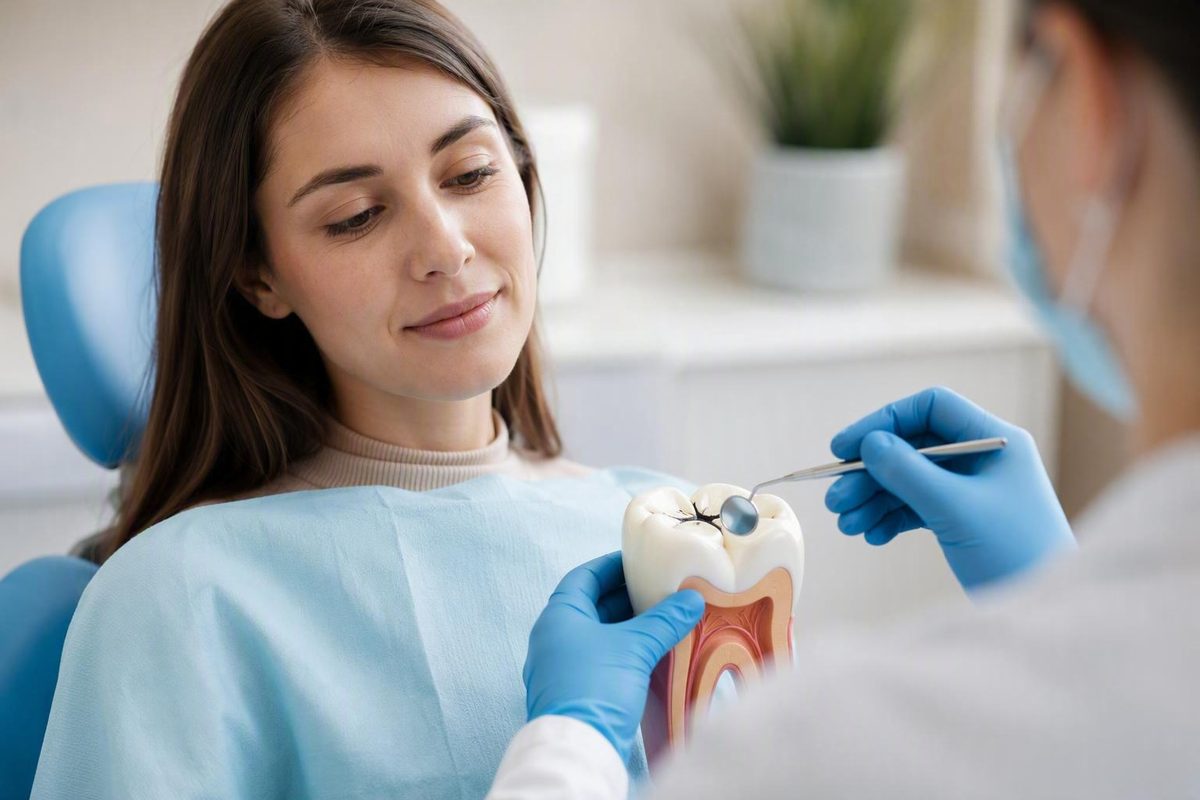
A black line on a molar can be alarming, especially when it appears despite regular brushing. But a dark groove is not automatically a cavity, and a painless tooth is not automatically healthy. The mark could be ordinary staining, an early enamel lesion without a physical hole, an established cavity, or deeper decay.
The distinction matters because management changes with the condition of the tooth. A sound but vulnerable groove may benefit from prevention. A selected noncavitated lesion may be monitored, supported with fluoride, or sealed. A physical defect or deeper structural involvement is more likely to require restorative assessment. If the pulp is affected, more extensive care may be considered.
Appearance and symptoms alone cannot settle which situation applies. A dentist must examine the tooth and consider lesion depth, surface breakdown, symptoms, remaining tooth structure, cavity history, and whether the tooth can be restored.
What a pit and fissure cavity is—and where it forms
Pits are small natural indentations in a tooth, while fissures are longer grooves. They are most prominent on the chewing surfaces of back teeth, particularly molars and premolars.
These features are normal anatomy, not defects by themselves. A deep groove may be completely sound. A pit and fissure cavity is decay that develops in one of these areas and damages the tooth.
A simple description of tooth anatomy helps explain how decay can progress:
- Enamel is the hard outer covering of the tooth’s crown.
- Dentin lies beneath the enamel and makes up much of the tooth.
- Pulp is the soft central tissue containing nerves and blood vessels.
- The root anchors the tooth below the gumline.
A pit-and-fissure lesion begins at a grooved surface, usually on the biting surface of a back tooth. Mineral can be lost from enamel before an obvious hole appears. If the process continues, the surface may break down and decay may extend into dentin or toward the pulp. Cavities can affect different parts of the crown or root, and a person may not feel one until deeper structures are involved (Cleveland Clinic’s overview of cavities).
Location distinguishes pit-and-fissure decay from two other broad categories:
- Smooth-surface decay develops on flatter tooth surfaces, including areas between neighboring teeth.
- Root decay develops on an exposed root surface.
- Pit-and-fissure decay can form on a tooth’s chewing surface and can also affect the front side of back teeth Cavities (Tooth Decay): Symptoms, Causes & Treatment.
These are location-based descriptions. They do not determine how quickly a particular lesion will progress or which treatment it will need.
It is also useful to distinguish several related terms:
- Natural pit or fissure: normal tooth anatomy.
- Caries lesion: an area affected by the tooth-decay process.
- Cavitation: physical breakdown of the tooth surface.
- Cavity: commonly used for a hole or damaged area produced by decay.
People often use “cavity” for any suspected decay. Clinically, however, whether the surface is still intact is important because a noncavitated lesion may be managed differently from an established physical defect.
Why back-tooth grooves can decay despite regular brushing
The chewing surface of a molar may look broadly flat, but it can contain a network of narrow grooves, branches, and indentations. Some fissures are broad and easy to clean. Others narrow into spaces that toothbrush bristles cannot enter effectively.
Food debris and plaque can remain in these protected areas. Plaque is a bacterial film. When its bacteria metabolize sugars from foods and drinks, they produce acids that weaken and demineralize enamel. If repeated mineral loss exceeds repair, an early lesion can develop and may eventually become cavitated.
This explains why someone can brush carefully and still develop a pit and fissure cavity. Brushing disrupts plaque on accessible surfaces, but it cannot change the microscopic shape of a narrow fissure. A clinical review notes that complex groove anatomy impairs cleaning and encourages plaque accumulation. It also reports that topical fluoride protects pits and fissures less effectively than smooth surfaces, helping explain why sealants can offer targeted protection (NCBI clinical review of pit and fissure sealants).
Deep grooves are not destined to decay. Their anatomy increases susceptibility, but risk also reflects factors such as:
- Groove depth and complexity
- Plaque retained in the area
- Frequency of sugary foods and drinks
- Fluoride exposure
- Previous or current cavities
- Whether the surface can be cleaned effectively
- Access to preventive care and follow-up
Sugar frequency deserves particular attention. Each sugary exposure gives plaque bacteria another opportunity to produce acid. Reducing how often teeth encounter sugar may be more practical than attempting to eliminate every sweet food.
Previous cavity experience also matters when a dentist assesses risk. Two people can have similarly shaped grooves but different histories and different preventive needs. Teeth in the same mouth can differ as well: one molar may have a broad, cleansable groove, while another has a narrow fissure that retains plaque.
Brushing remains essential, but a cavity is not proof that somebody failed to brush. The more accurate explanation combines groove anatomy, plaque, sugar exposure, fluoride protection, and individual cavity history.
Dark groove, early lesion, or physical cavity?
A dark line often prompts the question, “Is this a cavity?” Color alone cannot answer it. A brown or black fissure can be stained but structurally sound. It can also occur with inactive or active decay. White, chalky, or brown changes may reflect mineral alteration, but appearance does not establish whether a lesion is progressing.
Possible changes that justify attention include:
- A persistent or changing dark line in a groove
- A white, brown, or chalky change near a fissure
- A persistent change in surface contour or roughness
- A visible depression or broken surface
- Sensitivity to cold, heat, or sweets
- Pain while biting or chewing
- Toothache
- Food repeatedly lodging in the same defect
Each finding has more than one possible explanation. Sensitivity and pain, for example, are not unique to cavities.
The possibilities can be organized into three practical categories.
1. A sound or stained groove
The fissure is natural and its surface remains healthy. Pigments may collect in the groove and make it appear brown or black. Depending on the groove’s anatomy and the person’s cavity risk, a dentist may recommend routine prevention, monitoring, or a preventive sealant.
2. A noncavitated enamel lesion
Mineral has been lost from the enamel, but there is no established physical hole. The surface may appear white, brown, or otherwise altered, or the change may be difficult to see. Selected lesions may sometimes be arrested with fluoride, improved plaque control, dietary changes, monitoring, and possibly sealing.
“Noncavitated” does not mean unimportant. It means the surface has not undergone the type of physical breakdown that automatically points toward a conventional filling. The lesion’s depth and likelihood of progression still matter.
3. A cavitated lesion
The surface has physically broken down. The defect may be limited or may extend through enamel into dentin. A confirmed hole or deeper structural involvement generally makes restorative assessment more likely because the area can retain plaque and be difficult to clean.
Pain is not a dependable boundary between these categories. Early enamel disease may cause no symptoms, so the absence of discomfort does not rule out decay.
A dentist examines the tooth and may use dental radiographs when appropriate. The examination and images help the dentist judge whether decay is present and whether deeper structures may be involved.
How pit and fissure decay can progress
Tooth decay begins with mineral loss. Demineralization occurs when acids produced by plaque bacteria weaken enamel. This can happen before there is an established hole.
At this early point, management aims to change the conditions around the lesion. Fluoride can support remineralization and make enamel more resistant to acid. Better plaque control reduces the bacterial film producing acid, while less frequent sugar exposure reduces repeated acid production. A sealant may also be considered for a suitable noncavitated fissure.
That is why some early enamel lesions can be arrested without conventional drilling. It would be inaccurate, however, to promise that every white or brown mark can be reversed. The discoloration may not be decay, or the lesion may be too advanced for noninvasive care.
If mineral loss continues, the enamel can break down. Once there is an established surface defect, the area may retain more plaque and become harder to clean. Restorative assessment then becomes more likely.
Dentin beneath the enamel is softer, and decay that reaches it can progress more rapidly. Dentin involvement may also be associated with sensitivity, although symptoms do not provide a precise measurement of lesion depth.
If decay progresses toward the pulp, pain, inflammation, infection, or swelling may occur. Treatment depends on examination and diagnosis.
Diagrams often present tooth decay as a sequence from demineralization to enamel decay, dentin decay, pulp damage, and abscess. That sequence can be a useful teaching model, but it is not a home diagnostic rule. Lesions may become inactive, and symptoms or discoloration can have causes other than decay.
Management therefore depends on a combination of findings:
- Whether the surface is intact or cavitated
- How deeply the lesion extends
- Whether it appears likely to progress
- How much sound tooth structure remains
- Whether symptoms or pulp involvement are present
- Whether the tooth can be predictably restored
- The person’s broader cavity risk
- Whether reliable monitoring is possible
Waiting for pain is a poor strategy. Early lesions may be silent, while pain can mean that the assessment and treatment decision have become more complex.
Treatment options matched to the condition of the tooth
Treatment should respond to the actual condition of the tooth, not merely the color of a groove or the label attached to a “stage.”
The options below are possibilities rather than automatic prescriptions. General cavity care can range from fluoride for selected early disease to fillings, root-canal treatment, or extraction, depending on severity and clinical circumstances.
| Approach | Purpose | Situations in which it may be considered | Invasiveness | Follow-up needs |
|---|---|---|---|---|
| Fluoride, plaque control, dietary changes, and monitoring | Support remineralization and reduce repeated acid challenges | Sound high-risk surfaces or selected noncavitated enamel lesions | Noninvasive | Reassessment based on risk and observed change |
| Pit and fissure sealant | Create a barrier over a susceptible groove or selected noncavitated lesion | Sound high-risk fissures and some intact early lesions | Microinvasive surface conditioning without conventional cavity preparation | Check retention and repair or reseal if defective |
| Preventive resin restoration | Remove localized damaged tissue while preserving sound structure, then restore and seal adjacent grooves | Small localized cavitation or limited deeper involvement when clinically suitable | Minimally invasive | Monitor the restoration and remaining sealant |
| Conservative filling | Restore a physical defect and return the tooth to usable form | Confirmed cavitation, dentin involvement, or a lesion unsuitable for sealing alone | Restorative; extent varies | Monitor margins, wear, symptoms, and recurrent decay |
| Inlay or onlay | Replace a larger damaged portion while retaining suitable remaining tooth structure | Structural loss that exceeds what a small direct filling can manage | More extensive restorative care | Periodic assessment of the restoration and tooth |
| Crown | Cover and protect a substantially weakened tooth | Extensive structural loss, elevated fracture concern, or restoration after some pulp treatments | More extensive | Ongoing assessment of the crown, margins, tooth, and symptoms |
| Root-canal treatment | Treat diseased pulp while retaining a restorable tooth | Diagnosed pulpal disease or infection when the tooth can be retained | Endodontic treatment followed by restoration as appropriate | Review healing and the final restoration |
| Extraction | Remove a tooth that cannot be predictably retained | Non-restorable damage or other case-specific reasons | Surgical removal | Healing review and discussion of replacement when appropriate |
Noninvasive care
For a selected early, noncavitated lesion, the initial plan may include fluoride, more effective plaque control, less frequent sugar intake, and observation. This is active management rather than neglect. It requires reassessment and a willingness to change course if the lesion progresses.
A dentist may recommend professional fluoride according to the person’s risk and clinical findings. The product and frequency should be individualized rather than chosen from an online description.
Sealing versus restoring
Sealing is often described as microinvasive because the surface is cleaned and conditioned so that a material can bond, but conventional cavity preparation is avoided. Its principal advantage is the preservation of tooth structure. For a suitable early lesion, the goal is to isolate the fissure and stop further progression.
A preventive resin restoration or conservative filling is minimally invasive rather than noninvasive. Localized damaged tissue is removed while sound enamel and dentin are preserved where possible. A sealant may then be placed over adjoining vulnerable grooves.
The dividing line is not simply “small versus large.” Surface breakdown, lesion depth, the ability to obtain a reliable seal, remaining structure, symptoms, and capacity for follow-up all affect the choice.
An older systematic review and network meta-analysis evaluated noninvasive care, sealing, and minimally invasive restoration for shallow or moderately deep pit-and-fissure lesions in permanent teeth. Sealing and minimally invasive restoration reduced later invasive treatment compared with no treatment, but the certainty of evidence was low or very low. Sealing also led to more follow-up procedures such as resealing, so the review did not establish one universally superior strategy (PubMed-indexed systematic review).
Those findings should not be automatically extended to primary teeth, deep lesions, pulpal disease, or every visibly cavitated tooth. They instead illustrate a practical tradeoff: preserving more tooth initially may require closer maintenance, while restoration removes some tissue but provides a different form of structural control.
More extensive restoration or pulp treatment
As structural damage increases, a small direct filling may not provide adequate restoration. An inlay, onlay, or crown may then be considered. These treatments are possibilities, not automatic consequences of reaching a named stage; treatment descriptions from patient-education sources likewise vary according to the amount of damaged tooth structure (overview of cavity types and restorative options).
If the pulp is diseased or infected and the tooth remains restorable, root-canal treatment may be considered. If the tooth cannot be predictably restored, extraction may enter the discussion. The decision depends on pulp status, remaining structure, restorability, symptoms, and clinical judgment.
The central question is not, “Which procedure treats every pit and fissure cavity?” It is, “What is the least invasive approach likely to control this lesion and preserve a functional tooth in its actual condition?”
Dental sealants: prevention, early-lesion management, and maintenance
A dental sealant is a material placed over susceptible chewing-surface grooves to create a physical barrier. It limits access by plaque, microorganisms, and food particles.
Sealants have two uses that should not be confused:
- Preventive sealing covers a sound but high-risk groove before decay develops.
- Therapeutic sealing covers a selected noncavitated lesion with the aim of arresting it.
Support for sealing is stronger for sound high-risk surfaces and appropriately selected noncavitated lesions than for established cavitated disease. A physical defect, deeper structural involvement, or inability to achieve a reliable seal makes restorative assessment more likely. “Putting a sealant over a cavity” is therefore too broad: an intact early lesion is different from an open, plaque-retentive defect.
Who may be a candidate?
Eligibility should be based on the tooth and the person’s cavity risk, not on age alone. Relevant considerations include:
- Groove anatomy
- Whether the surface is sound, noncavitated, or physically broken
- Previous or current cavity experience
- Fluoride exposure
- Whether the surface can be cleaned
- Whether the tooth has erupted far enough to isolate
- Whether saliva can be controlled during placement
- Whether the sealant can be monitored afterward
Newly erupted permanent molars often receive attention because their anatomy can make them vulnerable soon after eruption. Adults may also be candidates. An adult with an unfilled, susceptible groove may benefit, while a child with broad, easily cleaned grooves and low cavity risk may not need every tooth sealed. Clinical guidance recommends basing sealant decisions on individual cavity risk and tooth condition rather than eruption age alone (NCBI guidance on sealant indications and materials).
How a sealant is placed
Although materials and protocols vary, placement generally involves:
- Examining the tooth and deciding whether sealing is appropriate
- Cleaning the chewing surface
- Isolating the tooth from saliva
- Conditioning or otherwise preparing the enamel
- Applying the sealant into the pits and fissures
- Hardening or curing the material
- Checking coverage, retention, and the bite
Moisture control is important because saliva contamination can interfere with bonding. A partly erupted molar can be harder to isolate because gum tissue may still cover part of the crown.
Resin-based versus glass-ionomer materials
Resin-based sealants generally retain better when the tooth can be isolated and prepared properly. Glass-ionomer materials may be considered when moisture control is difficult, including on some partially erupted teeth.
Neither material should be treated as universally best. A material with strong theoretical retention may perform poorly if the working surface is contaminated during placement.
What a sealant does not do
A sealant protects only the area it covers. It does not protect the sides of the tooth, spaces between teeth, exposed roots, or every other surface in the mouth. It does not replace:
- Fluoride toothpaste
- Thorough brushing
- Interdental cleaning
- Attention to sugar frequency
- Professional assessment
- Broader cavity-risk management
A sealant is not necessarily permanent. It can wear, chip, detach, or lose its seal at an edge. A partially missing or defective sealant should be assessed rather than assumed to remain protective. Repair or resealing may be appropriate.
There is no universal lifespan or fixed replacement date for every sealant. Longevity varies with the material, placement quality, tooth, bite, moisture control, and continued retention. Maintenance is therefore part of treatment: a sealant is most useful when tooth selection, placement, retention, and follow-up all align.
A practical prevention plan for cavity-prone grooves
Home care cannot guarantee that every deep fissure will remain decay-free, but it can reduce the conditions that allow decay to develop or progress.
Brush twice daily for about two minutes with fluoride toothpaste. Clean the outer, inner, and chewing surfaces, deliberately pausing over molars and premolars. Consumer oral-health guidance recommends twice-daily, two-minute brushing with specific attention to back-tooth chewing surfaces (Colgate’s pit-and-fissure prevention guide).
Technique matters, but it has a physical limit. Bristles may clean the opening of a groove without reaching its narrowest depth. Scrubbing harder does not alter the fissure’s anatomy.
Use fluoride consistently. Fluoride toothpaste supports enamel’s resistance to acid. Additional fluoride products or professional applications should be chosen according to individual risk and professional advice rather than added indiscriminately.
Clean between teeth. Pit-and-fissure decay occurs on chewing surfaces, but decay can also develop between neighboring teeth. Brushing molar grooves does not replace interdental cleaning.
Reduce the frequency of sugary exposures. Avoid repeatedly sipping sweet drinks or grazing on sugary snacks throughout the day. Fewer exposures mean fewer opportunities for plaque bacteria to produce acid.
Treat sealants as an added layer, not a substitute. A sealant may protect a high-risk groove, but it does not compensate for frequent sugar exposure or protect surfaces it does not cover.
Arrange individualized assessment. A suspicious groove, previous cavities, repeated sensitivity, or questions about sealant eligibility are reasons to consult a dentist. Follow-up timing should reflect individual risk and clinical findings rather than a universal six-month schedule.
Myth: Every black groove is a cavity. Fact: A fissure may be stained, sound, noncavitated, inactive, or actively decaying. Color cannot reliably distinguish among these possibilities.
Myth: A cavity always hurts. Fact: Early enamel disease may produce no symptoms, so lack of pain does not rule out decay.
Myth: Sealants are only for children. Fact: Children and adults may be candidates when tooth condition and cavity risk support their use.
Myth: Better brushing can reverse any cavity. Fact: Plaque control can help manage selected early lesions, but it cannot rebuild every established physical defect.
A practical prevention plan is layered: fluoride toothpaste, careful daily cleaning, fewer repeated sugar exposures, interdental care, and professional measures selected according to risk. No single layer makes the others unnecessary.
When to arrange a dental evaluation
Arrange a dental evaluation if you notice a persistent or changing dark groove, an apparent hole, a rough or broken area, repeated sensitivity, toothache, or pain with chewing. These findings can have causes other than decay, but they merit assessment if they persist or worsen.
An asymptomatic mark also merits dental assessment because early enamel disease may not hurt. Timing can reflect whether the mark is changing, the person’s cavity history, other symptoms, and access to professional advice.
An article cannot establish the cause, assess the pulp, or provide a complete urgency judgment for an individual.
The practical decision framework is straightforward:
- A natural or stained groove is not automatically disease.
- A noncavitated lesion may sometimes be managed without a conventional filling.
- An established defect or deeper decay needs restorative assessment.
- A sealant is useful only when matched to tooth condition, cavity risk, placement quality, and follow-up.
- Pain is not a reliable dividing line between early and advanced disease.
Only a dental assessment can determine whether a particular groove is sound, stained, demineralized, cavitated, or suitable for a sealant. Decay Guide is an independent information publisher, not a dental practice or diagnostic service; it provides general reference information rather than individual treatment advice.
Frequently asked questions
Is every black line in a molar groove a cavity?
No. Pigments can stain a naturally deep fissure even when the surface is sound. A black line can also occur with inactive or active decay, so appearance alone cannot distinguish among them.
Pain is not a reliable test either. Early enamel decay may be painless. A dentist can examine the surface and use radiographs when appropriate, but a photograph, mirror check, or tongue test cannot reliably determine lesion activity or depth.
Can a pit and fissure cavity be reversed without drilling?
Sometimes, if “cavity” is being used to describe an early, noncavitated enamel lesion rather than an established hole. Selected lesions may be arrested or remineralized with fluoride, plaque control, fewer sugar exposures, monitoring, and sometimes sealing.
Once the surface has physically broken down or deeper structures are involved, restorative assessment becomes more likely. Treatment may still be conservative when the findings permit preservation of sound tooth structure.
Can a dentist place a sealant over early decay?
Yes. A dentist may seal a selected noncavitated pit-and-fissure lesion. Surface integrity, lesion depth, the ability to achieve a reliable bond, cavity risk, and the possibility of follow-up all influence the decision.
That is different from routinely covering an open cavity. Established breakdown or deeper structural involvement may require a minimally invasive restoration instead. Any sealant placed over an early lesion must be monitored because loss of retention can undermine the barrier.
Are dental sealants useful for adults as well as children?
They can be. Newly erupted permanent molars are commonly considered because of their groove anatomy and early vulnerability, but age alone should not determine eligibility.
An adult may benefit when an unfilled molar or premolar has susceptible grooves and the person’s cavity risk supports sealing. Conversely, a sound, easily cleaned tooth in a low-risk person may simply be monitored. Tooth condition, moisture control during placement, cavity history, and follow-up matter more than membership in a particular age group.
How long do sealants last, and can they need replacement?
Sealants can remain effective for years, but there is no guaranteed lifespan or universal replacement schedule. Longevity depends on the material, tooth, bite, placement quality, moisture control, wear, and whether the sealant remains fully bonded.
Sealed surfaces should be inspected during risk-based follow-up. If a sealant is partly missing, worn, or defective, it may need repair or resealing. An intact sealant can continue protecting the covered groove, but a damaged one should not be assumed to provide the same barrier.