
How Tooth Decay Can Look—and Why a Dark Spot Is Not a Diagnosis
Some decay is pale and subtle, some dark and obvious, and some cannot be seen at home at all, so a normal mirror check cannot rule it out.
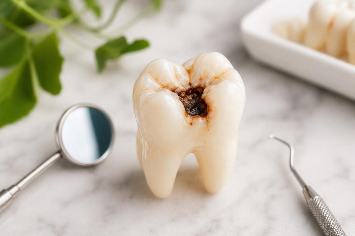
The short answer: cavities do not all look like black holes
A possible cavity can look like:
- A faint, chalky-white or milky patch
- Localized yellow, tan, brown, gray, or black discoloration
- A rough-looking area
- A small pit or indentation
- A discolored line or dot in a tooth groove
- A visible opening or hole
- A larger crater with missing or broken-looking tooth structure
These are reasons to pay attention—not a way to diagnose a tooth. No color, shape, photograph, or mirror check can confirm that a particular mark is decay. Staining, natural grooves, enamel changes, existing dental work, and inactive decay can all alter a tooth’s appearance.
There is no single look shared by every cavity. Appearance varies according to the stage of the decay process, its location, how easily the surface can be seen, and whether the tooth’s structure has broken down. Some decay is pale and subtle; some is dark and obvious; some cannot be seen at home at all.
It also helps to separate tooth decay from a formed cavity. Tooth decay is a process involving mineral loss and damage to tooth structure. Early demineralization can occur while the enamel surface remains intact, sometimes producing a chalky-white area rather than a hole. A cavity commonly refers to an opening or structural breakdown caused by decay. Early disease may not be painful or readily visible, and a dentist may detect it during an examination or on an X-ray (Cleveland Clinic’s overview of cavities).
The familiar image of a painful black hole represents only one possible presentation. Early mineral loss may be painless, and hidden decay may produce no visible sign during a home inspection. Conversely, a dark mark may be a stain or a naturally colored groove rather than active disease. Appearance can help you notice a change, but it cannot tell you what that change is.
How the appearance may change as decay progresses
Descriptions of decay are sometimes divided into numbered stages, but those labels are not necessary for understanding what may be visible. A more practical framework is early mineral loss, surface breakdown, and advanced damage. These are broad possibilities, not a rigid visual sequence that every tooth follows.
Early mineral loss
Before an opening forms, mineral loss in enamel may appear as a:
- Faint white patch
- Dull or opaque area
- Milky-white change
- Chalky spot that contrasts with the surrounding enamel
The change may be smooth, subtle, and painless. It does not necessarily contain a hole. A white spot is also not automatically decay because other enamel changes can look pale or opaque. Early mineral loss may be difficult to see, and a professional assessment is needed to determine whether the surface remains intact and whether the change represents active decay (Harvard Health’s explanation of cavity appearance and detection).
These changes can be especially easy to miss on back teeth, on the side of a tooth, or in places that are difficult to illuminate. Whiteness alone does not reveal the cause, activity, or likely outcome of a mark.
Surface breakdown
As decay progresses, an affected area may show yellow, tan, light-brown, dark-brown, gray, or black discoloration. Its texture or shape may also change, producing:
- A rough area
- A small pit
- An indentation
- A narrow opening
- A visible hole
- A spot where food repeatedly catches
These features may occur together, but they do not have to. A tooth can have discoloration without an obvious hole or a small defect without dramatic discoloration. Dentists assess the overall pattern—including visible changes, structural defects, clinical findings, and imaging when appropriate—rather than treating one color or shape as conclusive.
Advanced damage
More extensive structural loss may look like a larger dark opening or crater. The edges may appear irregular or jagged, and part of the tooth may look broken or missing. An opening may repeatedly trap food, although food trapping is nonspecific and can occur for reasons other than decay. Advanced damage can include a deep hole or missing tooth structure, but its true extent still cannot be measured in a mirror (Tulsa Dental Center’s visual overview of tooth decay).
Darkness and visible size do not reliably measure depth, activity, or severity.
Not every lesion visibly changes from white to brown and then black. Some remain difficult to see, some develop a structural defect without following a clear color sequence, and some dark areas are not active decay at all.
Symptoms are a separate issue. Sensitivity, pain, swelling, or an unpleasant taste may accompany later damage, but these symptoms do not reveal how deep a cavity is and are not required for decay to be present.
| Broad point in the process | Possible appearance | Possible sensations | May it be visible at home? | What appearance cannot establish |
|---|---|---|---|---|
| Early mineral loss | Dull, opaque, milky, or chalky-white patch; often no hole | Frequently none | Sometimes, although it may be extremely subtle | Whether the spot is decay, active, or capable of remineralization |
| Surface breakdown | Yellow, tan, brown, gray, or black area; roughness, pit, indentation, or small opening | None, sensitivity, discomfort, or pain | Sometimes | The depth of damage or treatment needed |
| Advanced damage | Larger opening, crater, irregular edge, or missing tooth structure | Pain, lingering sensitivity, biting pain, or no pain | Often, if the surface is accessible | Whether the pulp is involved or infection is present |
| Hidden decay | No visible change or an inconclusive shadow | None or nonspecific sensitivity | Often not | Whether decay exists or how far it extends |
The table is a comparison guide, not a diagnostic system. A person may see one of these features and have no decay—or have decay without seeing any of them.
What a possible cavity can look like in different parts of a tooth
Location affects both appearance and visibility. Looking at the type of tooth surface is therefore more useful than expecting every cavity to resemble the same dark hole.
Chewing surfaces of molars and premolars
Back teeth contain natural pits and fissures. In these areas, possible decay may resemble:
- A localized dark dot
- A brown or black line in a groove
- A rough-looking pit
- A small indentation
- An opening where food repeatedly catches
Healthy molars can also have deep, narrow, or stained grooves. A new structural defect deserves professional attention, but it still cannot be identified confidently at home.
Smooth front or side surfaces
On a relatively smooth surface, early mineral loss may appear as a localized chalky-white or opaque patch. Later changes can include yellow, brown, gray, or dark discoloration, with or without an indentation.
A smooth-surface mark may be easier to photograph than a molar groove, but visibility does not make it diagnostic.
Between teeth
Decay between two teeth may produce no visible sign during a home inspection. At most, someone might notice an indistinct shadow or discoloration, but that observation is inconclusive. Floss catching or food becoming lodged in the area is also nonspecific.
When clinically appropriate, dental X-rays can reveal mineral loss between teeth or in other locations that cannot be viewed directly. Whether an image is warranted depends on the dentist’s professional assessment, not simply on the presence of any mark.
Near the gumline or on an exposed root
A suspicious area near the gumline or on an exposed root may appear as localized discoloration or a surface defect. It is not reliable to assign a particular color to a stage or to assume that every darker root area is decayed.
Because these areas may be difficult to inspect, a persistent change is better assessed professionally than classified by color.
Around fillings, crowns, or other dental work
Decay can develop near or beneath existing restorations, where it may not have a useful visual signature for someone checking at home.
Professional examination—and sometimes imaging—is needed to investigate suspected decay around dental work. There is no dependable visual self-check that distinguishes a harmless restoration margin from active damage.
Across these locations, a cavity does not have to show both discoloration and a hole. One may be present without the other, or neither may be visible. Cavities can occur on chewing surfaces, between teeth, and on exposed root surfaces, and some are found during examination or on X-rays before they become apparent at home (Cleveland Clinic on cavity locations and diagnosis).
Cavity, stain, or natural groove? What appearance can—and cannot—tell you
No—a brown or black spot is not automatically a cavity. White, brown, gray, and black discoloration can occur with decay, but color is only one observation. Surface staining, naturally dark pits and grooves, and inactive or arrested lesions can resemble active decay.
A formed structural change may raise more concern than color alone. Examples include:
- A new pit or visible hole
- A broken or undermined-looking edge
- An area that appears to be losing tooth structure
- A place where food repeatedly becomes trapped
None confirms decay at home.
Several tempting shortcuts are unreliable:
- “One isolated dark spot is probably a cavity.” An isolated mark can still be staining or natural anatomy.
- “Stains always affect several teeth.” Staining can be localized.
- “If it remains after brushing, it must be decay.” Persistence means only that brushing did not remove it.
- “The darker it is, the deeper it goes.” Color does not reliably measure depth or activity.
- “If it does not hurt, it is only a stain.” Cavities can be painless.
- “If it is sensitive, it must be a cavity.” Sensitivity has causes other than decay.
Brushing may remove food debris or some surface material, making an area easier to see. It cannot turn a persistent mark into a diagnosis.
Photographs have similar limits.
Pain does not settle the comparison either. Some cavities cause no pain, while sensitivity or toothache can arise from conditions other than decay. Color overlap and the limits of visual self-checking make professional assessment necessary when a mark remains concerning (Putney Dental Care’s comparison of tooth staining and cavities).
The practical boundary is simple: you can document a persistent change, including its location and whether its shape appears to be changing. A dentist must distinguish staining, natural anatomy, an inactive lesion, an enamel variation, a restoration-related change, and active decay.
Why a normal-looking, painless tooth can still have decay
A normal appearance and lack of pain do not rule out tooth decay.
Early enamel decay often causes no symptoms and may be difficult or impossible to see. Decay can also remain hidden because it develops:
- Between adjacent teeth
- Beneath or around a filling, crown, or other restoration
- Below the gumline
- Within a deep chewing groove
- On a surface that cannot be viewed directly in a mirror
A dental examination resolves uncertainty by combining information rather than relying on one mark. A dentist can inspect the tooth under suitable lighting, assess discoloration and visible defects, examine restoration margins, and use appropriate instruments as part of the evaluation.
X-rays are not automatically necessary for every stain or groove. When indicated, however, they can reveal changes between teeth or in other locations that are not accessible to direct visual inspection. Professional examination and imaging can detect decay that a home mirror check misses (Harvard Health on hidden decay and dental X-rays).
On an X-ray, an area affected by mineral loss may appear darker than healthier tooth structure because the less-mineralized area allows more of the X-ray beam to pass through.
The purpose of professional assessment is not to declare every unusual mark diseased. It is to determine whether decay is present and what treatment, if any, is appropriate.
Visible changes versus symptoms that may accompany decay
Visual findings and physical symptoms should be considered separately. A sensation may increase concern, but it is not visual proof of a cavity.
Possible symptoms that can accompany decay include:
- Sensitivity to hot or cold foods and drinks
- Sensitivity to sweet foods or drinks
- Pain when biting or chewing
- Toothache
- Lingering pain after a trigger is gone
- Repeated food trapping
- Bad breath or an unpleasant taste in some cases
These findings are nonspecific. Sensitivity may occur without a cavity. Pain can come from several dental conditions. Roughness and food trapping may reflect tooth shape, structural damage, or a restoration problem. Bad breath and an unpleasant taste have many potential causes. Early decay may cause little or no pain, while temperature, sweet, pressure, or biting sensitivity may accompany more developed disease (LISTERINE’s overview of possible cavity symptoms).
A visibly damaged tooth may not hurt, while a sensitive tooth may show no obvious defect and may not be decayed.
There is an important distinction between suspected uncomplicated decay and signs that may indicate infection. Severe or throbbing pain, pus, a pimple-like bump on the gum, fever, or swelling of the gum, face, or jaw warrants urgent professional attention because these findings may accompany an abscess (Healthline’s overview of advanced decay and abscess signs).
How to respond
- Persistent visual change without severe symptoms: Arrange a dental evaluation, particularly for a new discoloration, pit, hole, broken-looking area, or repeated food trap.
- Pain or sensitivity: Seek timely assessment rather than using the sensation to diagnose the cause.
- Possible infection signs: Follow the urgent-care guidance above.
Chips, cracks, bleeding gums, swollen glands, or a metallic taste should not be treated as characteristic proof of a cavity. Depending on the circumstances, they may still warrant professional attention, but they do not identify decay by themselves.
The important boundary between an early white spot and a formed cavity
The difference between intact early demineralization and a formed opening matters because they do not have the same potential outcome.
When mineral loss is confined to enamel and the surface has not structurally broken down, the process may sometimes be stopped or remineralized under professional guidance. That does not mean every white spot is early decay or that every early lesion can be reversed. A dentist must consider whether the surface is intact, what caused the mark, and whether any lesion appears active.
Once tooth structure has broken down into an established hole, the missing structure does not regrow on its own. Restorative treatment is generally required to repair the defect. A filling is commonly used after cavity formation, while more extensive damage may require a crown, root-canal treatment, or extraction, depending on the clinical findings (Crest’s reviewed overview of cavity development and treatment).
Fluoride, brushing, dietary changes, and oral-care products should not be described as healing an established hole or regrowing missing tooth structure. Their preventive or early-lesion roles are different from restorative treatment.
A mirror cannot reliably identify which side of this boundary a spot falls on. Two pale marks may have different causes. Two dark marks may differ in activity. A small-looking defect may extend farther than expected, while a conspicuous stain may require no restoration. Visual appearance alone therefore cannot determine whether someone needs monitoring, support for remineralization, a filling, or another treatment.
A safe, non-diagnostic way to respond to a suspicious mark
After noticing a possible cavity, focus on documenting the change rather than trying to name it. A bounded observation checklist can help:
- Note the location. Is it on a chewing surface, smooth side, near the gumline, between teeth, or beside existing dental work?
- Consider whether it is new or persistent. A previous photograph taken under similar conditions may help you describe a change to a dentist, but it cannot diagnose it.
- Look for an obvious structural defect. Record whether there appears to be a pit, opening, broken edge, or missing tooth structure.
- Notice repeated food trapping. This is useful context even though it does not prove decay.
- Record symptoms. Note sensitivity, toothache, lingering discomfort, or pain when biting.
- Avoid estimating depth or activity. Color, apparent size, and photographs cannot provide that information.
Arrange a dental evaluation for persistent discoloration, a visible pit or hole, sensitivity, toothache, pain when biting, broken-looking structure, or another concerning change. A normal-looking tooth and lack of pain do not exclude hidden decay, particularly between teeth or around dental work.
If you have severe or throbbing pain, fever, pus, a pimple-like gum bump, or swelling of the face or jaw, seek urgent dental or medical care rather than treating the problem as a question about tooth color.
Decay Guide is an independent general-information publisher, not a dental practice. It does not diagnose, treat, or provide individual advice about a tooth. Use a visual change as a reason to seek clarification—not as a basis for declaring that a tooth does or does not have a cavity.
Is a tiny brown or black dot always a cavity?
No. A tiny brown or black dot can be decay, but it may also be staining, a naturally dark pit or groove, or an inactive area. This is particularly difficult to judge on molars, where normal fissures can look like fine dark lines or dots.
A new pit, an obvious defect, repeated food trapping, or associated symptoms may increase concern, but none confirms a cavity at home. Persistence after brushing does not prove decay. Professional examination is needed to determine what the dot represents.
Can a cavity be white instead of brown or black?
Early tooth decay or mineral loss can look like a dull, opaque, milky, or chalky-white patch before an opening forms. An intact white spot, however, is not necessarily a formed cavity—or even decay.
Other enamel changes can also appear white or opaque. Visual inspection cannot establish whether a lesion is active or whether the enamel surface is intact.
Can I have a cavity if my tooth looks normal and does not hurt?
Yes. Early decay may cause no symptoms, and decay between teeth, below the gumline, in deep grooves, or around existing dental work may not be visible at home.
A normal mirror check cannot exclude hidden decay. A dental examination and, when appropriate, X-rays can investigate areas that cannot be viewed directly.
What does a cavity between teeth look like on an X-ray?
An area of decay between teeth may appear darker than the surrounding healthy tooth structure because mineral loss allows more of the X-ray beam to pass through. Its shape and visibility depend on its location, extent, image angle, and neighboring structures.
That description should not be used to interpret an image at home. Overlapping teeth, restorations, shadows, and image quality can affect what appears dark. A dentist must interpret the X-ray alongside the clinical findings.
Can an early cavity go away without a filling?
Early mineral loss may sometimes be stopped or remineralized if the enamel surface remains intact. That stage is different from an established cavity with structural breakdown.
Once a formed opening exists, missing tooth structure does not grow back by itself, and restorative treatment is generally required. Because a mirror cannot reliably show whether the surface is intact, a suspicious white or dark spot should not be assumed to need—or not need—a filling based on appearance alone.
Cavities have no single reliable appearance. They may be pale or dark, smooth-looking or pitted, obvious or completely hidden. A persistent change is worth noticing, but neither color nor pain can confirm or exclude decay. Seek dental confirmation for suspicious changes. Severe throbbing pain, fever, pus, a gum bump, or facial or jaw swelling warrants urgent professional attention (Healthline on advanced decay and abscess signs). Decay Guide provides general information, not an individual diagnosis.