
How Early Tooth Decay Can Look, Feel—or Go Completely Unnoticed
Early mineral loss can be invisible. The earliest visible change may be a chalky white area. Stains and sensitivity cannot confirm decay at home.
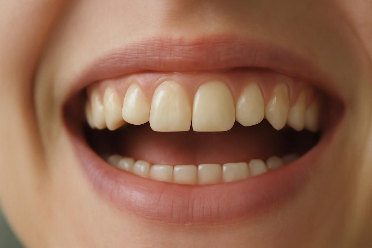
The first sign may be no sign at all
The first sign of a cavity is often no noticeable sign whatsoever. A tooth can look normal, feel normal and cause no pain or sensitivity while decay is beginning. Symptoms generally become more likely as the damaged area grows or reaches deeper tooth layers, so pain is neither necessary nor reliably early. Mayo Clinic notes that a cavity may cause no symptoms when it is just beginning.
It helps to separate two terms that are often used interchangeably:
- Tooth decay is a process in which acids contribute to mineral loss and damage in tooth structure.
- A cavity, in the narrower sense used here, is a permanent opening or hole produced after the tooth surface has broken down.
That distinction explains why advice about “reversing an early cavity” can sound contradictory. An intact area of enamel that has lost minerals—sometimes called a white-spot lesion or non-cavitated lesion—may still have an unbroken surface. A cavitated lesion has undergone structural collapse, leaving a pit or hole that cannot grow back.
The process begins when oral bacteria use sugars and produce acids. Teeth lose and regain minerals throughout the day, but repeated acid exposure can shift that balance toward continuing mineral loss. The earliest visible change may be a chalky white area, although early mineral loss can remain invisible. If the process continues until the enamel surface breaks, permanent structural damage has formed. The National Institute of Dental and Craniofacial Research explains the progression from early mineral loss to a permanent cavity.
This is why regular dental examinations matter even when your mouth feels fine. Small lesions can develop between teeth, in grooves or in other places that are difficult to inspect at home. Waiting for pain may mean waiting until a problem has become deeper.
Important: This article provides general education, not a diagnosis. A tooth’s color, texture, sensitivity, appearance in a photograph—or lack of pain—cannot establish whether that tooth has early mineral loss, a formed cavity, staining, a crack or another condition. Decay Guide states that its material is written by a health writer, has not been reviewed by a clinician and cannot replace an examination, as explained in its editorial and clinical-review disclosure.
Possible early clues you may see or feel
No single observation at home confirms tooth decay. Certain new or changing findings are nevertheless reasonable reasons to arrange an assessment, particularly when several affect the same tooth.
What you may see
A localized chalky white or off-white area can represent enamel mineral loss. It may look duller or less translucent than the enamel around it. This can be an early visible change, but it is not always present, and not every white mark is decay.
A brown, dark brown or black mark can also occur with tooth decay. Darker discoloration is more commonly described as decay advances, but color cannot reveal how deep a lesion is. Nor can it reliably distinguish decay from staining or another cause of discoloration.
A tooth may therefore:
- Have early decay without a visible color change.
- Have a white, brown or dark mark that is not decay.
- Contain a cavity without an obvious hole.
- Look normal from the visible side while decay develops elsewhere.
What you may feel
New, localized sensitivity can be a clue. Possible patterns include:
- A brief twinge after something sweet.
- Sensitivity to cold air, food or drinks.
- Discomfort with hot food or drinks.
- An intermittent dull ache.
- Pain or tenderness when biting or chewing.
Sensitivity becomes more likely when damage reaches the dentin beneath the enamel, but it can have other causes and cannot identify decay by itself. A painless tooth can still contain decay. Cleveland Clinic describes early outer-enamel decay as potentially symptom-free and lists temperature sensitivity, toothache, mouth pain, pits and discoloration among possible signs as damage progresses in its medically reviewed cavity guide.
Biting discomfort also deserves attention. Record whether it happens when you first bite, throughout chewing or when you release the bite. That description may help a dentist evaluate the problem, but it is not a reliable home test and does not establish that decay is responsible.
Changes in tooth structure
A rough patch, dent, small pit, sharp-feeling edge or visible hole is more suggestive of established structural damage than of the earliest intact white-spot stage. Even then, what feels like a hole could instead be a natural groove, chip, damaged restoration or another irregularity.
Some people also notice:
- Food repeatedly catching in one new area.
- Floss becoming newly difficult to pass.
- Floss snagging or fraying in the same place.
- A spot that feels sticky, uneven or sharp.
These are secondary, nonspecific observations. When new or persistent—especially alongside sensitivity or a visible change—they may justify an evaluation, but they do not prove that a cavity is present.
Most importantly, the absence of every clue on this list does not exclude decay.
How signs change as decay moves deeper
Symptoms can indicate that a tooth needs attention, but they cannot precisely stage decay. A small lesion in a hidden location may be invisible, while a shallow surface change may look prominent. Location and individual circumstances can affect how a lesion presents and progresses.
The following table is a simplified framework, not a timeline or self-diagnostic test. It reflects the general progression described in Cleveland Clinic’s overview of cavity stages, symptoms and complications.
| Stage | What is happening | What a person might notice | Limits of that clue |
|---|---|---|---|
| Mineral loss in intact enamel | Acids draw minerals from enamel, but the surface has not collapsed into a hole. | Often nothing. A localized chalky white or off-white area may appear. | The area may be invisible, and a white spot can have causes other than decay. |
| Enamel breakdown | Continuing mineral loss weakens and eventually breaks the enamel surface. | A localized color change, rough area, pit, dent or small hole may appear. The tooth can remain painless. | Color and texture do not reveal depth reliably. Natural grooves and stains can resemble damage. |
| Dentin involvement | Decay extends into the softer layer beneath enamel. | Sensitivity to sweets, cold, heat or pressure becomes more likely. Biting discomfort or an intermittent ache may occur. | These symptoms can also arise from non-cavity conditions. |
| Decay near or in the pulp | Damage approaches or affects the inner tissue containing nerves and blood vessels. | Toothache may become more persistent. Temperature discomfort may linger, and the tooth may be tender when biting. Swelling can develop. | Symptoms cannot establish the exact depth or cause without an examination. |
| Advanced complication | Infection may extend beyond the tooth and form an abscess. | Severe or escalating pain, gum or facial swelling, or pain spreading toward the jaw or face may occur. | These are advanced warning signs rather than typical first signs, and decay is not their only possible cause. |
The sequence helps explain why pain often appears late: changes limited to outer enamel may cause no sensation, while deeper involvement makes sensitivity and toothache more likely. It should not be used to conclude that a painless pit is shallow or that severe sensitivity proves the pulp is involved.
There is no universal progression schedule. One lesion may remain limited for a considerable period, while another may advance under different conditions. Decay between teeth, on an exposed root, in a deep chewing groove or around a restoration may also present differently.
An abscess, severe pain, facial swelling and pain spreading toward the jaw or face are therefore better understood as possible advanced complications, not as first signs of a cavity.
Cavity, stain, or another cause?
No. A white, brown, gray or black spot does not automatically mean you have a cavity. Tooth decay and non-cavity discoloration can overlap in color, and a mirror cannot show what is happening beneath the surface.
The comparison below describes clues rather than rules:
| Observation | May be more concerning for decay | May be more consistent with staining | Why it is inconclusive |
|---|---|---|---|
| Distribution | A localized mark affecting one area | Broader discoloration across part of a tooth or several teeth | Both decay and staining can be localized or widespread. |
| Change over time | A spot that appears to enlarge, darken, deepen or become rough | A stable mark without an apparent structural change | Subtle progression can be difficult to judge at home. |
| Surface | A pit, dent, rough patch or visible defect | A surface that appears smooth and intact | Natural grooves can feel rough, and a smooth-looking surface can conceal decay. |
| Symptoms | Localized sensitivity, biting discomfort or toothache | No pain or sensitivity | Cavities can be painless, while non-cavity conditions can cause pain. |
| Location | Grooves, pits, spaces between teeth, the gumline or beside a restoration | Areas exposed to pigments or deposits | Location may affect suspicion but cannot provide a diagnosis. |
A localized, changing, rough or deepening mark may deserve more concern than broad, stable discoloration, but it remains only a clue. A smooth or painless mark is not automatically harmless.
Sensitivity is similarly nonspecific. Possible explanations other than decay include:
- Exposed dentin.
- Gum recession.
- Enamel wear or erosion.
- Tooth grinding or clenching.
- A crack or chip.
- Pressure during biting.
- A problem involving an existing restoration.
Discoloration also has multiple possible causes. Surface stains can be associated with foods, drinks or tobacco exposure. Other color changes may be associated with deposits, aging, medication, trauma, fluorosis or differences in enamel. A lower-authority dental-practice overview lists these possibilities while also emphasizing that discoloration alone cannot identify a cavity.
Do not try to settle the question by pressing a tool into the area, scraping away discoloration or biting a hard object to see whether the tooth hurts. Flossing forcefully against a suspicious edge is not a valid diagnostic test either.
A changing mark, visible structural defect or discoloration accompanied by sensitivity, biting pain or toothache deserves professional evaluation. The purpose is not simply to confirm decay, but to consider other plausible causes.
Why home inspection can miss a cavity
Small areas of decay may be too subtle to see in a bathroom mirror. They may also be impossible to detect with the tongue, which cannot reliably measure enamel changes or reveal damage below the visible surface.
Common hard-to-see locations include:
- Between adjacent teeth.
- Inside deep grooves and pits on chewing surfaces.
- Near the gumline.
- On exposed root surfaces.
- Around or beneath an existing filling or other restoration.
Lighting, saliva, viewing angle, tooth anatomy and lesion location all limit home inspection. A phone photograph may provide a clearer view of an accessible surface, but it cannot show the depth of a mark or reliably reveal what is happening between teeth or beneath a restoration.
During an assessment, a dentist may ask:
- Which tooth or area seems affected.
- Whether the tooth reacts to sweets, cold, heat or pressure.
- How long discomfort lasts after a trigger is removed.
- Whether pain occurs while biting or releasing the bite.
- Whether a mark or rough area appears to be changing.
- Whether the tooth has an existing restoration or previous injury.
The dentist may then inspect the tooth for discoloration, surface breakdown, restoration problems or other structural changes and assess suspicious areas. Dental X-rays may be used when appropriate to reveal decay that is not visible during ordinary inspection, including some lesions between teeth. Penn Dental Family Practice notes that small cavities may be painless, invisible and undetectable with the tongue, and that dentists may need an examination or X-rays to identify hidden decay.
This does not mean that every suspicious spot requires imaging. The clinician decides whether an X-ray would be useful in the circumstances.
Color charts, phone photographs, mirror checks, tongue checks, symptom quizzes and online images may help you describe what you noticed. They cannot confirm or exclude decay.
When early decay may be reversible—and when it is not
Whether an “early cavity” can be reversed depends on what that phrase means.
If the enamel surface remains intact and has only begun to lose minerals, the process may sometimes be arrested or reversed through remineralization. Saliva supplies minerals, fluoride can support mineral replacement, plaque control reduces the environment in which bacteria produce acids, and reducing frequent sugar exposure limits repeated acid attacks.
A white-spot lesion may represent this non-cavitated stage. But not every white spot is decay, and a person cannot reliably determine from appearance alone whether the surface is intact or how far a change extends.
Once enamel has collapsed into a hole, the situation is different. Fluoride cannot regrow missing enamel or close a structural opening. Brushing remains important for the tooth and the rest of the mouth, but it does not rebuild lost tooth anatomy. The National Institute of Dental and Craniofacial Research distinguishes potentially reversible early mineral loss from a permanent cavity requiring dental repair.
The practical distinction is:
- Intact, non-cavitated enamel mineral loss: may sometimes be arrested or remineralized.
- A pit or hole caused by structural collapse: cannot be closed with toothpaste or home care.
- Decay extending into dentin: generally requires professional dental treatment rather than waiting for it to resolve.
These are examples, not recommendations for a particular tooth. Delta Dental describes treatment as varying according to the stage and extent of decay, with professional treatment required once decay has extended into dentin.
Do not assume that a painless white spot can safely be managed at home. Equally, do not assume that every white spot is already a permanent hole. An examination is needed to evaluate the surface and extent of the change.
What to do if you suspect early decay
Arrange a dental assessment if you notice a new or worsening tooth spot, persistent localized sensitivity, pain while biting, an intermittent or ongoing toothache, a rough area, a pit or a suspected hole. The purpose of the appointment is to identify the cause rather than merely confirm your own suspicion.
Before the visit, make a short symptom record. Note:
- Which tooth or side seems affected.
- Whether the trigger is sweet, cold, heat, touch or pressure.
- Whether pain occurs while biting, chewing or releasing the bite.
- How long discomfort lasts after the trigger ends.
- Whether discomfort occurs without an obvious trigger.
- Whether a mark, pit, rough edge or food-trapping area is changing.
- Whether the tooth has a restoration or history of injury.
Avoid repeatedly testing the tooth. A description of what happens during ordinary eating, drinking or cleaning is more useful than deliberately provoking pain.
While waiting for a routine appointment:
- Continue gentle brushing with fluoride toothpaste. Do not stop cleaning the area solely because it is sensitive.
- Continue ordinary interdental cleaning unless it causes substantial pain or a dental professional has given you different instructions.
- Reduce frequent sugary snacks and drinks. Repeated sugar exposure allows repeated acid attacks on enamel.
- Avoid scraping or probing the spot. Do not place pins, picks or household tools into a suspected hole.
- Do not test the tooth with hard food. This cannot diagnose decay and may aggravate existing damage.
- Contact the dental office if symptoms intensify or swelling develops.
Mayo Clinic identifies frequent snacking and sugary drinks among contributors to cavities, recommends regular dental care because decay may develop unnoticed, and advises contacting a dentist for toothache or mouth pain.
If sensitivity improves, do not treat that change as proof that a suspected cavity has disappeared. Early decay can be painless, and symptoms alone cannot confirm whether structural damage is present. Conversely, continuing sensitivity does not prove that decay is the cause.
Regular checkups remain important even after a symptom settles because decay may be detected before it becomes painful or visible at home.
When to book promptly and what counts as an advanced warning sign
Not every possible cavity is a medical emergency. The useful distinction is between an uncertain early change that should be discussed at an examination, symptoms that justify contacting a dentist and advanced warning signs that need prompt professional attention.
Discuss at a dental examination
A painless white spot or uncertain stable mark still deserves mention. It may represent staining, an enamel difference, early mineral loss or another change. Visual inspection at home cannot settle the question.
If you already have an appointment scheduled, tell the practice what you have noticed so its staff can decide whether the existing timing is appropriate. Do not wait for the area to hurt before mentioning it.
Contact a dentist rather than waiting for severe pain
Reasons to call include:
- New or persistent localized sensitivity.
- Pain or tenderness when biting.
- A visible pit, dent or suspected hole.
- A dark spot that appears to be changing.
- A rough or damaged-feeling area.
- Intermittent or persistent toothache.
- Other mouth pain associated with a particular tooth.
When calling, describe the severity, triggers, duration, pain pattern, swelling and whether the symptoms are worsening. The dental team can use that information to determine appropriate timing.
Seek prompt professional attention for advanced warning signs
Severe or escalating tooth pain, an apparent abscess, gum or facial swelling, or pain spreading toward the jaw or face can accompany advanced decay or infection. These are not typical first signs, and a cavity is not their only possible cause. They nevertheless warrant prompt professional attention rather than continued home monitoring. HealthPartners describes infection and abscess as possible consequences when decay progresses toward the nerve in its guide to cavity symptoms and progression.
Tell the dental office clearly if there is swelling, rapidly worsening pain or substantial difficulty eating or carrying out normal activities.
The decision framework is straightforward:
- Do not wait for pain before taking a new tooth change seriously.
- Do not assume every sensitive or discolored tooth is a cavity.
- Continue routine fluoride-based care while awaiting assessment.
- Avoid scraping, probing, pressure tests and other unreliable home experiments.
- Arrange an examination for new, persistent or worsening changes.
Professional examination—with dental X-rays when the clinician considers them appropriate—is the suitable way to evaluate early mineral loss, an established cavity, staining, restoration problems, cracks and other possible causes. Earlier assessment may reduce the chance that unnoticed decay progresses into deeper damage requiring more extensive treatment.
Can you have a cavity without any pain?
Yes. Early tooth decay and even a formed cavity can be painless. Symptoms become more likely as damage enlarges or reaches dentin and pulp, but pain is not required. HealthPartners notes that many people experience no symptoms at the beginning and that symptoms become more likely as decay grows or approaches the tooth’s nerve in its cavity symptom guide.
A normal-feeling tooth therefore cannot be assumed to be free from decay. Regular examinations matter because some cavities are identified before a person notices sensitivity, a visible mark or a hole.
Is a chalky white spot always the first sign of a cavity?
No. A chalky white or off-white area can be an early visible sign of enamel mineral loss, but early decay may produce no visible change at all. A white spot may also have another explanation.
Do not assume that the spot is harmless, but do not label it a cavity from appearance alone. An examination is needed to assess whether the surface is intact and whether further evaluation is appropriate. A dental-practice comparison likewise describes white spots as possible early changes while warning that cavities and stains can be difficult to distinguish without professional assessment.
Does sensitivity to cold or sweets mean I have tooth decay?
Not necessarily. New localized sensitivity to cold, heat or sweets can accompany decay, particularly when deeper tooth layers are involved. But sensitivity can also occur with gum recession, exposed dentin, enamel wear, pressure, grinding or other dental problems. Colgate’s overview similarly cautions that sensitivity can have causes other than a cavity and recommends professional assessment when cavity symptoms are suspected.
Arrange an assessment if sensitivity is new, persistent, worsening, limited to one tooth or accompanied by biting pain, discoloration or a structural change.
Can fluoride toothpaste heal a cavity?
It depends on the stage. Fluoride toothpaste can support remineralization when enamel has begun losing minerals but its surface remains intact. It cannot regrow missing tooth structure or close a formed hole.
Continue brushing with fluoride toothpaste, but do not rely on it as a substitute for assessment of a suspected pit or cavity. Delta Dental distinguishes early demineralization from the stage at which a hole has formed and explains that some very early enamel damage may be remineralized, while deeper decay requires treatment.
How can a dentist detect a cavity I cannot see?
A dentist can ask about symptoms and triggers, inspect tooth surfaces and existing restorations, and assess areas that are difficult to view at home. Dental X-rays may be used when appropriate to reveal decay between teeth or in other hidden locations.
Imaging is not automatically required in every case. The clinician decides whether it would be useful based on the examination and suspected location. Penn Dental Family Practice explains that small cavities may not be visible or perceptible with the tongue and that dentists may use examination and X-rays to find decay.