
When See-Through Tooth Edges Are Normal—and When to Have Them Checked
Stable, symmetrical front-tooth edges may be natural; new, spreading or uneven changes with sensitivity, pain or chips warrant a dental assessment.
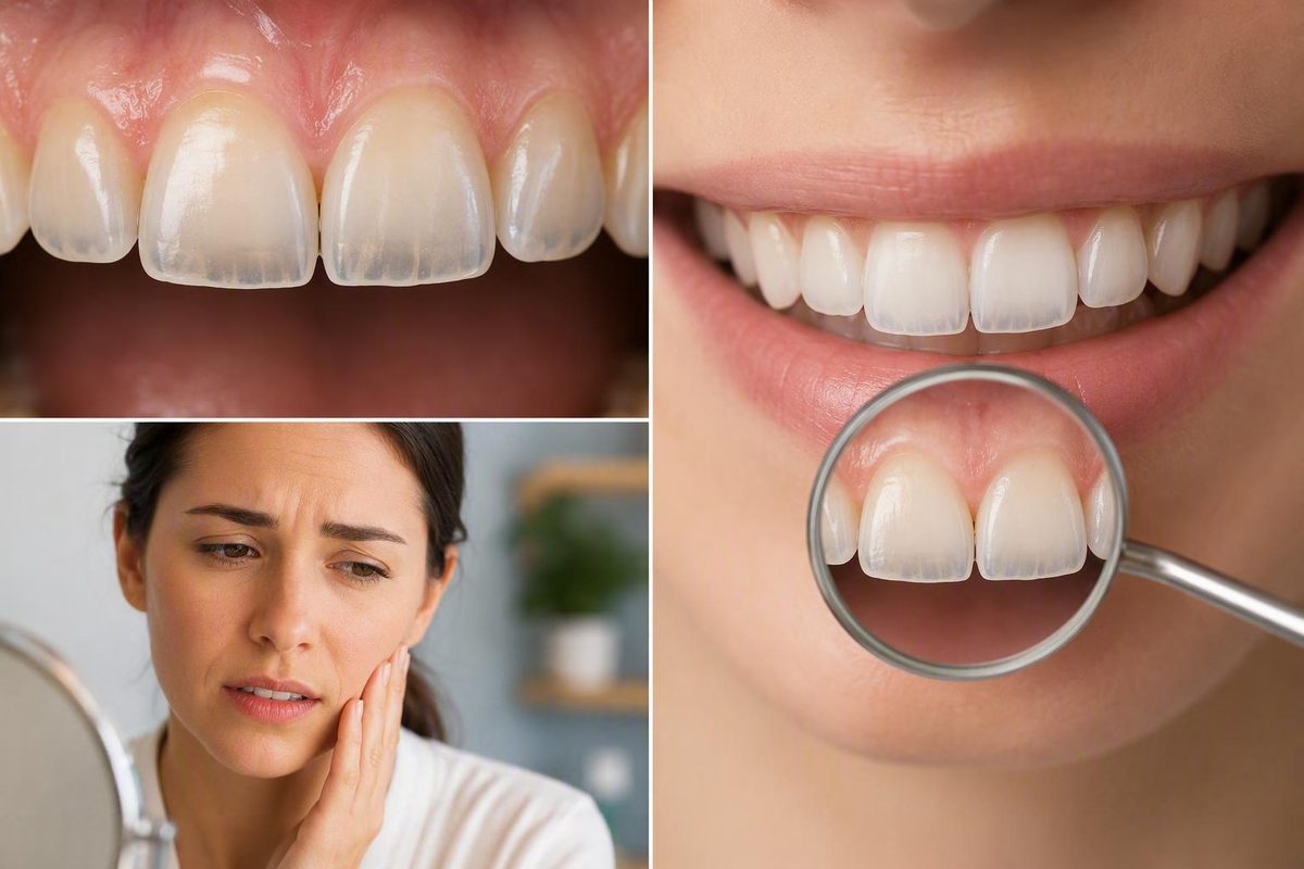
A recent, spreading, uneven, or worsening change may instead reflect enamel thinning, acid erosion, mechanical wear, a developmental enamel difference, or another dental problem. Transparency is an appearance, not a diagnosis, so neither a mirror nor a photograph can confirm enamel loss. Arrange a dental assessment if the change is new or progressing, especially if you also have sensitivity, pain, discoloration, chips, cracks, or a change in tooth shape. These distinctions are consistent with Cleveland Clinic’s clinical overview of translucent teeth.
Quick answer: normal edge translucency versus a concerning change
Front teeth do not have to be completely opaque to be healthy. Stable translucency in the same place on matching front teeth, without discomfort or surface damage, is generally less concerning.
The key question is whether the appearance is changing. Increasing transparency can accompany thinning enamel, while irregular edges may reflect wear or damage. Developmental differences can also affect enamel thickness and appearance without indicating active erosion.
| Lower-concern pattern | Arrange an evaluation |
|---|---|
| Slight translucency confined to front-tooth tips | A recent, increasing, spreading, or uneven change |
| Similar appearance on matching teeth | New or spreading yellow, gray, or bluish-gray discoloration beyond the thin edge |
| Appearance has remained unchanged | New sensitivity or pain |
| No chips, cracks, or altered contour | Chips, cracks, shortening, or changing tooth shape |
| No unusual surface change | Rough, unusually glossy, flattened, or “melted” surfaces |
A stable bluish cast limited to thin biting edges may be a normal optical effect; new, asymmetric, or broader discoloration is different. A dentist must consider the pattern, affected surfaces, symptoms, and exposure history before identifying a cause.
Arrange a routine dental assessment for a recent or progressive change. Seek faster care for substantial pain, swelling, a fracture, or rapid structural change. This article provides general information, not an individual diagnosis or treatment plan.
Why thinner enamel can make a tooth look clear and yellow at the same time
Enamel is the highly mineralized outer covering of the dental crown. It protects the dentin beneath it and the pulp farther inside from mechanical, chemical, and temperature-related stresses. Its appearance depends on its structure, thickness, and the tissues underneath.
Enamel is not perfectly opaque. At a front tooth’s biting edge, where there may be little or no dentin directly behind it, transmitted light can produce a clear, glassy, or blue-gray effect. This may be normal when mild and stable. If enamel becomes thinner, however, more light can pass through the edge. Elsewhere, thinning may allow the naturally yellowish dentin underneath to influence the tooth’s color more strongly. The result can be clearer edges alongside a yellower-looking central body.
Laboratory research supports the narrower biological point that demineralization can change enamel’s optical properties. One in-vitro study of extracted premolars found changes in translucency under experimental demineralizing conditions. It was an extracted-tooth experiment using artificial exposures, so it cannot explain why a particular person’s teeth look transparent or diagnose erosion in a living mouth.
Once mature enamel has been physically lost, the body cannot grow it back. The cells that produce enamel are no longer present after formation, and mature enamel is acellular and unable to remodel, as described in a scientific review of enamel formation. This is different from remineralizing a vulnerable surface that remains: minerals may strengthen existing enamel, but they cannot recreate missing thickness or replace a worn edge.
Possible causes: a cause-and-clue guide
Several processes can create overlapping changes. Acid exposure may come from the diet or from gastric acid associated with reflux or recurrent vomiting. Saliva normally helps clear and buffer acids, so persistent dry mouth may reduce that protection. The American Dental Association’s overview of dental erosion emphasizes that diagnosis requires both a clinical examination and an assessment of contributing factors.
The clues below are details to mention at an appointment, not proof of a diagnosis.
| Possible contributor | Clues to mention at an appointment | Appropriate follow-up |
|---|---|---|
| External acid exposure | Frequent soft drinks, sports drinks, fruit juice, citrus, or acidic sweets; prolonged sipping, holding, or swishing | Review the frequency and drinking pattern with a dentist; reduce repeated exposure |
| Internal acid exposure | Reflux, sour taste, recurrent vomiting, or frequent nausea | Seek dental assessment and discuss the underlying symptoms with an appropriate medical professional |
| Grinding or clenching | Flattened or shortened edges, chips, or suspected nighttime grinding | Ask a dentist to evaluate the wear pattern and whether protective treatment is appropriate |
| Brushing-related abrasion | Firm pressure, a hard brush, or vigorous scrubbing, particularly near the gumline | Show the dental team your technique; use gentle brushing rather than assuming brushing caused the change |
| Developmental enamel differences | Teeth have looked this way since eruption; longstanding pits, grooves, or thin areas | Ask whether the pattern is natural anatomy or enamel hypoplasia and whether it only needs monitoring |
| Reduced salivary protection | Persistent dry mouth, thirst, or onset after a medication change | Discuss dry mouth with a dentist or clinician; do not stop prescribed medication independently |
| Frequent whitening | The change or sensitivity appeared during repeated use of strips, pens, gels, or trays | Review continued use and bring the product details to the appointment |
Grinding, forceful brushing, naturally thin enamel, enamel hypoplasia, dry mouth, and some medications are among the possible contributors described in clinical guidance on translucent teeth and thin enamel. None can be confirmed from appearance or history alone.
Whitening requires particular caution in how the evidence is interpreted. An ADA news summary reports sensitivity, increased permeability, erosion, and translucency as possible effects of continuous or excessive use, but it does not establish a universally harmful concentration, dose, or schedule. Its discussion of frequent teeth whitening supports reviewing use with a dentist, not assuming whitening caused the change or that every user must stop.
Mechanical and chemical wear can also overlap. A developmental enamel difference can coexist with acquired wear, so a longstanding pattern does not automatically rule out a newer process.
Transparent teeth are not the same thing as tooth decay
Dental erosion and tooth decay can both damage hard tooth tissue, but they are different processes. Erosion is chemical loss caused by acids that do not originate from oral bacteria. Tooth decay involves plaque bacteria damaging the tooth surface over time. Either problem can coexist with wear, cracks, or developmental enamel differences.
Transparency alone is not identified as a defining symptom of decay. Recognized decay-related findings include:
- White, brown, or black spots
- Toothache
- Sharp pain with hot, cold, or sweet foods and drinks
- A visible hole or cavity
Early decay may cause no symptoms at all, according to NHS tooth-decay guidance. The presence or absence of transparent edges therefore cannot rule decay in or out.
Sensitivity is also nonspecific. It may occur when enamel loss exposes dentin, but pain can also be associated with decay, a cracked or chipped tooth, pulp inflammation, exposed roots, defective restorations, or leakage around a restoration. A dental examination is needed to distinguish normal anatomy from erosion, decay, grinding-related wear, fractures, and developmental differences.
How to protect the enamel that remains
You do not need to identify the exact cause before adopting low-risk protective habits. The priority is to reduce repeated acid exposure and unnecessary mechanical stress without neglecting routine cleaning.
After acidic food or drink:
- Finish it rather than sipping repeatedly over a long period.
- Do not hold or swish it around your teeth.
- Rinse your mouth with water afterward.
- Do not brush immediately after substantial acid exposure.
- Choose plain water more often as an everyday drink.
After vomiting, rinse rather than brushing immediately. The American Dental Association lists water, milk, or an appropriate sodium bicarbonate rinse as options and also notes that sugar-free gum may stimulate saliva, which helps buffer and clear acids. Recurrent vomiting or reflux needs attention to the underlying medical issue as well as dental protection; medically driven acid exposure is not a failure of discipline.
Brush gently with fluoride toothpaste and a soft-bristled toothbrush. Fluoride can help strengthen vulnerable enamel that remains, but toothpaste cannot rebuild enamel that has already been physically lost. Do not try to scrub away a clear or yellow appearance, because brushing harder will not restore enamel thickness.
Stay adequately hydrated to support normal saliva production. Persistent dry mouth deserves professional discussion, particularly if it began after a medication change. Do not stop or adjust prescribed medicine on your own.
If you suspect grinding or clenching, ask a dentist to examine the wear pattern. A protective appliance may be appropriate in some cases, but that decision should follow an assessment rather than an assumption that every worn or transparent edge is caused by grinding.
Can transparent teeth be reversed or restored?
Physically missing mature enamel does not naturally grow back. Fluoride and saliva may support remineralization or improve the acid resistance of enamel that is still present, but they cannot recreate enamel that is gone or replace a worn biting edge.
Management depends on the cause, activity, symptoms, and amount of structural change:
- Monitoring: Mild, longstanding, symmetrical edge translucency may need only observation and routine dental care.
- Professional preventive support: A dentist may recommend targeted fluoride measures or sensitivity management where remaining enamel or exposed dentin needs protection.
- Grinding protection: If examination confirms relevant grinding or clenching, a professionally recommended night guard may help reduce further mechanical wear.
- Dental bonding: Tooth-colored resin can add material to selected worn or translucent areas to improve contour, protection, or appearance.
- Veneers or crowns: These may be considered for selected teeth with greater cosmetic or structural needs, but they are not necessary for every transparent edge.
These options are among the cause-dependent approaches described in dental guidance on transparent tooth edges and enamel loss. Their suitability cannot be determined from appearance alone.
The treatment sequence matters.
When to see a dentist and what to mention
Arrange a dental evaluation if the transparency is recent, progressing, spreading, or uneven, or if it occurs with notable sensitivity, pain, new discoloration, chips, cracks, roughness, unusual glossiness, or changing tooth shape.
Seek prompt assessment rather than treating the change as purely cosmetic if you have:
- Rapid visible or structural change
- Substantial or persistent pain
- A broken or fractured tooth
- Swelling around a tooth or gum
- Pus or another possible sign of a dental abscess
- Difficulty biting because the tooth’s shape has changed
Pain, swelling, and a buildup of pus can accompany dental infection and warrant dental attention, while fractures and worsening changes also require professional assessment.
Before the appointment, note:
- When you first noticed the appearance
- Whether it is changing and how quickly
- Which teeth and surfaces are affected
- Any sensitivity, pain, chips, or cracks
- How often you consume acidic foods or drinks
- Whether you sip, hold, or swish acidic drinks
- Any reflux, nausea, or vomiting
- Persistent dry mouth and current medications
- Whitening products and frequency of use
- Toothbrush type, pressure, and brushing habits
- Possible daytime clenching or nighttime grinding
A dentist may combine this history with an examination of enamel texture, tooth contour, exposed dentin, decay, fractures, and existing restorations before recommending monitoring, prevention, cause control, or restorative care.
Dated photographs taken from a similar angle and in similar lighting may help you describe suspected progression. They cannot diagnose enamel loss: lighting, camera exposure, and angle can all change how transparent an edge appears.
Unchanged translucency limited to front-tooth tips may simply be normal anatomy, but a new or worsening change should not be self-diagnosed. Reduce repeated acid exposure, rinse after acid, brush gently with fluoride toothpaste, and discuss reflux, vomiting, dry mouth, grinding, or whitening with the appropriate professional. Do not stop medication or alter treatment for another health condition without qualified medical guidance. A dentist can determine whether the appearance needs monitoring, cause control, protective care, or restoration.