
How Worn Enamel Can Change the Feel, Color, and Shape of Your Teeth
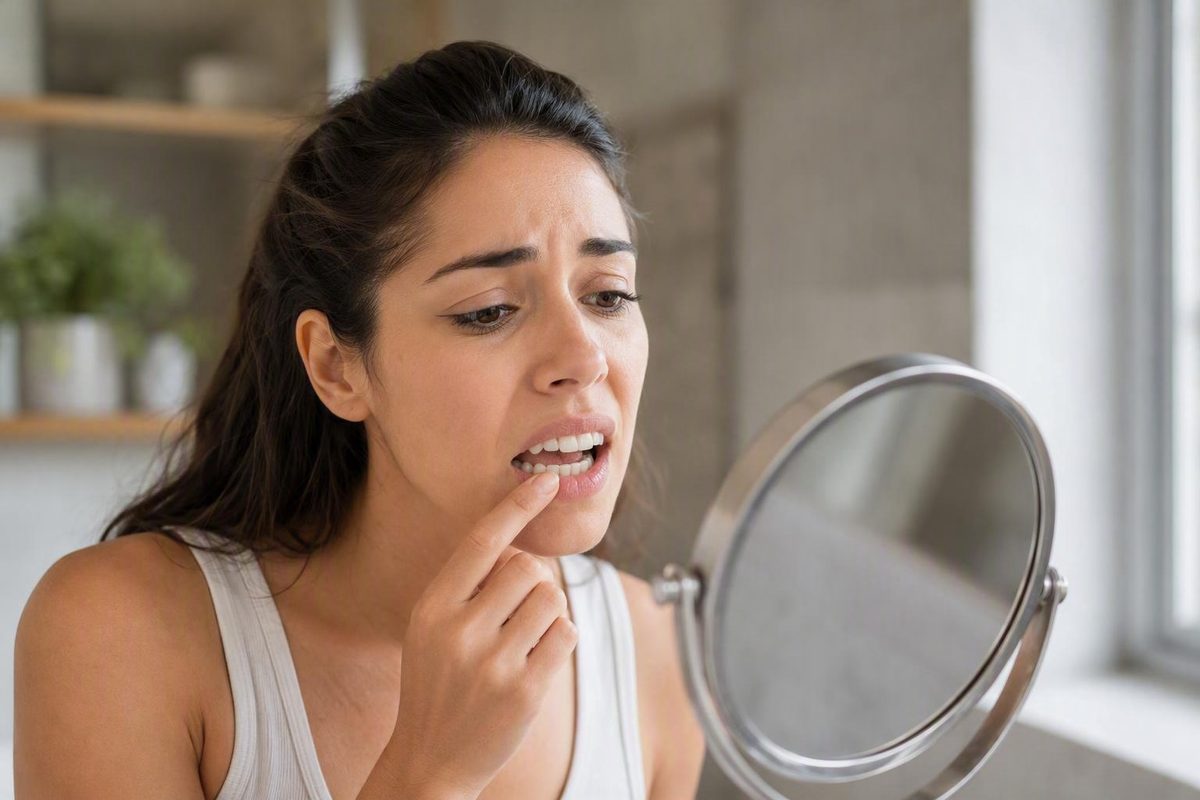
The short answer: common signs that enamel may be wearing away
Possible signs of tooth enamel loss include:
- Sensitivity to cold, heat, sweets, or acidic foods and drinks
- Teeth becoming increasingly yellow
- Translucent or glassy biting edges, especially on front teeth
- Smooth, shiny, dull, rough, or locally thinned surfaces
- Rough, sharp, uneven, or chipped edges
- Smooth depressions or “cupping” on chewing surfaces
- Teeth that look flatter, shorter, rounder, or less even
- Discomfort when biting or chewing
- Cracks, fractures, recurring cavities, or a changing bite
Enamel erosion generally progresses slowly and may not be obvious at first. Symptoms can include sensitivity, discoloration, chips, pitting, rough edges, cracks, and increased vulnerability to cavities, but they vary from person to person (Cleveland Clinic’s overview of tooth erosion).
Rather than expecting everyone to follow the same sequence, look for changes across several categories:
| What changes | Subtle possible clues | Changes that may accompany more extensive damage |
|---|---|---|
| Sensation | Brief twinges with cold, heat, sweets, or acidic items | Stronger or more frequent sensitivity, toothache, pain with chewing |
| Color | Gradual yellowing or more translucent front edges | More obvious dentin exposure or marked discoloration |
| Texture | Smooth or shiny spots, dullness, slight roughness | Cupping, deeper depressions, jagged areas, visible defects |
| Shape | Slight rounding or unevenness | Flattening, shortening, repeated chips, cracks, fractures |
| Function | No functional change or occasional discomfort | Difficulty chewing comfortably or a bite that feels different |
Sensitivity, discoloration, shiny areas, rough or chipped edges, cupping, cavities, and fractures have all been reported with enamel loss, but the findings do not establish a fixed stage or sequence (Penn Dental Family Practice’s guide to enamel loss).
These are descriptive clues, not a home diagnostic system. A mild twinge does not prove early erosion, and strong sensitivity does not reveal exactly how much enamel is missing.
Most importantly, no single sign confirms enamel erosion. Cavities, stains, cracks, gum recession, grinding, developmental enamel differences, trauma, and dental restorations can produce similar changes. Home observation is useful for noticing what is new or progressing—not for determining the cause.
What enamel loss can feel like
Enamel-related sensitivity may feel like a brief twinge, sharp sensation, or discomfort when a tooth encounters:
- Cold water, ice, or chilled air
- Hot food or drinks
- Sweet foods
- Acidic foods or beverages
Enamel forms the protective outer covering of the visible tooth. Beneath it is dentin, which is more sensitive. As enamel becomes thinner or is physically lost, dentin may be less insulated from temperature and taste-related triggers. Foods and drinks that were previously comfortable may then become noticeable.
Discomfort can become more pronounced when damage extends into deeper tooth structures.
Sensitivity also has many possible causes. Cavities, exposed roots from gum recession, cracks, grinding, and recent dental treatment can all make a tooth react to temperature or sweets. Early cavities may produce no symptoms; as decay progresses, possible symptoms include sensitivity, toothache, visible pits or holes, and pain when biting (Mayo Clinic’s guide to cavities and tooth decay).
The reverse is equally important: a lack of sensitivity does not rule out enamel loss. Slow erosion may alter tooth contour, translucency, or surface texture before discomfort develops.
If you are monitoring sensitivity, note:
- Which tooth or area responds
- Whether the trigger is cold, heat, sweetness, acidity, or pressure
- Whether the sensation stops quickly
- Whether it is becoming more frequent or intense
- Whether biting or chewing also hurts
What thinning enamel can look like
Increasing yellowing
A tooth can look more yellow as enamel becomes thinner and allows more of the naturally yellowish dentin underneath to show through. The change may affect one area or become noticeable across several teeth.
Yellowing alone is weak evidence of enamel loss. Natural tooth color varies, and surface stains, decay, aging, or other changes can also affect color. The more useful clue is a new or progressive change, especially when it occurs alongside sensitivity, translucent edges, or altered tooth shape.
Translucent or glassy edges
The biting edges of front teeth may look increasingly translucent, clear, or glass-like where enamel has thinned locally. Some natural translucency is normal, so compare like with like: the relevant clue is a change from the tooth’s previous appearance, not translucency by itself.
Smooth, shiny, dull, or rough areas
Enamel wear can produce several seemingly contradictory surface changes:
- A localized area may look unusually smooth or shiny.
- A broader surface may appear dull, matte, or worn.
- Edges may become rough, sharp, or uneven.
- A small area may feel different to the tongue.
These descriptions are not mutually exclusive. Different tooth surfaces and different combinations of acid and mechanical wear can produce different textures.
A mirror cannot show whether a changed surface is active erosion, old wear, a developmental difference, decay, or damage around a restoration. What matters most is whether the area is new, expanding, or accompanied by other symptoms.
Cupping and depressions
“Cupping” describes a smooth, bowl-like depression in a tooth surface, often on the chewing surface of a back tooth. It may appear as a shallow hollow or feel like a change in contour.
Not every pit, dent, dark point, or visible hole is cupping. Natural grooves, developmental pits, cavities, trauma, and damaged fillings can all create localized defects. Shape can provide a clue, but it cannot establish the cause.
Rounding, flattening, shortening, and chipping
Progressive loss of tooth structure can change the outline of a tooth. Possible changes include:
- Rounded corners
- Flatter biting or chewing surfaces
- Shorter-looking teeth
- Uneven biting edges
- Small chips
- Sharp or jagged edges
- Cracks or teeth that fracture more readily
More than one process may also be involved—for example, repeated acid exposure may weaken a surface that is also subjected to grinding.
One less familiar observation is a filling that appears raised above the surrounding tooth. The filling may not have moved; the natural structure around it may have worn down. This sign has been described in dental-practice guidance, but it is not specific to erosion and requires professional interpretation because restorations can also become loose, damaged, or uneven for other reasons (discussion of erosion and existing restorations).
Natural tooth color, ridges, translucency, and shape vary widely. A mirror check is most useful when it identifies a change over time.
Subtle changes versus more extensive damage
There is no universal sequence that every person follows. Describing findings as subtle or more extensive is more accurate than trying to assign yourself to a fixed stage.
Subtle possible changes include:
- Mild, brief sensitivity
- Slowly increasing yellowing
- Greater translucency at front-tooth edges
- A small smooth or shiny area
- Slight rounding, dullness, or roughness
- Shallow surface depressions
More extensive loss may be accompanied by:
- Stronger or more frequent sensitivity
- Toothache or chewing discomfort
- More visible dentin
- Clearly shortened or flattened teeth
- Repeated chipping
- Cracks or fractures
- A bite that feels different
- Recurring cavities
These findings do not mean that infection, tooth loss, or a particular procedure is inevitable. They indicate that more tooth structure may be involved—or that another dental problem needs to be excluded.
Erosion and decay should not be treated as the same process. Erosion can weaken the tooth surface and increase susceptibility to cavities, but a cavity is bacterial tooth decay. A person can have erosion, a cavity, or both.
Appearance and symptoms vary according to which teeth are involved, how quickly the change is occurring, saliva levels, previous dental work, grinding, acid exposure, and other factors. A dental examination—not a checklist—determines the likely process and its extent.
Arrange a dental evaluation for persistent or increasing sensitivity, visible thinning, changing tooth shape, cracks, chips, pain when biting or chewing, or recurring cavities. If you have toothache or mouth pain, Mayo Clinic advises seeing a dentist as soon as possible (when to seek care for tooth pain).
Enamel erosion, cavities, stains, cracks, and grinding can overlap
Dental erosion is acid-related softening and loss of tooth structure. The acid may come directly from food, drinks, or stomach contents. Tooth decay develops differently: bacteria in plaque use carbohydrates and produce acids that demineralize enamel and may eventually create a cavity.
That distinction explains why clean teeth can still experience erosion. Plaque control helps reduce bacterial disease, but it does not prevent direct, repeated contact between enamel and dietary or stomach acid. Acid erosion may affect a broader exposed surface, whereas cavities are areas of bacterial decay that become localized openings or holes (comparison of acid erosion and cavities).
Appearance can sometimes provide clues:
- Erosion may affect a broader surface and can look smooth, thinned, shiny, or cupped.
- Decay may produce a localized opening, pit, or hole.
- Grinding or mechanical wear may flatten biting surfaces or chip edges.
- A crack may affect one tooth and cause sensitivity or discomfort with pressure.
- Gum recession may expose a sensitive root surface.
- Staining may alter color without representing missing enamel.
These distinctions are concepts, not a home diagnostic test. Both erosion and decay can cause sensitivity, discoloration, pain, and visible defects. A dark spot, white spot, pit, groove, or depression should not automatically be labeled as either erosion or a cavity.
Overlap is also possible. Repeated acid exposure may soften a surface, mechanical forces may remove additional structure, and the weakened area may become more susceptible to decay. Conversely, a person may have sensitivity from gum recession and unrelated surface staining without significant enamel erosion.
The practical question is therefore not simply, “Does this look like erosion?” It is, “What process—or combination of processes—is changing this tooth?” Answering that requires examination of the defect, surrounding teeth, bite, gums, restorations, and exposure history.
What the location and pattern of wear may suggest
The location and distribution of wear can help a dentist form a hypothesis about its cause. They cannot prove the cause on their own.
Dental erosion commonly appears on biting surfaces, inside surfaces, and top edges of teeth. Wear on inner surfaces may prompt questions about exposure to stomach acid from reflux or repeated vomiting, although the location does not establish that either condition is responsible (Healthdirect’s guidance on dental erosion).
Other potentially informative patterns include:
- Front-tooth edges: Increasing translucency may reflect localized thinning.
- Back-tooth chewing surfaces: Smooth, bowl-like depressions may represent cupping.
- Several teeth changing gradually: This may suggest repeated acid exposure, grinding, or another recurring wear pattern.
- One tooth changing: Possibilities include a crack, trauma, localized decay, a restoration problem, or another defect.
Potential contributors to tooth wear include:
- Acidic foods and drinks
- Sipping an acidic drink repeatedly over a long period
- Reflux
- Repeated vomiting
- Reduced saliva or persistent dry mouth
- Grinding or clenching
- Forceful brushing
- Other abrasive or mechanical wear
Not every contributor produces the same pattern, and several can operate together. Acid may soften a surface that is then exposed to brushing, chewing, or grinding forces.
“Sugar-free” does not necessarily mean safe for enamel. An acidic drink can contribute to erosion without containing sugar because direct acid exposure does not require plaque bacteria.
Reflux, repeated vomiting, eating disorders, and dry mouth should be handled sensitively and without blame. Repeated vomiting is a health symptom, not a hygiene failure. Dental assessment can address the effects on the teeth, while an appropriate medical professional can investigate and manage the underlying condition.
How a dentist assesses suspected enamel loss
A dentist evaluates more than an isolated symptom. The examination may consider:
- Tooth color and whether dentin appears more visible
- Translucency at tooth edges
- Smoothness, shine, dullness, or roughness
- Overall tooth contours
- Cupping or other depressions
- Chips, sharp edges, and cracks
- The distribution of wear around the mouth
- Whether teeth look shortened or flattened
- Whether the bite appears to have changed
- How restorations relate to the surrounding tooth surface
The dentist may also assess sensitivity patterns, gum recession, cavities, restoration damage, and signs of grinding or other mechanical wear. The overall pattern may suggest that acid, friction, decay, or several processes are involved, but no individual pattern is definitive.
Imaging is not required for every case of possible enamel wear; its use depends on the visible findings and the clinical question (overview of examination findings and possible imaging).
Before an appointment, it can help to note:
- What triggers sensitivity
- When the change began
- Whether one tooth or several seem affected
- Whether the change is progressing
- How often acidic drinks are consumed or sipped
- Whether the mouth often feels dry
- Whether clenching or grinding may occur
- Whether reflux or repeated vomiting is present
- Whether a restoration looks or feels different
The cause matters because preventing further loss depends on the process involved. Repairing a worn area without addressing repeated acid exposure, reduced saliva, grinding, or another contributor may leave the underlying risk unchanged.
Decay Guide publishes general dental-health reference information. It is not a dental practice and does not diagnose, treat, or recommend a procedure for an individual. Personal symptoms and treatment choices should be discussed with a qualified dental professional.
What can and cannot be restored—and what to do meanwhile
Two different situations are often confused:
- Early mineral weakening in enamel that is still physically present
- Enamel structure that has already been dissolved, worn, chipped, or otherwise lost
Remaining weakened enamel may sometimes be strengthened through remineralization. Fluoride toothpaste and professionally selected fluoride-based measures may support that process.
Physically missing enamel is different. It does not naturally regrow, and toothpaste, mouthwash, fluoride, supplements, or home remedies cannot recreate the original missing contour. Fluoride may strengthen enamel that remains, but it cannot replace enamel that has already been lost (enamel erosion management and limits).
While waiting for an assessment, conservative general measures include:
- Reduce repeated sipping of acidic drinks.
- Choose plain water more often.
- Use fluoride toothpaste.
- Brush gently with a soft-bristled brush.
- Clean between the teeth regularly.
- Avoid chewing ice, pens, or other hard objects.
After an acidic drink or vomiting, rinse with water rather than brushing immediately. Healthdirect advises waiting at least 30 minutes before brushing because the tooth surface may remain temporarily softened. Its guidance also recommends discussing reflux or another contributing medical condition with a doctor when appropriate (brushing and medical guidance after acid exposure).
Brushing harder does not rebuild enamel and may add mechanical wear. For related general reading, see What Daily Brushing Cannot Fix, which explains why plaque control and acid-related wear require different approaches.
Professional options vary according to the cause, location, symptoms, amount of remaining structure, and condition of the bite. Depending on those findings, a dentist may discuss prevention, monitoring, sensitivity management, or restoration. Procedures such as bonding, fillings, veneers, crowns, or root canal treatment are not interchangeable, and inclusion on a general list does not mean any one of them is appropriate for a particular tooth.
Persistent reflux, repeated vomiting, an eating disorder, or ongoing dry mouth may require medical support as well as dental care. Addressing the source of acid exposure or reduced saliva can be an important part of limiting further damage.
The practical takeaway is to use the signs checklist to notice change, not diagnose its cause. A new pattern of sensitivity, yellowing, translucency, cupping, roughness, shortening, or chipping may indicate enamel wear, but similar findings can result from decay, stains, cracks, gum recession, or grinding. Because physically missing enamel does not regrow, persistent symptoms or progressive structural changes are worth discussing with a dentist.
Frequently asked questions
Can you have enamel loss without pain or sensitivity?
Yes. Enamel loss can develop gradually without immediate discomfort. Visible thinning, translucency, smooth wear, or altered tooth shape may become noticeable before sensitivity begins.
Pain also varies with the location and cause of damage. A small crack or cavity may be more symptomatic than gradual wear across a broad surface. An absence of pain does not prove that enamel is intact.
Does a yellow tooth mean the enamel is gone?
No. A tooth may look more yellow when thinner enamel reveals more of the yellowish dentin beneath it, but yellowing does not mean that all enamel is gone.
Natural tooth color, surface stains, decay, and other changes can also cause discoloration. A new change affecting one tooth—or yellowing accompanied by sensitivity, translucency, or shape changes—is worth showing a dentist rather than assuming erosion is responsible.
Are pits or dents in teeth always signs of enamel erosion?
No. Smooth, bowl-like depressions on chewing surfaces may be erosion-related cupping, but pits and dents can also reflect natural anatomy, developmental differences, decay, trauma, or damage around a restoration.
Cavities may form visible openings or holes and can cause sensitivity or pain, but early decay may also be symptom-free. Appearance alone is not reliable enough to classify a defect.
Can lost tooth enamel grow back?
Physically lost enamel does not naturally grow back. Toothpaste, mouthwash, fluoride, supplements, and home remedies cannot recreate missing tooth structure.
Early mineral weakening is different. If the enamel structure remains, remineralization may strengthen it and help limit further damage. A dental professional can determine whether the concern is early mineral change, structural loss, decay, or a combination.
When should possible enamel loss be checked by a dentist?
Arrange an evaluation for persistent or increasing sensitivity, visible thinning, new translucency, changing tooth shape, smooth depressions, repeated chips, cracks, chewing pain, or recurring cavities. A change affecting only one tooth also deserves assessment because a crack, localized decay, trauma, or restoration problem may be involved.
Toothache or mouth pain should be assessed as soon as possible. Even without pain, a progressive change in tooth structure is worth checking because physically missing enamel does not regrow.