
A White Mark Can Signal Mineral Loss—or Simply How the Enamel Formed
They may mean active mineral loss to control, a structural defect to protect or only an appearance concern to monitor.
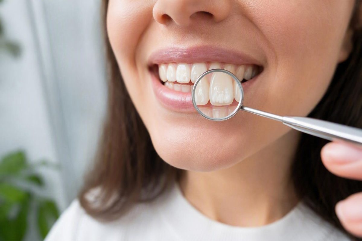
The short answer: white spots do not all mean the same thing
Some white spots are an early, noncavitated stage of tooth decay. Others reflect dental fluorosis, enamel hypoplasia or another developmental enamel defect, acid-related enamel damage, or a temporary change caused by the tooth drying out. A white spot is therefore a sign, not a diagnosis.
When enamel becomes more porous beneath its surface, it scatters light differently from the surrounding enamel. The affected area may look milky, chalky, or opaque even though no visible hole has formed. Plaque-related mineral loss is one way this porosity develops, but enamel that formed differently can produce a similar optical effect. Different causes may also occur together, so color alone cannot establish what a spot is according to a clinical review of white-spot lesions.
A useful first distinction is timing:
- Changes that developed after the tooth erupted may reflect plaque-related demineralization, repeated acid exposure, or—in short-lived cases—dehydration.
- Changes built into the enamel before eruption may reflect fluorosis, hypoplasia, hypomineralization, or another developmental defect.
That distinction is not always obvious in a mirror or photograph. The table summarizes patterns a dentist may consider; none of its features is a diagnostic home test. The comparisons reflect descriptions of developmental, plaque-related, acid-related, and temporary white areas in dental and medically reviewed overviews of common white-spot causes and enamel changes.
| Possible explanation | Typical timing | Pattern or location a dentist may consider | Possible texture | Relevance to decay | Potential reversibility | Usual management goal |
|---|---|---|---|---|---|---|
| Plaque-related demineralization | Develops after eruption | May occur near the gumline, between teeth, or around orthodontic brackets where plaque remains undisturbed | May be dull, chalky, or rough; an early lesion can still have an intact outer surface | Can represent an early stage of the caries process; continued mineral loss may lead to surface breakdown | Selected intact lesions may sometimes be arrested or remineralized; physically lost enamel does not regrow | Control the disease process, protect the surface, and address appearance separately if desired |
| Dental fluorosis | Develops while teeth are forming, before eruption | May appear as diffuse, bilateral, or symmetrical streaks and opacities | Often intact, although appearance and severity vary | It is a developmental change rather than active decay, but decay can occur independently on the same teeth | The developmental pattern does not “remineralize away” | Monitor sound enamel or reduce the visible contrast if the appearance is bothersome |
| Enamel hypoplasia or another developmental defect | Forms before eruption | May affect one tooth, several teeth, or corresponding teeth; the pattern and developmental history matter | Hypoplasia may leave enamel thin, pitted, grooved, or partly missing; other mineralization defects may produce poorly mineralized enamel | Structurally deficient enamel may be more susceptible to sensitivity, staining, wear, or decay | Missing or structurally defective enamel does not regenerate | Protect vulnerable areas, manage disease risk or sensitivity, and restore defects when necessary |
| Acid-related enamel damage | Develops after eruption with repeated non-bacterial acid exposure | Distribution may reflect where acid repeatedly contacts teeth, but location alone cannot establish the cause | Enamel may become altered, softened, worn, or eroded; appearance varies | It is not necessarily plaque-driven caries, although damaged tooth structure may still require protection | Exposure can be reduced, but enamel physically lost through erosion does not grow back | Identify and reduce the relevant exposure and preserve the remaining tooth structure |
| Temporary dehydration | Appears while the tooth surface is dry, sometimes after sleeping with the mouth open | May be noticed on waking or when teeth have been held dry | The contrast generally fades as saliva rehydrates the surface | Temporary whitening alone is not proof of decay | The appearance may disappear after rehydration | Confirm that the change is genuinely temporary and assess a mark that persists or changes |
These are probabilities, not visual rules. A symmetrical streak may suggest a developmental cause, but it does not prove fluorosis. A white band beside the gums may raise concern for plaque-related mineral loss, but its location does not confirm active decay. Timing, distribution, texture, surface integrity, symptoms, and change over time all matter.
When a white spot may be early enamel demineralization
Plaque bacteria metabolize sugars and produce acids. Repeated acid challenges can remove minerals from enamel, creating microscopic pores beneath the surface. The altered enamel then scatters light differently and may become visibly opaque while its outer surface remains intact. Continued mineral loss can eventually lead to roughness, discoloration, surface collapse, and a cavity, but that progression is not inevitable in every lesion as outlined in a dentist-authored explanation of demineralization.
A lesion may be more concerning for active demineralization when several clues occur together:
- It is in a plaque-stagnation area, such as beside the gumline, between teeth, or around an orthodontic bracket.
- It looks dull, matte, chalky, or opaque rather than glossy.
- Its surface appears rough or shows early breakdown.
- It is new, enlarging, or becoming more noticeable.
- It is becoming sensitive or changing toward brown or gray.
By contrast, a spot that is smooth, shiny, hard, intact, and unchanged may be inactive, arrested, or developmental. That distinction still requires professional judgment. A dentist considers the combination of location, surface condition, history, plaque distribution, symptoms, and the person’s wider caries risk rather than treating any one feature as conclusive.
Location is useful as context. A chalky band following the gumline can correspond to an area where plaque has remained undisturbed. Pale marks tracing the margins of former orthodontic brackets may follow the same pattern because brackets and wires make consistent cleaning more difficult. A faint symmetrical streak that has looked the same since the tooth erupted has a different history and is less typical of newly active plaque-related mineral loss.
These descriptions should not encourage readers to scrape, probe, or repeatedly dry a tooth. It can be difficult to judge subtle hardness, roughness, or surface breakdown accurately at home. A photograph also cannot show every feature a dentist uses to assess whether a lesion is active.
If demineralization is identified while the surface remains intact, conservative management may still be possible. The aim is to change the conditions that favor mineral loss and, where appropriate, support remineralization. Once the surface has physically collapsed, the options may shift toward restoring lost structure.
Developmental causes: fluorosis and enamel hypoplasia
Developmental white spots differ from plaque-related demineralization in one central respect: the enamel formed differently before the tooth entered the mouth.
Dental fluorosis develops when excessive fluoride is ingested while teeth are forming. After eruption, the enamel may show faint white streaks, lacy markings, diffuse opacities, or more extensive discoloration. A bilateral or symmetrical pattern can be more consistent with fluorosis than a single new patch beside accumulated plaque, but appearance is not definitive.
Fluorosis is not active tooth decay. That does not mean every white mark thought to be fluorosis is harmless, or that a person with fluorosis cannot also develop caries. It means the enamel pattern originated during tooth development rather than from a currently advancing plaque lesion.
This resolves the apparent contradiction around fluoride. Excessive swallowed fluoride during tooth development can contribute to fluorosis, while appropriately selected topical fluoride after eruption can help prevent caries and support remineralization of suitable early lesions. Timing, route, amount, and purpose differ as explained in a medically reviewed overview of white spots. Fluoride is neither uniformly harmful nor a guarantee that every visible white area will disappear.
Enamel hypoplasia is a developmental structural defect in which less enamel forms than expected. It may leave enamel thin, pitted, grooved, irregular, or partly missing. A related but distinct developmental mineralization defect may leave enamel present but poorly mineralized. Using the broader term developmental enamel defect is therefore more accurate when softness or weak mineralization is involved.
Hypoplasia can affect baby teeth, permanent teeth, or both. Where enamel is thin or absent, the tooth may be more sensitive or susceptible to staining and decay. These defects differ from a purely optical opacity because the amount or contour of enamel may be altered.
A dentist may ask about premature birth, significant childhood fever or infection, trauma to a baby tooth that could have affected a developing permanent tooth, medical treatment, medication history, or genetic conditions. These details can help establish when enamel development may have been disturbed. Even after a detailed history, however, the specific cause often remains unknown. Pediatric dental material distinguishes hypoplasia from decay that develops after eruption and describes possible findings such as pits, grooves, thin enamel, and missing structure in primary and permanent teeth.
An individual white spot does not prove a nutritional deficiency, fluoride problem, medication effect, genetic disorder, or systemic condition. Such factors may be relevant to a broader developmental history, but none should be treated as the default explanation without an examination.
Why spots appear after braces—or only in the morning
Timing can narrow the possibilities, although it rarely settles the diagnosis by itself.
Orthodontic brackets, wires, and attachments create sheltered areas where plaque can remain and where toothbrush bristles may not clean effectively. Localized demineralization can develop around bracket margins or near the gumline during treatment. Once the appliances are removed, the contrast between affected enamel and the surrounding tooth may become much easier to see.
The spots may therefore seem to have appeared on the day the braces came off even though mineral loss occurred while the brackets were attached. A pattern resembling pale outlines around former brackets is consistent with this process, but not every white area first noticed after orthodontic treatment is decay.
A different timing pattern can occur after sleep. Teeth that have dried because the mouth remained open may look temporarily whiter or patchier. As saliva returns and rehydrates the enamel, that contrast may fade. A genuine enamel lesion can also become more conspicuous when dry, so disappearance after rehydration is a clue rather than proof that the tooth is healthy.
A short timing pathway can help organize what to tell a dentist:
- Present since eruption: A developmental change such as fluorosis, hypoplasia, or another enamel defect may be considered.
- Newly developed near plaque: Demineralization becomes more concerning, particularly near the gumline or in another hard-to-clean area.
- First obvious after braces: Mineral loss may have developed around the appliances, although other causes remain possible.
- Visible only when the teeth are dry: Temporary dehydration may contribute, especially if the mark reliably fades once saliva returns.
Noting timing is useful. Repeatedly drying the teeth or postponing an examination to conduct home experiments is not.
What a dentist uses to narrow down the cause
Because several conditions can produce white, milky, or chalky enamel, a dentist combines appearance with history and clinical context.
Relevant assessment questions include:
- When was the spot first noticed?
- Was it visible when the tooth erupted, or did it develop later?
- Has it changed in size, color, texture, or sensitivity?
- Is it isolated or present on corresponding teeth?
- Does it follow the gumline, surround a former bracket, or occur where plaque collects?
- Is the surface intact, hard, smooth, shiny, dull, rough, pitted, or broken?
- Is there sensitivity, pain, swelling, or irritation?
- Are there other signs of plaque-related disease, erosion, or developmental enamel differences?
Distribution can be informative. A diffuse, symmetrical pattern present since eruption can suggest a developmental cause. A localized, dull lesion in a plaque-stagnation area can suggest active demineralization. Neither observation establishes the diagnosis by itself.
The history may include previous orthodontic treatment, fluoride exposure while the teeth were developing, dental trauma, significant childhood illness or infection, premature birth, medication history, medical treatment, and other developmental factors. Pediatric dental descriptions specifically note that texture, location, pattern, timing, and developmental history can help distinguish developmental enamel defects from later decay without making appearance alone diagnostic.
More than one explanation may apply. Someone can have mild developmental opacities and later develop plaque-associated mineral loss on the same or different teeth. Repeated acid exposure can also affect teeth that already have developmental defects. This is why a photograph, color description, or single location rule cannot reliably separate all causes.
An examination does not automatically mean that every person needs imaging or an identical diagnostic protocol. The next steps depend on the lesion’s location, surface integrity, symptoms, suspected cause, and the person’s overall risk.
Decay Guide publishes general reference information and does not diagnose or treat individual readers. The descriptions here can help someone prepare useful observations, but they cannot determine what a particular spot is or which treatment is appropriate.
Can white spots be reversed?
“Reversed” can mean three different things, and separating them prevents unrealistic expectations:
- Arresting further mineral loss: The conditions driving the lesion are brought under control so it no longer progresses.
- Remineralizing an intact early lesion: Minerals are added back into porous enamel while the surface remains unbroken.
- Making the color less noticeable: The optical contrast is reduced through cosmetic or minimally invasive treatment.
Selected early demineralized lesions with intact surfaces may sometimes be arrested or remineralized. Whether that is realistic depends on the cause, lesion activity, surface integrity, plaque control, exposure to sugar or acid, and overall caries risk. It does not mean every chalky area will vanish or return to exactly the same appearance as the surrounding enamel. Consumer oral-health material also distinguishes potentially manageable early demineralization from developmental defects that cannot simply be brushed away when discussing white-spot reversibility.
A successfully arrested lesion may remain visibly white. Controlling the disease process and producing a uniform color are separate outcomes. A stable mark does not necessarily mean management failed if the surface is now hard, intact, and inactive.
Enamel that is pitted, cavitated, physically missing, or developmentally deficient does not regenerate. Remineralization may strengthen suitable porous enamel, but it cannot rebuild a missing contour or fill a hole. Structural loss may require a protective or restorative approach.
Likewise, fluorosis or hypoplasia should not be described as “reversed” merely because its appearance can be blended, reduced, or covered. Cosmetic treatment changes how the spot looks; it does not change how the enamel originally formed.
For a professionally assessed spot that is stable, inactive, structurally sound, and not bothersome, monitoring without cosmetic treatment may be reasonable. Avoiding unnecessary alteration of sound tooth structure can itself be a valid goal. The decision depends on biological risk, structural integrity, and the person’s preferences.
Treatment depends on whether the goal is health, appearance, or both
Treatment should begin by defining the problem. Is there active mineral loss to stop? An intact early lesion that may benefit from remineralization? A structural defect requiring protection? Or a stable color difference that the person would simply like to make less visible?
The options below are organized by purpose rather than as a strict ranking. Some appearance treatments remove enamel, while others cover it or change its shade, so “least to most invasive” is not a single straightforward sequence.
1. Monitoring and disease control
If a spot is stable and inactive, monitoring may be enough. When demineralization is active or plausible, the first goal is to change the conditions favoring mineral loss. That may include more effective plaque removal, especially along the gumline and around orthodontic appliances, and reducing how frequently teeth are exposed to sugar or acid.
This is not an instruction to brush harder. Excessive force does not compensate for areas that are repeatedly missed. The aim is gentle, consistent, thorough cleaning supported by care appropriate to the person’s assessed risk.
2. Professionally selected remineralization
Topical fluoride has the strongest support among the remineralization approaches described in the detailed clinical review. Product selection and application should reflect lesion activity and caries risk rather than an assumption that every white mark needs the same treatment.
Standard fluoride toothpaste supports prevention, but consumers should be cautious about claims that a particular over-the-counter “remineralizing” product will erase visible spots. An intact early lesion may become stronger without disappearing. Developmentally missing or physically lost enamel cannot be rebuilt by toothpaste.
3. Resin infiltration
Resin infiltration is a minimally invasive option for selected porous lesions. The optical change may help the lesion blend with surrounding enamel.
It may therefore address both lesion management and appearance in carefully selected cases. It is not suitable for every white spot, does not rebuild missing enamel, and cannot substitute for controlling plaque-related disease.
4. Professionally monitored whitening
Whitening changes the shade of the teeth around a spot and may sometimes reduce contrast. Results cannot be predicted from color alone. Some white areas remain visible, while others can appear more prominent, including some fluorosis-related marks. Whitening does not treat active decay or regenerate enamel and may make certain spots more noticeable.
Whitening should not be used as a diagnostic experiment. The cause and surface condition should be assessed before it is chosen to address appearance.
5. Microabrasion
Microabrasion removes a controlled, superficial amount of enamel to reduce the visibility of a shallow defect. It is primarily an appearance-altering procedure and permanently removes some tooth structure.
Deep lesions, opacities extending well below the surface, and defects involving dentin are not appropriate targets for simple superficial abrasion. The lesion’s depth, cause, and structural condition must be assessed first.
6. Composite bonding
Composite bonding places tooth-colored material over or within a defect. It can mask discoloration, replace a limited amount of missing structure, or improve contour. Bonding covers or restores the area; it does not remineralize enamel or resolve an uncontrolled caries process.
7. Veneers
Veneers cover the visible surface of a tooth and may conceal persistent defects. The health of the tooth and the extent of the defect should therefore be considered first.
Veneers are a coverage strategy, not a treatment for active mineral loss by themselves.
8. Structural restoration
A filling, crown, or another restorative approach may be considered when there is surface breakdown, substantial missing enamel, dentin involvement, or a lesion that cannot be managed adequately through conservative methods. At that point, the purpose shifts from blending color to restoring or protecting compromised tooth structure.
A clinical review describes this general progression from diagnosis and disease control to remineralizing, optical, enamel-removing, or restorative options according to lesion depth and activity rather than recommending one universal treatment.
White spots on children’s teeth
A white spot on a child’s tooth may represent early plaque-related demineralization, fluorosis, enamel hypoplasia, another developmental enamel defect, or an acid-related change. Parents should not automatically label it a cavity, but they should not assume it is harmless fluorosis either.
This applies to baby teeth as well as permanent teeth. A white area on a baby tooth can be the first visible stage of plaque-related mineral loss, particularly when it develops near the gumline or another plaque-retentive area. It can also reflect enamel that formed differently before eruption. The tooth’s age does not settle the question.
In children, hypoplasia may leave enamel thin, pitted, grooved, irregular, or partly absent. A different developmental mineralization defect may leave enamel poorly mineralized. Depending on the defect, affected teeth may be sensitive or more susceptible to staining, wear, and cavities.
Fluorosis also develops before eruption. Excessive fluoride ingestion while teeth are forming may later appear as faint streaks, lacy markings, or patches when those teeth enter the mouth. This developmental process is different from a new plaque-related lesion forming on an erupted tooth.
Parents should supervise young children’s toothpaste use so an appropriate amount is used and toothpaste is not routinely swallowed. That does not mean fluoride toothpaste should generally be avoided. Appropriate topical use helps prevent decay; unnecessary ingestion during tooth development is the separate concern.
Management depends on the cause, location, depth, and structural effect of the defect. It may range from monitoring and fluoride-based protection to sealants, bonding, or restoration. Pediatric dental material similarly lists early decay, fluorosis, and developmental enamel defects among possible causes and emphasizes matching management to the underlying problem rather than the color alone.
Parents can help by noting whether the spot was present at eruption, whether it is changing, and whether the child reports sensitivity. They should avoid picking at the area or beginning cosmetic treatment without a dental assessment.
Prevention and signs that deserve timely assessment
General preventive care cannot prevent every developmental enamel defect, but it can reduce plaque-related mineral loss and help protect erupted teeth:
- Brush twice daily with fluoride toothpaste using a gentle, thorough technique.
- Clean between teeth with an appropriate interdental method.
- During orthodontic treatment, clean carefully above, below, and around brackets and along the gumline.
- Limit frequent sugary snacks and drinks that repeatedly support acid production by plaque bacteria.
- Limit frequent acidic drinks and foods that repeatedly expose enamel to acid.
- Supervise young children’s toothpaste use so they use it appropriately rather than swallowing it.
With braces, consistent plaque removal around each bracket margin and along the gumline matters more than simply applying more pressure. A dental or orthodontic team can demonstrate tools and techniques suited to the appliance. These preventive measures—including fluoride toothpaste, interdental cleaning, careful cleaning around braces, and limiting frequent sugar or acid exposure—are also reflected in dental-practice guidance on white-spot prevention and progression.
Do not pick, scrape, or aggressively scrub a white spot. Do not begin whitening merely to see whether the mark disappears. These experiments do not identify the cause and may alter the appearance before the area is assessed.
The timing of an appointment depends on individual findings, but a new or changing area warrants dental attention. Do not delay assessment when a spot is:
- spreading or becoming more opaque;
- rough, soft, or no longer intact;
- sensitive or painful;
- pitted or visibly breaking down;
- turning brown or gray;
- associated with swelling or irritation.
A painless new white spot is not automatically a dental emergency, but early assessment can matter. If the area is an intact demineralized lesion, conservative disease control may be possible before structural breakdown occurs.
The safest sequence is straightforward: notice when and where the mark appeared, continue gentle plaque control with fluoride toothpaste, avoid experimenting with whitening, and have the cause assessed before choosing either remineralizing or cosmetic treatment.
A white spot may be an early warning of mineral loss, a stable mark left by tooth development, or a temporary optical change. Color alone establishes neither decay nor harmlessness. The central question is not simply whether the spot can be hidden, but whether there is active mineral loss to control, a structural defect to protect, or only an appearance concern to monitor.
Frequently asked questions
Are white spots on teeth always cavities?
No. Some white spots are early noncavitated caries lesions, but others result from fluorosis, enamel hypoplasia, another developmental defect, acid-related change, or temporary dehydration.
A spot is more concerning for active demineralization when it is new, dull or chalky, rough, changing, or located where plaque accumulates. A smooth, shiny, hard, stable spot may be inactive or developmental. These features help a dentist assess the lesion but cannot confirm or exclude decay at home because several causes can look similar.
Why did white spots become visible after my braces were removed?
Brackets and wires create plaque-retentive areas that are more difficult to clean. Mineral loss can develop around bracket margins during orthodontic treatment and become obvious only after the brackets are removed and the full enamel surface is visible.
Marks near former bracket outlines or along the gumline are consistent with localized plaque-related demineralization. However, not every spot first noticed after braces has that cause; developmental and other enamel changes remain possible when assessing post-orthodontic white spots.
Can fluoride both cause and help prevent white spots?
Yes, because the timing and type of exposure differ. Excessive swallowed fluoride while teeth are developing can cause fluorosis, which may appear as white streaks or opacities after eruption.
Appropriately selected topical fluoride on erupted teeth can help prevent caries and support remineralization of suitable intact early lesions. It cannot remove a developmental defect or rebuild missing enamel simply because both situations involve fluoride.
Can teeth whitening make white spots stand out more?
It can. Whitening may reduce contrast in some cases by lightening the surrounding enamel, but it may fail to conceal the spot or make the difference more obvious. Results depend on the cause and depth of the mark.
Whitening does not diagnose a white spot, treat active decay, or regenerate enamel. The cause and surface condition should be assessed before whitening is chosen, particularly because some fluorosis-related marks can become more conspicuous after whitening.
Why do white spots sometimes appear in the morning and then disappear?
Sleeping with the mouth open can dry the enamel surface. Dry enamel scatters light differently, so some areas may temporarily look whiter. Once saliva rehydrates the teeth, the contrast may fade.
A mark that reliably disappears after the mouth becomes moist is more consistent with temporary dehydration than a persistent structural lesion. It is not definitive proof, because genuine enamel defects may also look whiter when dry. A spot that remains visible or changes should be assessed rather than diagnosed from rehydration alone.