
Treating Gum Disease Without Surgery: Options, Limits, and What to Expect
Scaling and root planing should not be expected to predictably regenerate substantial gum tissue or supporting bone that has already been lost.
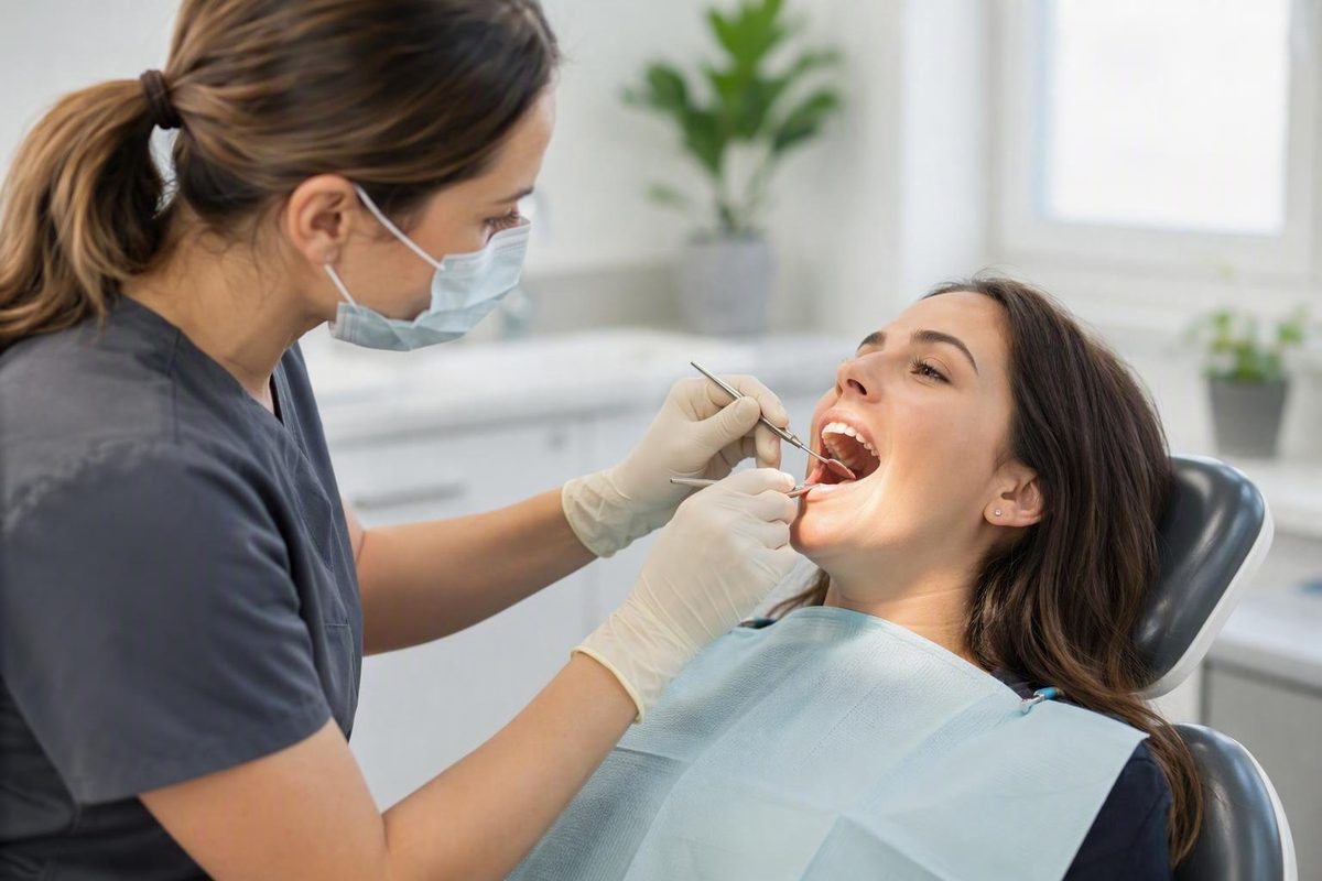
Non-surgical gum disease treatment can include a professional cleaning for gingivitis, scaling and root planing for periodontitis, and selected additions such as topical or oral antibiotics, antimicrobial rinses, medication trays, bite management, or certain laser-assisted procedures. For active periodontitis, scaling and root planing is usually the foundation of initial non-surgical care. Its purpose is to clean periodontal pockets, reduce plaque, calculus, bacteria, and inflammation, and help prevent further damage to the gums and supporting bone (American Academy of Periodontology).
These treatments are not interchangeable. Medication or technology may supplement mechanical cleaning when there is a defined reason, but neither automatically replaces it. The appropriate plan depends on the diagnosis, distribution of disease, pocket measurements, bleeding, attachment and bone loss, tooth stability, health and behavioral risk factors, and response after healing.
Non-surgical treatment can often control gum disease. It should not, however, be expected to predictably rebuild substantial gum tissue or supporting bone that has already been lost.
Important: Decay Guide is an independent information publisher, not a dental practice. It cannot diagnose gum disease, determine whether you are suitable for a procedure, or provide individualized treatment advice. Those decisions require an examination by a dentist or periodontist.
What counts as non-surgical gum disease treatment?
The term covers several levels of care:
- Professional preventive cleaning: Removes plaque and tartar from accessible tooth surfaces. This may be sufficient for plaque-related gingivitis when paired with effective daily plaque control.
- Scaling and root planing: Treats root surfaces below the gumline and within periodontal pockets. It is commonly the initial non-surgical treatment for periodontitis.
- Antibiotic or antimicrobial treatment: A locally placed gel, prescribed rinse, or oral antibiotic used selectively to support mechanical treatment.
- Medication trays: Custom trays used at home to deliver a product prescribed by a dental professional.
- Laser-assisted treatment: A broad category covering different devices and protocols. A laser may supplement mechanical cleaning or be used differently under a particular treatment protocol.
- Bite-related measures: A guard, limited bite adjustment, or stabilization of selected mobile teeth when grinding, clenching, uneven forces, or mobility are separate concerns.
A simple disease-based pathway is:
- Gingivitis without attachment or bone loss: Professional cleaning and better home plaque control may allow the inflammation to resolve.
- Periodontitis that is not advanced or highly complex: Scaling and root planing is generally the starting treatment.
- Persistent, advanced, or structurally complex disease: Further non-surgical treatment, referral to a periodontist, or surgery may be appropriate after the complete findings and response to initial care are considered.
Candidacy cannot be determined from symptoms or a stage label alone. Two people described as having “moderate” disease may have very different problems. One may have a few isolated pockets; another may have generalized bone loss, difficult root anatomy, smoking exposure, loose teeth, or a health condition that affects healing. Initial treatment and the need to escalate therefore depend on the complete clinical picture.
The main objective is disease control: clean the affected root surfaces, reduce the burden that sustains inflammation, make the area easier to maintain, and limit additional damage. Non-surgical treatment has important limitations, and surgery may still be recommended if it does not produce adequate periodontal health.
Diagnosis comes before choosing a treatment
A routine cleaning, deep cleaning, and periodontal surgery serve different purposes. Choosing among them begins with establishing whether the problem is gingivitis, periodontitis, recession from another cause, or a combination.
Gingivitis is gum inflammation without the supporting-tissue destruction that defines periodontitis. The gums may bleed, swell, or appear red, but there is no periodontitis-related loss of attachment or supporting bone.
Periodontitis damages the structures that support the teeth. The attachment around a tooth can change, periodontal pockets can deepen, and supporting bone may be lost. This distinction matters because a surface-focused cleaning may help control gingivitis but may not reach deposits within diseased periodontal pockets.
A periodontal assessment may include:
- Medical and dental history: Symptoms, medicines, tobacco exposure, relevant health conditions, previous periodontal treatment, and other risk factors.
- Oral examination: Plaque, calculus, swelling, recession, and other tissue changes.
- Bleeding and inflammation findings: Bleeding during probing can help identify inflamed sites, although it must be interpreted with other findings.
- Pocket measurements: A periodontal probe measures the space around each tooth at multiple locations.
- Attachment, mobility, and other findings: The clinician may evaluate recession, exposed roots, loose teeth, drainage, bite forces, furcation involvement, and tooth prognosis.
- Dental X-rays when indicated: Imaging can show the distribution and extent of supporting-bone loss.
- Staging and grading: If periodontitis is diagnosed, its severity, treatment complexity, risk factors, and anticipated progression may be incorporated into the classification.
For general context, healthy periodontal pockets are usually 1–3 millimeters deep. Pockets deeper than 4 millimeters may indicate periodontitis, while pockets deeper than 5 millimeters are difficult to clean through routine care. Diagnosis may also involve history, examination, probing, X-rays, and staging and grading (Mayo Clinic).
Those measurements are not a self-diagnosis tool. One deep reading does not by itself establish periodontitis, determine a tooth’s prognosis, prove that treatment failed, or make surgery necessary. Clinicians consider the pattern across the mouth, attachment and bone loss, inflammation, recession, anatomy, previous treatment, and other local factors.
Symptoms are prompts for evaluation rather than a way to choose treatment. Bleeding, swelling, persistent bad breath, sensitivity, recession, discomfort while chewing, and loose teeth can occur with periodontal disease, but an examination is needed to identify their cause and extent.
Recession requires particular care. A receding gumline may accompany periodontitis, but it can also be associated with forceful brushing, thin tissue, tooth position, tobacco use, grinding, or uneven bite forces. An exposed root does not necessarily mean that there is an infection requiring deep cleaning, and a person without visible recession may still have periodontal pockets.
A general dentist may diagnose and manage less complex disease. A periodontist—a dentist who specializes in the gums and tooth-supporting structures—commonly manages advanced, recurrent, difficult-to-access, or otherwise complex cases. Referral does not automatically mean surgery; it may be for diagnosis, prognosis, or specialist-delivered non-surgical care.
Scaling and root planing: the core non-surgical procedure
A routine preventive cleaning and scaling and root planing are not different names for the same service.
A routine cleaning is primarily preventive and surface-focused. It removes accessible plaque, stains, and calculus from teeth when treatment inside diseased periodontal pockets is not required.
Scaling and root planing, often called deep cleaning, is therapeutic. It reaches below the gumline to treat affected root surfaces and deposits within periodontal pockets.
What happens during scaling?
The clinician uses hand instruments, ultrasonic equipment, or a combination to remove plaque, hardened calculus, bacteria, and bacterial products from tooth and root surfaces. Treatment extends into the below-gum sites identified during the examination. A laser may be incorporated in some settings, but its use does not eliminate the need to establish how hardened deposits on the root will be removed.
What happens during root planing?
Root planing addresses the root surface. The clinician smooths rough or contaminated areas to make additional buildup more difficult and to create a surface compatible with healing and closer tissue adaptation.
Local anesthesia is commonly used. It can make treatment more manageable, but it does not justify a promise that every patient will feel nothing during or after the procedure. Treatment may involve one isolated area, several sections of the mouth, or a generalized pattern, and it may be completed in one or more visits according to disease extent, access, anesthesia needs, and provider protocol.
Mechanical treatment can be challenging even when performed appropriately. Furcations between the roots of multi-rooted teeth, root grooves, concavities, deep pockets, and the back surfaces of molars may limit access and visibility. Scaling and root planing reduces microbial load but does not necessarily eliminate every disease-associated microorganism (2024 comparative review).
Incomplete improvement at a difficult site therefore does not automatically mean that the initial treatment was inappropriate or poorly performed. The site may need more healing time, renewed instrumentation, improved home plaque control, a selective adjunct, specialist access, or surgery that permits direct access to the root.
The non-surgical treatment process
1. Examination and measurements Confirm the diagnosis and map pockets, bleeding, attachment, mobility, recession, and bone loss.
2. Anesthesia if needed Numb the treatment area according to its extent and the patient’s needs.
3. Scaling Remove plaque, calculus, bacteria, and bacterial products above and below the gumline.
4. Root planing Clean and smooth affected root surfaces.
5. Healing period Continue the recommended home care while inflammation settles and tissue response becomes assessable.
6. Objective reassessment Recheck bleeding, residual pockets, plaque control, tissue stability, mobility, and other relevant findings.
7. Maintenance or additional treatment Move to individualized periodontal maintenance if the disease is controlled, or address persistent active sites with further care.
The purpose is not to sterilize the mouth. It is to disrupt and reduce the deposits and microbial burden that sustain inflammation, support healing, and make long-term plaque control more achievable.
Antibiotics, rinses, trays, and bite management are selective additions
Deep cleaning removes deposits mechanically. Adjuncts are considered when a clinician expects a specific added benefit, not simply because a product or device is available.
Antibiotics and antimicrobial products
Possible delivery methods include:
- Antibiotic gel placed in selected periodontal pockets
- A prescribed antimicrobial or antibiotic mouth rinse
- Oral antibiotics in selected circumstances
These products do not scrape calculus from a root or correct inaccessible anatomy. They may help control selected bacterial concerns, but they are not universal substitutes for scaling and root planing. The evidence supplied here supports describing them as case-dependent options, not providing a do-it-yourself list of systemic-antibiotic indications, drug choices, or doses.
Drug selection and dosing require professional assessment. Short-term antimicrobial use is commonly emphasized in periodontal care, and the recommendation should be tied to a defined site or clinical finding rather than treated as an automatic part of every deep cleaning.
Useful questions include:
- Which site or finding is the medication intended to address?
- What benefit is expected beyond mechanical cleaning?
- Is the recommendation local or systemic, and why?
- What risks and alternatives should I discuss with the prescriber?
- How will the clinician determine whether it helped?
Custom medication trays
A medication tray is made from impressions of the mouth and used at home to deliver a prescribed product. The tray is only a delivery device; its regulatory status does not prove that the medicine placed inside it safely or effectively treats periodontitis.
The American Academy of Periodontology explains that tray systems were cleared by the US Food and Drug Administration based on their similarity to fluoride trays, but that this process did not establish the safety or effectiveness of any particular tray-delivered medicine for gum disease.
If a tray is proposed, ask for evidence concerning the specific medication, dose, diagnosis, intended outcome, and treatment protocol, rather than relying on the clearance status of the tray itself.
Bite adjustment, guards, and stabilization
Bite-related care addresses a different mechanism. When grinding, clenching, uneven contact, or excessive force contributes to mobility or tissue trauma, possible measures may include a bite guard, limited adjustment of selected contacts, or stabilization of certain mobile teeth (overview of non-surgical periodontal measures).
These approaches do not remove deep calculus or independently control periodontal infection. Conversely, an antibiotic does not correct grinding, and a desensitizing product does not rebuild lost attachment. Each recommendation should have a clear target:
- Scaling and root planing: Plaque, calculus, and affected root surfaces
- Antimicrobials or antibiotics: Selected bacterial concerns
- Desensitizing or fluoride products: Discomfort from exposed roots
- Bite guard or limited adjustment: Excessive or uneven forces
- Stabilization: Support for selected mobile teeth
Questions to ask before accepting an adjunct
- Why is this being recommended in my case?
- What measurable benefit is expected beyond scaling and root planing?
- Is it addressing infection, sensitivity, force, mobility, or another problem?
- What are the risks and realistic limitations?
- What conventional alternative is available?
- Could scaling and reassessment reasonably come first?
- Is the quoted fee separate from the deep cleaning?
- Has insurance coverage been confirmed, or is reimbursement uncertain?
Laser gum treatment: possible option, uncertain superiority
“Laser gum treatment” does not describe one standardized procedure. One clinician may use a laser to remove selected soft tissue alongside mechanical root cleaning; another may follow a different non-surgical or surgical protocol.
A laser may be:
- Added to scaling and root planing
- Incorporated into a non-surgical pocket-treatment protocol
- Used under a protocol classified as periodontal surgery
- Directed primarily at soft tissue rather than hardened calculus on the root
That distinction matters when evaluating claims. A laser may affect bacteria or selected tissue, but the patient should still ask how plaque and hardened calculus on the root will be removed.
Current evidence does not establish that laser treatment is generally superior to conventional scaling and root planing. The comparative literature includes randomized trials and reviews, but also in-vitro research, case series, and case reports. It includes both laser monotherapy and lasers added to scaling, and it provides no pooled estimate proving better outcomes across different patients, diagnoses, devices, and protocols (peer-reviewed comparative review).
Small numerical differences from promotional pages should not be treated as proof of a clinically meaningful advantage. Without adequate information about sample size, uncertainty, statistical analysis, patient selection, adverse events, and long-term follow-up, a reported difference in pocket reduction cannot establish broad superiority.
Safety and effectiveness can depend on:
- Wavelength and power
- Pulse and energy settings
- The tissue or deposit being targeted
- Whether roots are also mechanically instrumented
- Pocket and root anatomy
- Operator training and technique
- Diagnosis and disease severity
- Patient selection and health factors
The American Academy of Periodontology reports that controlled studies have found results similar to certain other non-surgical options, including scaling and root planing alone. It also warns that an inappropriate wavelength or power level can damage periodontal tissues. That safety limitation is one reason to ask about the exact procedure and the operator’s training rather than treating “laser” as a sufficient description.
Laser-assisted non-surgical cleaning and LANAP are not interchangeable terms. A periodontal practice comparison explicitly classifies scaling and root planing as non-surgical and LANAP as laser periodontal surgery (SRP and LANAP classification). The same page contains promotional claims that should not be generalized, but its classification illustrates why the exact procedure name matters.
Be cautious about promises that laser care will be painless, guarantee faster healing, predictably regenerate lost tissue, or eliminate any possibility of surgery. Improvement in inflammation or pocket measurements is not the same as rebuilding substantial lost gum or bone.
Before agreeing to a laser procedure, ask:
- Is the laser supplementing scaling or replacing part of it?
- How will hardened calculus on the root be removed?
- What is the exact procedure and device?
- Is the procedure classified as non-surgical care or surgery?
- What evidence applies to my diagnosis and disease severity?
- What training does the operator have with this device and protocol?
- What tissue risks are relevant?
- What conventional alternative is available?
- What additional fee is being charged?
- Has insurance coverage been verified?
Expected results, recovery, and the limits of treatment
The central expectation should be disease control, not restoration of the mouth to its condition before periodontitis developed.
After effective treatment, possible improvements include:
- Less inflammation and bleeding
- A lower bacterial and deposit burden
- Shallower or more maintainable pockets
- Closer tissue adaptation to cleaned roots
- Better access for daily cleaning
- Greater stability when treatment is followed by appropriate maintenance
Many patients may need no additional active treatment after scaling and root planing, although ongoing periodontal maintenance is commonly required. Others may have isolated residual sites or advanced structural damage that needs further care.
The essential distinction is between healing of inflammation and regeneration of lost structure. Swollen tissue may shrink and become firmer, and pocket measurements may improve. Those changes do not show that substantial lost bone, attachment, or receded gum tissue has predictably grown back.
Healing may also make recession more noticeable. That does not automatically mean the treatment damaged the gums, although an unexpected or progressive change should be assessed.
Short-term effects can include tenderness, bleeding, gum discomfort, and tooth sensitivity. Gum recession and infection are also recognized complications. Recovery varies with disease severity, procedure type, oral and overall health, and aftercare (Cleveland Clinic).
There is no responsible universal promise about pain, downtime, healing time, or when sensitivity will stop. Treatment of one isolated area may feel different from generalized treatment, and an exposed root may remain sensitive after inflammation improves. Follow the treating clinician’s written aftercare instructions and contact that office if symptoms are severe, worsening, or otherwise outside what the clinician told you to expect.
Recession is a separate treatment question
Treating active inflammation may help control periodontitis-related tissue damage, but it does not guarantee that an exposed root will be covered again.
When sensitivity is the main concern, a clinician may consider a desensitizing or fluoride product. If forceful brushing, tooth position, grinding, or bite forces contribute, management should address those factors rather than treating recession as infection alone.
Substantial recession may require a separate periodontal assessment. Selected cases may be considered for gum grafting, but the need and likely result depend on the cause, anatomy, tissue condition, and treatment goal (discussion of non-surgical recession management).
Numerical “success rates” from individual practices should be interpreted cautiously. Such figures may not disclose patient selection, comparison groups, statistical methods, independent validation, or long-term follow-up. A useful result is stable, measurable disease control in the individual patient—not a marketing percentage.
Reassessment: how clinicians decide whether non-surgical care was enough
A mouth that feels better may still contain active sites, while temporary sensitivity or more visible recession does not necessarily mean treatment failed.
At reassessment, the clinician may review:
- Bleeding and visible inflammation
- Residual pocket depths and their distribution
- Tissue attachment and stability
- Plaque levels and home-cleaning effectiveness
- Tooth mobility
- Drainage, discomfort, or other symptoms
- Furcation and root-anatomy challenges
- Risk factors that remain uncontrolled
- Evidence of continuing bone loss when new imaging is clinically indicated
A residual site may have several explanations:
- The original pocket was deep or difficult to access.
- A groove, concavity, furcation, or back-molar surface limited instrumentation.
- Deposits or affected root areas remain inaccessible.
- Daily plaque control has not been sufficient.
- Smoking, health factors, or damaging forces continue to affect stability.
- Structural damage is too advanced for closed, non-surgical access.
- The tooth has another problem that changes its prognosis.
Further care might involve renewed instrumentation, better home-care technique, a selective adjunct, periodontal maintenance, or specialist referral. Persistent deep pockets or inflammation, substantial bone loss, advanced recession or attachment loss, loose teeth, and inadequate response to properly delivered initial treatment can prompt consideration of a periodontist or surgery.
Escalation options address different problems:
- Flap or pocket-reduction surgery: Lifts the gum to improve access to deep root surfaces and may include reshaping or repositioning tissue.
- Gum grafting: Addresses selected soft-tissue deficiencies or exposed roots.
- Bone grafting: Treats selected bone defects.
- Guided tissue regeneration: Uses a barrier in an attempt to support regeneration in suitable defects.
These procedures are not interchangeable and do not guarantee complete restoration. Advanced periodontitis may require surgery, but the decision should integrate disease distribution, access, bone pattern, tooth prognosis, previous response, expected benefit, health factors, and patient preferences—not one pocket measurement alone (Mayo Clinic).
At the results visit, ask:
- How did bleeding change?
- Which pocket measurements improved, remained stable, or worsened?
- Are any sites still considered active?
- Is attachment stable?
- Has tooth mobility changed?
- Is the bone level believed to be stable?
- Could difficult sites improve with further non-surgical care?
- What is the specific goal of the proposed additional procedure?
- What alternatives exist, including monitoring or referral?
- What are the likely consequences of delaying treatment?
Maintenance keeps initial improvement from becoming temporary
Scaling and root planing is not a one-time guarantee. Periodontitis can remain active or recur if plaque accumulates, maintenance is missed, or relevant risk factors remain uncontrolled.
Professional care and home care have different roles:
- Professional treatment removes hardened deposits and treats periodontal pockets and root surfaces that home tools cannot adequately reach.
- Daily brushing disrupts new plaque on accessible surfaces.
- Interdental cleaning addresses areas a toothbrush does not clean effectively.
- A clinician-guided rinse may provide supplemental plaque control in selected circumstances.
- Periodontal maintenance permits recurring deposits to be removed and tissue findings to be monitored.
A rinse is not a substitute for brushing, interdental cleaning, or professional care. Undiluted hydrogen peroxide should not be presented as a treatment for gum disease because it can burn gum tissue. It also cannot remove established deposits from deep periodontal sites (Cleveland Clinic aftercare guidance).
Maintenance intervals are individualized. Some dental providers commonly describe three- or four-month schedules, but that is not a mandatory timetable for every patient. The interval may depend on residual pockets, plaque control, disease history, smoking, health factors, tooth prognosis, and how quickly deposits return.
Other relevant measures can include:
- Avoiding smoking and chewing tobacco
- Managing health conditions that affect dental health
- Cleaning daily with tools and techniques suited to your anatomy
- Using gentle rather than damaging brushing pressure
- Addressing grinding or clenching when it contributes to mobility or trauma
- Attending reassessment and maintenance visits even when symptoms improve
Brushing, flossing, mouthwash, peroxide, and consumer devices cannot remove established deep calculus or replace professional care for periodontitis. Decay Guide’s general-information feature on what daily brushing cannot fix explains the practical distinction between disrupting new plaque and trying to remove hardened or inaccessible deposits.
Appointment checklist
Take these questions to your dentist or periodontist:
- What is my exact diagnosis: gingivitis, periodontitis, recession, or a combination?
- Where are the affected sites?
- What do my pocket and bleeding measurements show?
- Is there attachment or bone loss, and how extensive is it?
- Are any teeth mobile or difficult to maintain?
- Why is a routine cleaning, scaling and root planing, or specialist care being recommended?
- Will treatment cover isolated sites, sections, or the full mouth?
- What anesthesia and instruments are planned?
- Is an antibiotic, rinse, tray, laser, or bite treatment optional?
- What additional benefit is expected from each adjunct?
- What are the risks and realistic limits?
- What improvement is expected, and what tissue is unlikely to return?
- When and how will the response be reassessed?
- What findings would lead to further non-surgical care, referral, or surgery?
- How often should periodontal maintenance occur in my case?
- What are the total fees, including adjunct and follow-up charges?
- What has the insurer confirmed, and what remains uncertain?
Frequently asked questions
Does scaling and root planing hurt, and how many visits does it take?
Local anesthesia is commonly used, so the treatment area can usually be numbed. You may still notice pressure or other sensations during care, and tenderness, bleeding, gum discomfort, or tooth sensitivity can occur afterward.
There is no universal number of visits. Treatment may cover one isolated area or multiple sections and may be completed in one or more appointments. The schedule depends on disease extent, anesthesia, access, provider protocol, and the amount of instrumentation required. Claims that every patient needs exactly two visits, has no downtime, or will be sore for a fixed number of days should be treated as generalizations rather than guarantees.
Can non-surgical treatment cure periodontitis permanently?
Non-surgical care can control periodontitis, reduce inflammation, improve pocket conditions, and limit further damage, but it does not provide permanent immunity from recurrence.
Some people need no further active treatment after scaling and root planing, yet ongoing plaque control and periodontal maintenance are still commonly required. Residual pockets, difficult anatomy, tobacco exposure, health factors, and renewed plaque accumulation can allow disease to persist or return. The long-term goal is stable, maintainable periodontal health—not a guarantee that future treatment will never be needed.
Can receding gums or lost bone grow back after deep cleaning?
Scaling and root planing should not be expected to predictably regenerate substantial gum tissue or supporting bone that has already been lost.
As inflammation subsides, gum tissue may become firmer and adapt more closely to a cleaned root. Pocket measurements may improve, but that is different from replacing missing structure. Recession may also look more obvious after swollen tissue heals.
Exposed-root sensitivity can be managed separately in some cases. Significant recession or selected bone defects may justify assessment for grafting or regenerative procedures, but suitability and expected results depend on the defect, anatomy, cause, and complete clinical findings.
Can brushing, flossing, mouthwash, or peroxide treat gum disease without a dentist?
Daily brushing and interdental cleaning reduce new plaque, can support the resolution of plaque-related gingivitis, and remain essential after professional periodontal treatment. A dentist-recommended rinse may sometimes be a useful supplement.
These measures cannot remove established calculus inside deep periodontal pockets. Mouthwash does not mechanically clean an affected root surface, and undiluted hydrogen peroxide can injure gum tissue. If periodontitis is present, home care remains essential but does not replace diagnosis, professional cleaning, reassessment, and maintenance.
When should gum disease be treated by a periodontist or considered for surgery?
A general dentist may manage gingivitis and less complex periodontal disease. Referral becomes more likely when there are persistent deep pockets, continuing inflammation after initial care, substantial bone loss, advanced recession or attachment loss, complex root anatomy, loose teeth, uncertain prognosis, or a possible need for grafting or regenerative treatment.
Surgery may be considered when non-surgical access cannot adequately clean a site or when a structural defect requires a procedure designed for that problem. It is not automatically required because of one pocket measurement. The decision should follow diagnosis, appropriate initial treatment, healing, and reassessment of the complete clinical picture.
The most defensible treatment sequence is straightforward: establish the diagnosis, begin with appropriate mechanical cleaning, allow healing, reassess objective findings, and add medication or technology only for a defined reason. If disease remains active or structural damage is advanced, specialist or surgical care may be appropriate.
Non-surgical treatment can be highly useful for controlling gum disease, but it is neither a home remedy nor a promise to restore lost tissue. Use the questions in this article to have a specific discussion with a qualified dentist or periodontist. Decay Guide provides general information only and cannot determine what treatment you need.