
How Gum Recession Changes—and Which Signs Need a Dentist
Early, moderate and advanced can help describe what you notice, but they cannot reveal the cause, amount of tooth support or whether the area is changing.
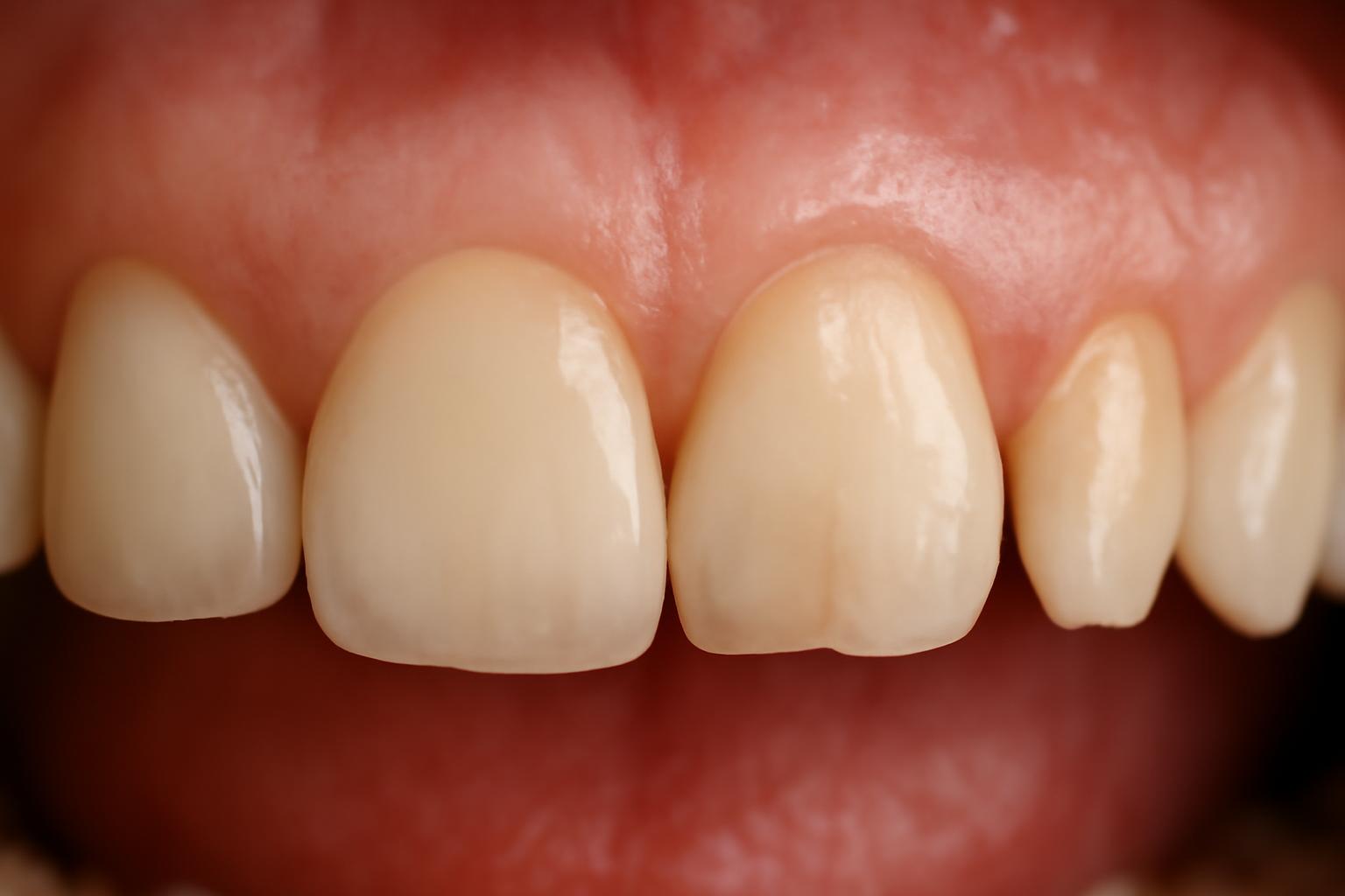
Gum recession occurs when the gum tissue pulls away from a tooth, exposing more of the tooth or the root beneath it. You may first notice sensitivity, a tooth that looks longer, a yellowish area near the gumline, or a notch that catches food. These changes can be useful clues, but they do not reveal why the gum moved or whether the bone supporting the tooth has been affected (Cleveland Clinic’s overview of gum recession).
Although people commonly search for “receding gums stages,” early, moderate, and advanced are best understood as descriptive labels—not a universal three-stage clinical system. A dentist assesses more than the visible gumline, including inflammation, periodontal pockets, root condition, tooth stability, bone levels, bite forces, and changes over time.
Are there official stages of receding gums?
The reviewed sources do not establish a universal early–moderate–advanced or numbered clinical staging system for gum recession. Some patient-education pages use terms such as “mild, moderate, and severe,” while others use “early, moderate, and advanced,” generally without agreed measurement thresholds. One dental-practice guide, for example, uses the latter framework to organize possible signs but does not identify it as a formal consensus classification (example of descriptive early-to-advanced terminology).
That distinction matters because gum recession is a clinical finding, not a complete diagnosis. It describes the position of the gum margin: the tissue has moved away from its previous position and exposed more tooth structure or root. It does not, by itself, establish:
- Why the recession occurred
- Whether gum inflammation is active
- Whether a periodontal pocket is present
- How much supporting bone remains
- Whether the area is stable or changing
- Whether surgery is needed
- Whether a similar-looking change has another explanation
Terms such as early, moderate, and advanced can still help organize what someone might see or feel. In this article, early means subtle or newly noticed changes, moderate means clearer root exposure or symptoms, and advanced means extensive visible exposure. Mobility, shifting, deep pockets, persistent inflammation, and bad breath are treated separately as warning signs of possible periodontal disease or compromised support; they do not determine the amount of recession.
These categories are an orientation guide, not stages that you can reliably assign to yourself. Not every case follows the same sequence. Recession can affect one tooth, several teeth, or a broader area, and a localized defect does not inevitably progress to bone loss, loose teeth, or tooth loss.
A photograph can help document that something appears different, but it cannot show bone levels, measure a periodontal pocket, determine disease activity, or establish the cause.
Editorial disclosure: Decay Guide is an informational publisher, not a dental practice. Its articles are written by a health writer, are not clinician-reviewed, and do not replace a dental examination, as explained in the site’s editorial disclosure.
A practical early-to-advanced gum recession guide
The following comparison describes possible patterns rather than official receding-gums stages. It deliberately avoids millimeter cutoffs because visible recession, periodontal-pocket depth, bone support, symptoms, and progression are separate findings. A person may also have features from more than one row.
| Descriptive level | Possible appearance | Possible symptoms | Associated findings to ask a dentist about | Recommended action |
|---|---|---|---|---|
| Early or subtle pattern | A tooth may look slightly longer; the gumline may appear subtly lower or less symmetrical; there may be no obvious visible change | No symptoms, mild cold sensitivity, or occasional tenderness | Whether the change is true recession; brushing technique; inflammation; tooth position; early root exposure | Arrange a routine dental examination if the change is new, persistent, or unexplained |
| Moderate or clearly visible pattern | Yellowish root exposure, a notch or ledge near the gumline, a more uneven gumline, or a larger visible portion of the tooth | Greater sensitivity to hot, cold, or sweet foods; discomfort during brushing; possible food trapping | Root wear or decay, plaque below the gumline, tooth position, local trauma, periodontal pockets, or change since a previous visit | Book a dental evaluation rather than selecting treatment from appearance alone |
| Advanced visible pattern | Extensive root exposure or a marked change in the gumline | Sensitivity may be present, although pain does not reliably show severity | Tooth mobility or shifting, deep pockets, persistent bleeding or swelling, persistent bad breath, bite changes, or suspected bone loss | Seek prompt professional care when tooth stability or ongoing inflammation is involved |
Early or subtle changes
Early recession may cause no noticeable symptoms. When symptoms occur, they can include mild sensitivity—often to cold—or occasional tenderness at the gumline. A tooth may appear slightly longer, or the gumline may seem less even than it used to.
None of these observations is specific to recession. Tooth sensitivity can have other causes, while differences in apparent tooth length may reflect normal anatomy, tooth wear, restorative work, or changes in nearby tissue. A dentist must identify the position of the gum margin and examine the tooth before confirming what changed.
The useful question is not simply, “What stage is this?” but, “Is this a genuine change, and what might be contributing to it?” Whether an early-looking defect needs monitoring or treatment depends on the examination rather than its label.
Moderate or more visible changes
As recession becomes more noticeable, part of the root may become visible. The exposed area can look yellower than the enamel-covered crown because the root surface has a different structure and appearance. A line, notch, or ledge may also be visible near the gumline.
Sensitivity may become more noticeable with hot, cold, or sweet foods and drinks. Food may repeatedly catch around the changed gum or root contour. Exposed root surfaces are less protected than enamel-covered crowns and may be vulnerable to sensitivity, wear, and root-surface decay, which is one reason a newly visible root deserves assessment (California Dental Association’s patient guide to receding gums).
A notch does not necessarily consist entirely of missing gum. Part of the visible defect may involve wear or damage to the tooth surface itself. A dental examination can distinguish movement of the gum margin from a root-surface defect that may require separate management.
Advanced changes and warning signs
Extensive root exposure can reasonably be described as advanced recession in everyday language. But loose or shifting teeth, deep periodontal pockets, persistent bleeding, swelling, and persistent bad breath should not be treated merely as evidence of “more recession.” These findings raise concern about concurrent inflammation, periodontal disease, or damage to the structures supporting the teeth.
This distinction is especially important when a tooth is mobile. The visible gum margin does not hold the tooth in place by itself. Periodontitis can damage the soft tissues and bone supporting teeth, and advanced disease can result in mobility or tooth loss. A loose or shifting tooth therefore requires evaluation of its supporting structures, not just the amount of root that can be seen (California Dental Association’s explanation of periodontal disease and tooth support).
Symptoms do not necessarily intensify in a neat sequence.
There is also no reliable universal timeline for moving from one descriptive pattern to another. Some defects may remain stable, while others change. A mirror or photograph cannot establish the rate of progression; comparable professional measurements over time are more informative.
Action summary: Arrange a dental examination for a newly visible change, a persistently longer-looking tooth, unexplained sensitivity, a gumline notch, or an exposed root. Seek prompt care for a loose or shifting tooth, persistent swelling or bleeding, ongoing pain, persistent bad breath with gum changes, or a gumline that appears to be changing rapidly (guidance on signs that warrant prompt assessment).
Gum recession is not the same as gingivitis or periodontitis
A receding gumline does not automatically mean that you have periodontal disease. Recession can occur alongside gum disease, but it can also have mechanical, anatomical, behavioral, orthodontic, or traumatic contributors. Appearance alone cannot identify which explanation applies.
| Finding or condition | What it describes | Typical concern |
|---|---|---|
| Gum recession | The gum margin has moved and exposed more tooth or root | Sensitivity, root wear or decay, food trapping, appearance, and whether the area is changing |
| Gingivitis | Inflammation limited to the gums | Redness, swelling, tenderness, or bleeding without the supporting-tissue destruction associated with periodontitis |
| Periodontitis | Disease affecting the tissues and bone supporting the teeth | Periodontal pockets, loss of support, bone loss, mobility, shifting, and possible tooth loss in advanced disease |
Bleeding and swelling mainly indicate inflammation, not the physical amount of recession. Someone can have inflamed, bleeding gums without visible recession. Conversely, a person can have an isolated exposed root without obvious bleeding, pain, or periodontal destruction. Gingivitis and periodontitis describe gum-disease processes, so their severity categories should not be substituted for descriptive recession labels (overview of recession, gingivitis, and periodontitis).
Recession and periodontal disease can coexist. Periodontal inflammation may accompany tissue loss, while an exposed root may create a contour where food collects. But the presence of recession alone does not establish that periodontitis is active.
Persistent bad breath, deep periodontal pockets, tooth shifting, mobility, or X-ray evidence of bone loss raises concern about concurrent periodontal disease or compromised support. These findings require their own assessment. A gumline photograph cannot establish whether the supporting bone around a tooth is intact.
It is also misleading to assume that recession always begins with gingivitis and then inevitably advances through periodontitis to tooth loss. That sequence concerns inflammatory periodontal disease and does not describe every recession defect. Forceful brushing, thin or susceptible tissue, tooth position, or local trauma may be relevant to an isolated area without evidence that it has followed a gingivitis-to-periodontitis pathway.
The practical approach is to keep several questions separate:
- Has the gum margin moved?
- Is the gum inflamed?
- Is there a periodontal pocket or loss of attachment?
- Has the supporting bone changed?
- Does the area appear stable or is it changing?
Only a clinical evaluation can put those answers together.
Why gums recede: the cause matters more than the label
The same visible recession pattern can arise from different factors, and more than one may be present at the same time. Potential contributors identified in the reviewed patient-education sources include:
- Plaque-related inflammation and periodontal disease
- Forceful brushing, long scrubbing strokes, or hard bristles
- Naturally thin or susceptible gum tissue
- Tobacco use
- Grinding or clenching
- Tooth or bite misalignment
- Gum trauma
- Lip or tongue piercings that contact the gum
- Orthodontic treatment or other tooth-movement factors
These possibilities do not mean that every person with recession has all—or any particular one—of them. Appearance alone cannot establish causation. A tooth may, for example, have susceptible surrounding tissue while also being exposed to excessive brushing force or plaque-related inflammation. Medically reviewed patient guidance similarly lists periodontal disease, aggressive brushing, tobacco, grinding, misalignment, and oral jewelry among possible contributors (WebMD’s overview of gum-recession causes).
The distribution can provide an investigative clue, but it does not prove the cause.
Localized recession: A single affected tooth does not prove brushing damage.
Recession around several teeth: Widespread recession does not prove periodontal disease.
Finding the cause matters because it changes management:
- If active periodontal disease is present, controlling inflammation and deposits above or below the gumline becomes a priority.
- If brushing pressure is contributing, technique and toothbrush choice need attention.
- If grinding, clenching, or concentrated bite forces are suspected, the dentist may assess wear, tooth contacts, and stability.
- If tooth position or bite alignment is relevant, selected cases may warrant orthodontic assessment.
- If a piercing or another object repeatedly contacts the gum, that local source of trauma must be considered.
Orthodontic treatment can be part of the dental history, and tooth-position concerns may require specialist input. Any proposed management must account for the surrounding gum and bone anatomy; the gumline alone is not enough to determine what should be done.
Recession often develops gradually and may not hurt. A lack of pain therefore does not establish that an area is stable or harmless. At the same time, visible recession does not prove that ongoing damage is occurring. Repeat measurements can help distinguish an old defect from one that appears to be changing.
How a dentist measures recession and checks tooth support
A professional assessment combines visual examination with clinical measurements. Symptoms help explain what is bothering you, but they cannot reliably establish the cause, periodontal status, bone support, or need for surgery.
A dentist or hygienist may record several distinct findings:
- Gum-margin position or recession: How far the gum margin has moved relative to a fixed point on the tooth
- Periodontal-pocket depth: The depth of the space between the gum and tooth when measured with a periodontal probe
- Root condition: Whether the exposed surface appears healthy, worn, restored, or decayed
- Bone level: The supporting bone visible on appropriate dental X-rays
Recession depth and periodontal-pocket depth are not the same measurement. Pocket measurements therefore should not be converted into early, moderate, or advanced recession stages. Cleveland Clinic’s diagnostic overview likewise describes recession and pocket depth as findings that are measured during a professional examination rather than as interchangeable self-staging thresholds (Cleveland Clinic’s diagnosis and testing guidance).
During the examination, the clinician may also assess:
- Redness, swelling, bleeding, or other signs of inflammation
- The amount and pattern of visible root exposure
- Root-surface wear, grooves, notches, restorations, or decay
- Plaque and tartar above or below the gumline
- Areas where food repeatedly collects
- Tooth mobility or migration
- Tooth position within the arch
- Signs associated with grinding, clenching, or uneven bite forces
- Contact from a piercing or another source of local trauma
- The anatomy and tissue around the recession defect
Dental X-rays may be recommended when bone loss, periodontal disease, tooth movement, or another structural concern is suspected.
Repeat professional measurements are particularly useful. A one-time examination describes the current condition; comparable findings recorded at later visits can help determine whether the gum margin appears stable or has changed.
Even measurements taken by a professional must be interpreted alongside the rest of the examination.
The assessment should ultimately answer more useful questions than “Which stage is it?”:
- Is this genuinely gum recession?
- Is inflammation or periodontal disease present?
- Is the exposed root healthy, worn, or decayed?
- Is the tooth stable?
- Is there evidence that the area is changing?
- Which contributing factors can be addressed?
- Is treatment being considered for disease control, comfort, root protection, tissue coverage, or a combination of goals?
Can receding gums grow back, and what can treatment accomplish?
Receded gum tissue does not ordinarily grow back on its own. That does not mean every exposed root requires surgery, nor does it mean nothing can be done. Treatment can pursue several different outcomes that are often confused with one another:
- Stopping or limiting further recession
- Reducing sensitivity
- Repairing or protecting an exposed root surface
- Surgically increasing tissue thickness or covering part of the root
Treatment is selected according to the cause, periodontal health, anatomy, symptoms, evidence of change, and the patient’s goals—not simply from an early, moderate, or advanced label. Available approaches can include sensitivity management, restorations, disease control, changes to mechanical forces, orthodontic assessment, or gum grafting in selected cases (WebMD’s medically reviewed treatment overview).
Reducing sensitivity
A dental professional may recommend desensitizing toothpaste, professional fluoride, or another desensitizing agent for an exposed and sensitive root. These approaches may reduce symptoms, but they do not restore the missing gum margin.
Sensitivity management should not substitute for identifying the cause. Because sensitivity is not specific to recession, persistent or unexplained symptoms warrant examination rather than an assumption that an exposed root is responsible.
Protecting a worn or decayed root
A restoration repairs or covers part of the tooth surface; it does not recreate gum tissue. Sensitivity products, root-surface fillings, grafting, and periodontal treatment may be considered for different problems after an examination identifies what is present (patient guidance on sensitivity and root-surface treatment).
The condition of the root can also affect treatment planning. A worn, restored, decayed, or irregular root is not equivalent to an undamaged root, even if the visible amount of recession appears similar.
Controlling plaque-related inflammation or periodontal disease
Scaling and root planing may be considered when periodontal disease and deposits below the gumline are present. It is not an automatic treatment for every exposed root or subtle recession pattern.
When periodontitis is present, disease control must be addressed rather than treating the visible gumline alone. Follow-up examination and measurements can then help assess the tissues and determine whether additional periodontal care is needed.
Removing mechanical contributors
If brushing pressure appears to be contributing, a dentist or hygienist may recommend a gentler technique and a soft-bristled toothbrush. The aim is effective plaque removal without repeated forceful scrubbing.
If grinding, clenching, or concentrated bite forces are suspected, the clinician may assess tooth wear, tooth contacts, and stability. A protective appliance may be considered when clinically appropriate, but it is not a universal treatment for recession.
Selected tooth-position or bite problems may warrant orthodontic or other bite assessment. This requires individualized planning because the gum and bone surrounding the tooth are relevant to any proposed tooth movement.
Increasing gum coverage or tissue thickness
Gum grafting is a professional surgical option that may cover part of an exposed root or reinforce susceptible tissue.
A graft should not be described as guaranteed restoration of the original anatomy. Complete coverage may not be achievable, and the result depends on the individual defect and surrounding structures. Patient guidance describes grafting as an option that may cover part of a root or strengthen tissue rather than as a guarantee of complete replacement.
The practical labels “early,” “moderate,” and “advanced” do not select treatment. A dentist must first determine what is causing the recession, whether periodontal disease is present, whether the root is damaged, and whether the area is stable or changing.
What to do now: self-checks, prevention, and appointment timing
A restrained home check can help you describe what has changed. In good light, notice whether:
- One tooth looks longer than before
- The gumline has become less symmetrical
- A yellowish root area has appeared
- There is a new line, ledge, or notch near the gum
- Hot, cold, sweet foods, brushing, or air trigger sensitivity
- Food repeatedly traps in one area
- The gum is red, swollen, tender, or persistently bleeding
- A tooth feels different when you bite
- A tooth appears to have shifted or feels loose
These observations can help you decide to make an appointment, but they cannot establish a clinical stage. Do not insert tools below the gumline, attempt to measure periodontal pockets, infer bone loss from appearance, or choose treatment from a photograph.
Earlier photographs may help show when you first noticed a difference, but changes in angle, lighting, and the appearance of surrounding tissue can be misleading. A photograph cannot replace clinical measurement or an X-ray when one is indicated.
Arrange a routine dental evaluation for:
- Newly visible recession
- A persistent change in apparent tooth length
- An uneven gumline that appears new
- An exposed root
- A new gumline notch
- Repeated food trapping around a changed gumline
- Unexplained or persistent sensitivity
Sensitivity can have causes other than recession, and bleeding can reflect inflammation even when the gum margin has not moved. An examination is therefore preferable to assuming the diagnosis.
Seek prompt dental care for:
- A loose or shifting permanent tooth
- Persistent bleeding or swelling
- Ongoing or worsening pain
- Persistent bad breath accompanied by gum changes
- A gumline that appears to be changing rapidly
These findings may indicate active inflammation, periodontal disease, or compromised tooth support rather than recession alone. They should not be used merely to assign an “advanced” label.
For day-to-day prevention:
- Brush gently with a soft-bristled toothbrush instead of using heavy pressure or long horizontal scrubbing strokes.
- Clean between the teeth daily using a method you can use safely and consistently.
- Attend professional examinations and cleanings at the interval recommended for your oral health.
- Avoid tobacco.
- Have grinding, clenching, tooth-position problems, or harmful bite forces assessed when present.
These measures may reduce repeated injury, plaque accumulation, or inflammation, but they do not make lost gum tissue regrow. Commercial products that promise to regenerate a receded gumline should not replace diagnosis and cause-specific care. Preventive guidance consistently emphasizes gentle brushing, interdental cleaning, professional care, tobacco avoidance, and attention to grinding or bite problems (prevention and professional-care guidance).
Gum recession does not inevitably lead to bone loss or tooth loss. The visible gumline is only one part of the picture. Early, moderate, and advanced labels can help describe what you notice, but they cannot reveal the cause, the amount of tooth support, or whether the area is changing.
Frequently asked questions
What does early gum recession look like?
Early gum recession may produce no obvious symptoms. Possible clues include a subtle change in the gumline, a tooth that appears slightly longer, mild cold sensitivity, or occasional tenderness.
These signs do not prove recession. Sensitivity can have other causes, and apparent tooth length can be affected by tooth shape, wear, restorations, or nearby tissues. A dentist can confirm whether the gum margin has moved and record a baseline for later comparison.
Is gum recession the same as periodontal disease?
No. Recession describes a gum margin that has moved and exposed more of a tooth or root. Periodontal disease is an inflammatory condition that can damage the tissues and bone supporting the teeth.
The two can coexist, and periodontal disease can contribute to recession. But recession may also be associated with brushing trauma, susceptible tissue, tooth position, grinding, tobacco, orthodontic factors, or local injury. Persistent bleeding, deep pockets, bad breath, mobility, shifting, or bone loss requires assessment for periodontal disease rather than being attributed to recession alone.
Can receding gums grow back naturally?
Receded gum tissue does not ordinarily grow back naturally. Better brushing technique, interdental cleaning, disease control, and removal of repeated trauma may help limit further damage, but they do not recreate the lost gum margin.
Treatment may still reduce sensitivity, protect a worn or decayed root, control periodontal disease, or surgically increase tissue thickness and cover part of an exposed root. The appropriate option depends on the cause, anatomy, progression, symptoms, and treatment goals.
When should I see a dentist about receding gums?
Arrange an evaluation when you notice a newly longer-looking tooth, an uneven gumline, visible root exposure, a gumline notch, repeated food trapping, or unexplained sensitivity. These changes are not necessarily urgent, but they should be examined rather than self-staged.
Seek prompt care if a permanent tooth feels loose or shifts, the gums bleed or swell persistently, pain continues or worsens, persistent bad breath accompanies gum changes, or the gumline appears to be changing quickly.
Can gum recession affect only one tooth?
Yes. Gum recession can affect one tooth, several teeth, or a broader part of the mouth. A single affected tooth may lead a dentist to investigate local brushing pressure, tooth position, trauma, a nearby piercing, or an area-specific periodontal problem.
The pattern is only a clue. Localized recession does not prove brushing damage, and widespread recession does not prove periodontal disease. A clinical examination is needed to identify likely contributors and determine whether the area appears stable or is changing.