
When a Decayed Wisdom Tooth Can Be Saved
A wisdom-tooth cavity may be arrested, filled or lead to extraction. Access, decay depth, tooth position and harm to the next molar guide treatment.
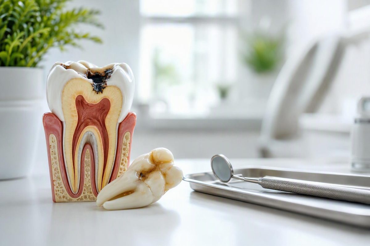
Decay in a wisdom tooth does not automatically require extraction. A fully erupted, useful tooth with accessible, limited decay may be repairable, while removal becomes more likely when the tooth cannot be restored or cleaned predictably, is impacted, repeatedly becomes infected or is damaging the neighboring molar.
The decay process is the same as elsewhere in the mouth: plaque bacteria use sugars and starches to produce acids, causing the tooth to lose minerals. The difference is the location. A third molar that is tilted, partly covered by gum or pressed against the second molar can trap food and be difficult to inspect, brush, floss, isolate and restore.
Choose the findings closest to your situation; the tool shows the appropriate next step and the factors that affect treatment.
Wisdom-Tooth Decay Decision Guide
Use dental findings if you have them. Choose “Unknown” rather than guessing about decay depth or tooth position.
No urgent symptoms are selected, but unknown decay depth and access mean a filling, monitoring or extraction cannot be chosen reliably. The wisdom tooth and second molar both need examination.
1. Current Symptoms
2. Decay Finding
3. Position and Access
Sources: NIDCR tooth-decay process; NHS wisdom-tooth removal and dental abscess guidance; ADA and NICE wisdom-tooth guidance. This guide does not determine restorability or diagnose infection.
The tool cannot establish how deep the decay is or whether the tooth is restorable. Those questions require a dental examination and, when appropriate, X-rays. If the tooth is partly erupted or angled against the second molar, the condition of both teeth matters.
Decay Can Be Present Without Obvious Pain
Possible signs of decay in a wisdom tooth include sensitivity to cold or sweets, pain when chewing, food repeatedly catching at the back of the mouth, or a visible hole or dark area. None of these signs establishes the depth of the damage. Early decay often causes no symptoms, while a deeper lesion may be hidden from view.
The cavity can occur on the chewing surface, on an exposed side of the wisdom tooth or at the contact with the second molar. A lesion at that contact is particularly difficult to see in a mirror. As with other decay between teeth, it can be difficult to detect without a proper examination.
Pain and swelling around a partly erupted wisdom tooth may instead come from pericoronitis, inflammation or infection in the gum around the tooth. A flap of gum can remain over part of the crown when the tooth lacks room to erupt completely. Decay and pericoronitis can occur at the same time, so the presence of inflamed gum does not rule out a cavity.
The NHS lists both gum infection and tooth decay among the problems associated with wisdom teeth that lack room to erupt fully. It also notes that a dentist may use X-rays to assess the position of the teeth (NHS). Symptoms and a mirror inspection therefore cannot reliably distinguish gum inflammation, decay in the wisdom tooth and damage to the tooth in front.
The Second Molar Needs Its Own Examination
A tilted lower wisdom tooth can create a difficult-to-clean contact against the back surface of the second molar. Food and plaque may collect at this contact even when the visible chewing surface of the wisdom tooth looks intact. The second molar is an important chewing tooth, so hidden damage there can have greater consequences than the appearance of the wisdom tooth alone suggests.
A systematic review and meta-analysis found that decay on the back surface of lower second molars was more common when lower wisdom teeth were horizontal or angled forward. Most included studies were cross-sectional, and the authors noted limitations in the imaging used. The finding shows an association; it does not predict that a particular angled wisdom tooth will cause decay (Journal of International Society of Preventive & Community Dentistry).
The examination therefore should not stop after finding a cavity in the third molar. The dentist also needs to assess the adjacent surface of the second molar and the gum and bone around the contact. A treatment plan can differ when both teeth are affected.
Removing the wisdom tooth does not repair decay already present in the second molar. That tooth needs its own assessment and, where appropriate, treatment. Conversely, treating the second molar does not make an inaccessible or repeatedly infected wisdom tooth easier to maintain.
An Intact Early Lesion May Be Arrested
If mineral loss is still confined to an intact enamel surface, the process may be arrested or partly reversed. Fluoride, saliva and fewer repeated acid attacks can help replace lost minerals. This is treatment of an active disease process, not regrowth of a missing piece of tooth.
Once the surface has broken down into a cavity, the missing tooth structure does not grow back. If the tooth is to be retained, a formed hole generally requires restorative treatment rather than home care alone (NIDCR).
The boundary between intact mineral loss and a cavitated surface cannot be judged reliably from pain, color or a photograph. A dark mark is not a measurement of lesion depth, and the absence of pain does not establish that the enamel is intact. It is the same distinction that applies to molar decay at different stages.
A dentist who identifies a non-cavitated lesion may recommend measures intended to arrest it and continued monitoring. Whether that is practical also depends on access. A surface that remains covered by gum or pressed tightly against another tooth may be harder to inspect and keep plaque-free than the same type of lesion on a fully erupted, upright tooth.
Restorability and Access Drive the Treatment Choice
The choice is not simply between filling every cavity and extracting every decayed wisdom tooth. The dentist weighs the amount of sound tooth, the position of the third molar, its usefulness, the possibility of maintaining it and the effects on nearby structures.
| Finding | Keeping May Be Reasonable | Removal Becomes More Likely |
|---|---|---|
| Eruption | Fully erupted and accessible | Impacted or partly under gum |
| Decay | Limited and restorable | Extensive or unrestorable |
| Maintenance | Can be cleaned reliably | Food and plaque repeatedly collect |
| Other teeth | Second molar remains healthy | Neighboring molar is being damaged |
A fully erupted tooth is generally easier to examine, isolate and restore than one partly under gum. Isolation matters because the dentist must be able to reach the damaged area and place a predictable restoration. A cavity on an accessible chewing surface presents a different problem from decay hidden low on the side of a tilted tooth.
The amount and location of sound tooth also matter. Limited cavitated decay may leave enough structure for restoration. Extensive damage, or damage in an area that cannot be reached and maintained predictably, supports removal. The draft evidence does not provide a universal cavity size or depth at which that decision changes; it must be judged for the individual tooth.
Function is another part of the assessment. A retained wisdom tooth may contribute to chewing when it is upright and meets an opposing tooth. A tooth with little functional role may offer less benefit when keeping it would require difficult treatment and continued monitoring.
The health of the neighboring second molar can change the balance. Decay, resorption or gum damage around that tooth may make continued contact with an angled wisdom tooth undesirable. The second molar still requires a separate plan for any damage already present.
A Filling Makes Sense Only When It Can Be Maintained
A filling is most plausible when the wisdom tooth has erupted fully, the damaged area is accessible, enough sound tooth remains and the person can clean around it reliably. The tooth should also have a useful or maintainable position rather than remaining trapped beneath gum or tightly angled against its neighbor.
Restoring the cavity repairs missing structure, but it does not straighten the tooth or remove an overlying gum flap. If the original position continues to trap food or prevent effective cleaning, decay can remain a maintenance concern around the restoration or on another surface.
A filling decision therefore includes what happens after treatment. The relevant question is not only whether material can be placed in the cavity, but whether the restored tooth can be inspected and kept clean over time. If access is poor, technically placing a restoration may not make retention predictable.
The American Dental Association lists decay as a reason removal may be considered when restoring the wisdom tooth is not possible or desirable. It also says wisdom teeth that are retained should continue to be monitored (ADA MouthHealthy). That wording does not treat decay by itself as an automatic extraction decision.
Extraction Is More Likely When Retention Is Unpredictable
Removal becomes more likely when decay is unrestorable, the tooth is impacted or partly erupted, infection repeatedly occurs, cleaning remains unreliable or the tooth is harming the second molar. Several unfavorable factors can occur together—for example, a forward-angled tooth may be difficult to restore while also creating an inaccessible contact with the neighboring molar.
NICE guidance says pathology-free impacted wisdom teeth should not be removed routinely, while unrestorable decay is an accepted indication for surgery (NICE). This distinguishes an impacted tooth without disease from one with damage that cannot be predictably repaired. It does not establish that every impacted or decayed wisdom tooth should be removed.
The difficulty and risks of surgery also belong in the decision. Possible complications include dry socket, infection and nerve injury. Nearby teeth, the jawbone or sinuses can also be damaged (Mayo Clinic). The relevance of each risk depends on the tooth’s position and surrounding anatomy, which is why the extraction assessment is individual.
Neither “remove every wisdom tooth” nor “always fill it” is a sound universal rule. Retaining a tooth avoids surgery but requires that the decay be treatable and the tooth maintainable. Extraction removes the problematic wisdom tooth but carries surgical risks and does not reverse damage already caused to the second molar.
Monitoring Is Active Follow-Up, Not Ignoring the Tooth
A wisdom tooth that is retained after early decay, a filling or an assessment without treatment still needs review. Its position can make changes difficult to see, and the contact with the second molar deserves attention. Monitoring is particularly relevant when a dentist has judged that immediate extraction is not warranted but access remains less than ideal.
At an appointment, the useful points to clarify are whether the surface is intact or cavitated, whether the lesion can be reached for restoration, whether the wisdom tooth is functional, and whether the second molar has decay or other damage. If extraction is proposed, the reason should be tied to findings such as unrestorable decay, repeated infection, poor access or harm to the neighboring tooth rather than the presence of a wisdom tooth alone.
If a filling is proposed, the maintenance question remains central: how the area can be cleaned and monitored after restoration. If observation is proposed, the plan should identify what is being watched—the decay, eruption position, gum around the tooth, second molar or a combination of these.
Care While Waiting for an Assessment
Brush twice daily with fluoride toothpaste and pay deliberate attention to the back tooth. Spit after brushing rather than rinsing immediately. If the space between the wisdom tooth and second molar is safely accessible, clean between them without forcing floss or sharp objects beneath a gum flap.
Reducing frequent sugary or starchy snacks and drinks lowers the number of acid attacks on the teeth. These steps can support an intact early lesion and reduce plaque accumulation, but they cannot rebuild a formed hole. Temporary improvement in discomfort also does not show that cavitated decay has healed.
Food catching repeatedly at the same contact, new sensitivity, pain on chewing or swelling around a partly erupted tooth are reasons to arrange an examination rather than relying on home cleaning. Because the source of symptoms may be the wisdom tooth, surrounding gum or second molar, treatment should not be chosen from symptoms alone.
Swelling or Systemic Symptoms Need Faster Care
Seek urgent dental care for severe or worsening pain, facial or jaw swelling, fever, pus, or a bad taste accompanied by feeling unwell. These findings require assessment for infection rather than waiting to see whether routine cleaning settles the problem.
Difficulty breathing, speaking or swallowing, major swelling in the mouth, swelling or pain around an eye, or marked difficulty opening the mouth requires emergency medical help (NHS dental abscess guidance). These warning signs take priority over deciding whether the wisdom tooth eventually receives a filling or is removed.