
How to Recognize Concerning Changes in Your Gums
A new red, pale, white or dark patch is more informative than gum shade alone, especially with swelling, soreness, bleeding, recession or altered texture.
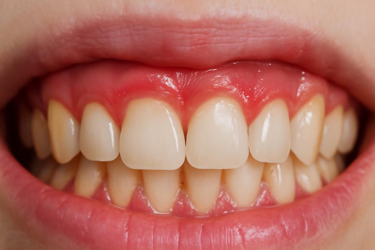
The short answer: a visual checklist of unhealthy gums
Potentially unhealthy gums may look swollen, puffy, smooth, glossy, shiny, red, reddish-purple, or darker than their usual color. They may bleed repeatedly during brushing or flossing. Other warning signs include gums pulling away from the teeth, exposed roots, longer-looking teeth, visible spaces at the gumline, pus, and teeth that have become loose or shifted. These findings are recognized signs of periodontal disease, but no single sign confirms a diagnosis. NIDCR describes swollen, red, bleeding or receding gums, persistent bad breath, sensitive or loose teeth, and painful chewing as possible signs.
These are warning signs, not a visual diagnosis. A mirror or photograph can help you notice a change, but neither can measure periodontal pockets, show supporting bone loss, or establish whether you have gingivitis or periodontitis.
A useful first check is to compare what you see with your own normal appearance:
| Generally healthy-looking gums | Common early warning signs | Potentially advanced warning signs |
|---|---|---|
| Firm and resilient | Puffy or enlarged around tooth margins | Marked recession or exposed roots |
| Closely fitted around each tooth | Smoother, stretched, or shinier than usual | Visible separation between a tooth and gum |
| Not visibly swollen | Red, reddish-purple, or darker than usual | Pus at or near the gumline |
| Not prone to recurrent bleeding | Bleeding that keeps returning during oral care | New gaps or noticeable tooth movement |
| Stable natural pigmentation | Possible tenderness or soreness | Loose teeth or a marked bite change |
You do not need to have every sign in the table. Early gingivitis commonly causes red, swollen or bleeding gums and may produce little or no pain. More advanced periodontal disease can involve recession, pus, loose teeth and changes in how the teeth fit together. Cleveland Clinic outlines this progression while emphasizing that professional examination, pocket measurements and dental X-rays may be needed to determine severity.
The visual distinction is not simply “pink means healthy and red means unhealthy.” Natural gum pigmentation varies. More useful questions include:
- Is the tissue puffier, smoother or shinier than before?
- Has its color changed from your usual shade?
- Does the gum still fit closely around each tooth?
- Does bleeding keep returning?
- Does a tooth look longer because the gumline has moved?
- Is there pus, an emerging gap or tooth movement?
Puffiness and easy bleeding commonly accompany early inflammation. Exposed roots, pus, shifting teeth and marked bite changes are more concerning because they may reflect changes beyond the visible gum surface. Appearance alone, however, cannot establish a disease stage or predict how a condition will progress.
Healthy gums are defined by more than color
Generally healthy gums are firm, resilient, nonswollen and closely fitted around the teeth. They should not look soft, enlarged or stretched away from a tooth, and they are not ordinarily prone to recurrent bleeding during routine brushing or flossing. Aspen Dental’s overview similarly describes healthy gums as firm, snug around the teeth and free from easy bleeding or swelling.
Color is only one part of that assessment. Healthy pigmentation can include shades of pink, brown, black or purple. A naturally dark area that has remained stable may be normal pigmentation rather than a sign of disease. What matters more is whether the color, texture or contour has changed.
Instead of asking only, “Are my gums pink?” consider:
- Firmness: Does the tissue look firm or soft and puffy?
- Swelling: Is the margin around one or more teeth enlarged?
- Surface: Has the gum become unusually smooth, taut or glossy?
- Fit: Does the tissue closely follow the tooth, or is there visible separation?
- Bleeding: Does bleeding repeatedly occur during ordinary oral care?
- Tenderness: Is there new soreness when brushing, eating or touching the area?
- Gumline position: Has more of a tooth or its root become visible?
A new red, pale, white, dark or otherwise changed patch is more informative than an absolute color label, particularly when it occurs with swelling, soreness, bleeding, recession or altered texture. Color alone should not be used to assign a cause: several conditions, local irritation and normal pigmentation can produce superficially similar appearances.
Natural gumline shape also varies. One person may have a more curved or uneven gumline than another, and some teeth naturally look longer. The relevant question is whether the contour has changed. Older photographs taken in clear, similar lighting may help reveal a difference, but they cannot identify its cause or prove that deeper supporting tissues are healthy.
Healthy-looking gums are therefore best recognized through a combination of stable pigmentation, firmness, close fit, lack of swelling and absence of recurrent bleeding.
Early warning signs: puffiness, redness, shine, and bleeding
Early gum inflammation often appears around the edges of the teeth. The gum margin may look thicker or rounder instead of closely following the tooth. Tissue between adjacent teeth can become enlarged, while the surface may appear smoother, stretched or glossier than usual.
Redness should be judged relative to the person’s baseline. Naturally pink gums may become red or deep red. Naturally brown, black or purple gums may look redder, darker or unevenly inflamed. Swelling and texture changes are often more useful than color alone when distinguishing possible inflammation from stable pigmentation.
Gingivitis commonly involves red or swollen gums that bleed easily during brushing or flossing. It may cause little or no pain, so the absence of soreness is not reliable reassurance. Cleveland Clinic identifies red, puffy and bleeding gums as typical signs of gingivitis and notes that painful symptoms may be absent early on.
Bleeding needs context. One light episode does not prove gum disease. Local irritation, brushing too forcefully or restarting flossing after a break can sometimes provoke bleeding. More concerning patterns include bleeding that:
- Returns repeatedly during brushing or flossing
- Persists rather than remaining an isolated event
- Occurs without brushing or flossing
- Appears with swelling, redness, recession or tenderness
- Comes from the same localized area again and again
There is no reliable mirror-based rule for how long bleeding may safely be ignored. If it persists or keeps returning, arrange a dental assessment rather than trying to compensate by brushing harder. Tenderness may accompany inflammation, but it is not required; equally, discomfort does not prove that gum disease is the cause.
Use good lighting when checking the gumline, and compare neighboring teeth as well as the right and left sides of the mouth. A localized shiny margin, ring of puffiness or enlarged area between teeth may be easier to recognize through comparison. The purpose of this check is to describe a change—not to determine its cause or stage.
Receding gums, pockets, and longer-looking teeth
Gum recession means that the edge of the gum has moved away from its former position, exposing more of the tooth and sometimes its root. The tooth has not grown; it looks longer because less of it is covered.
Possible visual clues include:
- One tooth appearing longer than neighboring teeth
- A visible notch or change in the gumline
- Part of the root becoming exposed
- A gumline that has moved when compared with an older photograph
- A developing space between the gum and tooth
Not every uneven gumline or naturally long-looking tooth indicates disease. The important finding is a new, persistent or progressing change. Recession may affect one tooth or several, and the amount visible in a mirror does not necessarily show what is happening beneath the gumline.
A periodontal pocket is a space between the tooth and gum where the tissue no longer fits as closely as expected. You might notice visible separation, but you cannot reliably determine the pocket’s depth by looking at it. Dental professionals measure the space with a periodontal probe. According to NIDCR, healthy gum pockets are usually 1 to 3 millimeters deep; deeper measurements may indicate periodontal disease when interpreted with the rest of the examination. NIDCR explains how pocket measurements and dental X-rays are used during diagnosis.
An exposed root may be accompanied by sensitivity to temperature, touch or certain foods. Sensitivity is not a visible sign, however, and it can have causes unrelated to recession or periodontal disease. It should be described as one part of the overall pattern rather than treated as proof of a diagnosis.
Arrange a dental assessment if recession appears to be progressing, a root has become exposed, or an apparent space has developed between a tooth and the gum. A dental professional can compare gumline levels, measure pockets and evaluate whether inflammation or supporting-tissue changes are present.
Signs that may accompany more advanced periodontal disease
Some findings are more concerning than mild redness or puffiness because they may indicate that the relationship between the gums, teeth and supporting structures has changed:
- Pus at or near the gumline
- Marked recession
- Clearly exposed roots
- Loose or noticeably shifting teeth
- New or widening spaces between teeth
- A change in how the upper and lower teeth meet
- A tooth that has moved from its former position
These are not routine early-gingivitis signs. Periodontitis affects the tissues and bone supporting the teeth. As that support is lost, teeth may eventually move, loosen or be lost. Pus, mobility and bite changes therefore warrant prompt dental contact rather than waiting for an unspecified future checkup.
Bone loss itself cannot be evaluated reliably in a bathroom mirror. Gum tissue may obscure changes around the roots, and a surface photograph cannot show the condition of the supporting bone. Dental X-rays may be needed as part of a professional evaluation.
Visible movement also cannot reveal the exact amount of support lost or establish a disease stage. Teeth may move for more than one reason. A professional examination is needed to interpret mobility, gaps, recession or bite changes together.
These warning signs do not make severe progression inevitable. Red or bleeding gums do not mean that tooth loss will occur, and not everyone with periodontal disease displays every advanced sign. The reason to act is that support already lost through periodontitis generally is not fully restored, even though professional care can manage active disease and help limit further progression.
Gingivitis versus periodontitis: what appearance can and cannot tell you
Gingivitis is inflammation limited to the gums. It commonly causes redness or darkening, swelling, puffiness and bleeding. Supporting bone has not yet been lost at this stage, and gingivitis is often reversible with appropriate professional care and effective oral hygiene.
Periodontitis affects deeper tissues and bone supporting the teeth. It may involve periodontal pockets, recession, pus, loss of attachment, bone loss, tooth movement, bite changes and eventually tooth loss. Professional care can address active disease and help limit progression, but structural support already lost generally is not fully reversible.
Appearance cannot reliably distinguish the two because their visible signs overlap:
- Redness does not prove that the condition is gingivitis.
- Bleeding does not show whether supporting bone has been affected.
- Recession does not reveal its cause or the precise disease stage.
- Lack of pain does not rule out early or more advanced disease.
- A photograph cannot measure pocket depth or show bone loss.
Plaque can contribute to inflammation along the gumline. If it remains, it can harden into tartar. Once established, tartar cannot be removed by ordinary brushing or flossing and requires professional removal. This is one reason better home care alone cannot be assumed to resolve every case of inflamed gums. The distinctions among gingivitis, periodontitis, tartar and supporting-bone loss are summarized in Aspen Dental’s comparison of healthy gums, unhealthy gums and gingivitis.
Home oral hygiene remains important, but it cannot measure pockets, determine whether tartar extends below the gumline or restore supporting structures already lost. Persistent bleeding, pockets, recession, pus or mobility require professional evaluation rather than self-diagnosis from a photograph, checklist or pain score.
Other symptoms that can accompany unhealthy-looking gums
Not every symptom associated with gum problems is visible. Alongside a change in appearance, a person may notice:
- Tenderness or soreness
- Bleeding during brushing or flossing
- Sensitivity around an exposed area
- Persistent bad breath
- An unpleasant or lingering taste
- Discomfort while chewing
- A feeling that a tooth is moving
- A change in how the teeth meet
These symptoms can accompany periodontal disease, but they are not specific to it. Bad breath, sensitivity, taste changes and pain can have several possible causes and should not be used alone to diagnose a gum condition.
Early gum disease may produce little or no pain. Painless swelling or recurrent bleeding can still warrant attention, while painful gums do not automatically indicate periodontitis or advanced disease.
A single localized swollen, sore, discolored or bleeding area may reflect irritation or another local problem rather than widespread gum disease. If it persists, worsens, repeatedly bleeds, develops pus or occurs with a changing gumline, have it assessed.
When describing the problem to a dentist, note:
- Where the change is located
- Whether it is localized or widespread
- When you first noticed it
- Whether it is changing
- What triggers bleeding
- Whether there is tenderness, sensitivity, odor or an unpleasant taste
- Whether a tooth feels mobile or the bite feels different
A clear description helps separate what you can observe from the clinical findings a dental professional must evaluate.
How dentists evaluate gum changes—and when to make an appointment
Arrange a dental assessment for gum changes that persist or keep returning, including:
- Recurrent bleeding during brushing or flossing
- Ongoing swelling, redness or tenderness
- Recession or newly exposed roots
- A persistent localized change
- Bad breath occurring alongside gum changes
- An apparent space between a tooth and gum
- A tooth that looks increasingly long because the gumline has moved
Contact a dentist promptly for pus at the gumline, a loose or noticeably shifting tooth, or a marked change in the bite. These findings may involve more than surface inflammation and should not be postponed on the assumption that brushing will resolve them.
During an examination, a dentist or dental hygienist may assess:
- The location and extent of inflammation
- Swelling and bleeding
- Gum recession
- Pocket depth around individual teeth
- Tooth mobility
- Changes in tooth position
- The way the teeth fit together
- Plaque and tartar above or below the gumline
Dental X-rays may be used to evaluate supporting bone that cannot be judged from the visible gum surface. A photograph can document how the tissue looked at a particular time, but it cannot rule out deep pockets, tartar below the gumline or bone loss. Established tartar requires professional removal.
Before an appointment, a simple monitoring framework may help:
- Compare with your baseline. Look for a genuine change in color, contour, firmness or gumline position.
- Track the bleeding pattern. Note whether it was isolated, keeps returning or occurs without oral care.
- Watch for structural change. Pay attention to exposed roots, widening spaces, recession or movement.
- Record associated symptoms. Mention tenderness, sensitivity, bad breath, unpleasant taste or chewing discomfort.
- Describe rather than diagnose. Report what changed and when instead of trying to assign a disease stage.
Dated photographs taken under similar lighting may help document progression, but they should supplement—not replace—an examination.
Decay Guide provides general reference information and is not a dental practice, diagnosis service or source of personalized dental advice, as explained in its publication scope and disclaimer. An individual gum change must be assessed by a qualified dental professional.
Frequently asked questions
Can dark brown, black, or purple gums be healthy?
Yes. Dark brown, black or purple pigmentation can be healthy when it is natural and stable and the tissue is firm, nonswollen, closely fitted around the teeth and not prone to recurrent bleeding. Healthy gum color can include pink, brown, black and purple shades, as described in this periodontal practice’s overview of normal gum-color variation.
What matters more is a new change from your baseline, especially when it is localized or accompanied by swelling, soreness, bleeding, recession or altered texture. Color alone cannot identify the cause.
Are bleeding gums always a sign of gum disease?
No. One light episode may follow local irritation, forceful brushing or the resumption of flossing after a break. Bleeding is more concerning when it persists, keeps returning, occurs without oral care or appears with swelling, recession, tenderness or another gumline change.
Bleeding alone cannot diagnose gum disease, but repeated bleeding should not be accepted as normal.
Can gum disease be present without pain?
Yes. Gingivitis and other periodontal changes can be painless, particularly during their early stages. The absence of soreness is therefore not reliable reassurance. Persistent swelling, bleeding, altered texture, recession or changes in how the gums fit around the teeth still deserve assessment.
Can unhealthy gums return to normal?
It depends on the cause and the structures affected. Gingivitis is often reversible with appropriate professional care and effective oral hygiene. Periodontitis involves deeper supporting tissues; active disease can be treated and managed, but bone and attachment already lost generally are not fully restored.
Home care remains important, but it cannot remove established tartar or reverse every cause of swelling, bleeding, recession or mobility. The likely outcome cannot be determined from appearance alone.
Can a photograph tell whether I have gingivitis or periodontitis?
No. A photograph may document redness, swelling, recession, exposed roots or a visible space at the gumline, but it cannot accurately measure periodontal pockets, detect tartar below the gumline or show supporting bone loss.
It can be useful for tracking a visible change over time, particularly when photographs are taken under similar lighting and from a similar angle. Diagnosis and staging still require a professional examination and, when appropriate, dental X-rays.
The practical takeaway
Healthy-looking gums are generally firm, nonswollen, closely fitted around the teeth and not prone to regular bleeding, regardless of their natural pigmentation. Puffiness, shine, recurrent bleeding, recession, exposed roots, pus or tooth movement should be taken seriously, but none establishes a diagnosis from appearance alone.
Compare what you see with your normal baseline and arrange a dental assessment for changes that persist or recur. Contact a dentist promptly for pus, loose or shifting teeth, or a marked change in how your teeth fit together.