
How to Recognize a Gumline That May Be Pulling Back
A longer-looking tooth is most informative when it represents a new change and appears with a shifted gum edge or newly exposed root.
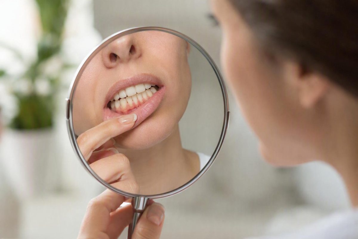
The short answer: three visual clues to gum recession
Receding gums usually look as though the edge of the gum has moved away from the visible crown of a tooth. That shift exposes more tooth surface and sometimes part of the root that would normally be covered. The three clearest visual clues are:
- A tooth that looks longer than it used to
- A visible yellowish or darker root surface near the gumline
- A new dip, notch, or uneven edge in the gumline
The tooth itself has not grown. It looks longer because less gum tissue covers its lower portion. Gum recession can affect one tooth or several and can range from a slight change to pronounced root exposure, according to Cleveland Clinic’s overview of gum recession.
The position of the gum edge matters more than color alone. Imagine tracing the gumline around each tooth. A potentially receding area may sit farther from the crown than it once did, leaving more of the tooth visible. Around one tooth, the change may resemble a shallow U- or V-shaped dip. Across several teeth, the margin may look irregular or stepped.
A line, groove, ridge, or notch near the gumline can support suspicion, but none proves that recession is present. A tooth may also have always had a different contour from its neighbors.
Recession can be mild and localized or involve a broader section of the gumline. One area may show only a slight dip, while another exposes a wider strip of root. For that reason, a photograph of “typical” recession is not a template for every mouth.
A mirror check can help you notice a possible change. It cannot confirm recession, identify the cause, show the condition of the supporting bone, or establish whether the gumline is progressing. Online image comparisons have the same limitations.
What an exposed tooth root may look and feel like
The visible portion of a tooth is commonly called the crown. The root extends below it and is normally covered by gum tissue. When the gum edge moves away from the crown, part of the root may become visible.
An exposed root may appear as a yellowish, cream, tan, or darker band at the base of a tooth. The contrast can be obvious when the crown is relatively bright, but it may also be subtle. Root exposure may follow the curved gum edge or appear beneath a localized dip. Some people notice a ridge where the enamel-covered crown meets the newly visible surface. Dental descriptions of recession include a differently colored area near the base of the tooth, an uneven margin, and grooves or notches around the gumline (visual indicators described by NEYO Dental).
Color alone is not diagnostic. Natural tooth shades vary, and lighting can exaggerate differences. A yellow, brown, dark, or rough-looking area near the gum may instead be:
- Surface staining
- Tartar attached to the tooth
- Tooth wear or a groove
- Tooth decay
- A shadow created by the shape of the tooth or gum
The most useful visual combination is position plus contrast. Likewise, a longer-looking tooth accompanied by a root-like band is more informative than either feature alone.
An exposed root may also be sensitive to cold, heat, sweets, brushing, flossing, or professional cleaning. Recession can nevertheless be present without discomfort, and sensitivity can have causes other than gum recession. Cleveland Clinic lists these forms of sensitivity among possible warning signs but advises professional examination and measurement to diagnose the condition (Cleveland Clinic’s symptoms and diagnosis guidance).
Rather than trying to identify the surface by touch, note what can be seen and felt during ordinary daily activities:
- Does the gum edge appear to have changed?
- Does the tooth look longer than before?
- Is a new band visible below the crown?
- Is the change limited to one tooth?
- Does cold, sweetness, brushing, or flossing trigger sensitivity in that area?
Those details may help a dentist evaluate the tooth, but they do not amount to a home diagnosis.
Subtle, obvious, and advanced-looking changes
There is no need to assign a home severity category based on millimeters. Clinical measurements must be taken from consistent anatomical reference points and interpreted alongside the rest of a periodontal examination. For visual recognition, descriptive categories are more useful.
A subtle-looking change
A subtle change may appear as:
- A slight shift in the gum edge
- One tooth beginning to look longer than a neighboring tooth
- A small localized dip around a canine or lower front tooth
- A thin yellowish or darker band at the tooth’s base
- Minimal visible root surface
- A faint line or ridge near the gumline
Early recession often develops gradually and may cause little or no pain. A slow change can be difficult to recognize because the eye adapts to it. It may first become noticeable during brushing, after sensitivity begins, or in a close-up photograph, but none of those circumstances establishes when the recession started.
An older personal photograph may help you ask whether the gum position has changed. It cannot provide a valid measurement.
A more obvious-looking change
A more obvious change may include:
- A clearly uneven or shifted gum margin
- A broader area of yellowish or darker root surface
- One tooth looking substantially longer than nearby teeth
- A distinct notch or groove near the gumline
- Increasingly visible space near the bases of adjacent teeth
- Food repeatedly collecting in newly noticeable spaces
- Several teeth showing reduced gum coverage
Color contrast can make root exposure look dramatic even when the gumline shift is less obvious.
Concerning or advanced-looking findings
Pronounced root exposure deserves professional assessment, particularly when it appears with food-trapping spaces, a loose or shifting tooth, tooth movement, or a bite that no longer comes together in the usual way. Loose or shifting teeth and bite changes may indicate a problem involving the tissues that support the teeth rather than the visible gum edge alone (advanced findings described by Las Colinas Dental Group).
Do not assume that recession by itself caused mobility or a changed bite. The stability of a tooth depends on structures that cannot be evaluated in a mirror. Periodontal disease, loss of support, trauma, and other dental problems may need to be considered.
Appearance cannot establish:
- Periodontal pocket depth
- The amount of attachment loss
- The condition of the supporting bone
- Whether the change is progressing
- Why the recession occurred
- Whether a dark or worn area is decay, tartar, stain, wear, or root surface
Pain is not a dependable severity scale. A small exposed area may be highly sensitive, while a more visible area may cause little discomfort. A modest-looking change can still deserve attention when it is new or worsening.
Healthy variation versus a gumline that has changed
There is no single healthy gum color that applies to everyone. Natural pigmentation, contour, and tissue appearance vary. Gum shape is also affected by tooth position, tooth shape, tissue thickness, and individual anatomy. Descriptions that treat light or “coral pink” gums as a universal standard are therefore misleading; pigmentation can be lighter, darker, mottled, or more pronounced (discussion of gum contour and natural pigmentation).
When looking for possible recession, focus on four questions:
- Has the position of the gum edge changed?
- Is more tooth or root surface visible than before?
- Has a new dip, notch, or irregular contour appeared?
- Does the difference appear to be increasing over time?
Gums without visible recession generally fit around the teeth without exposing the roots. The gumline is not normally ruler-straight, however. It follows the curved shape of each tooth, and one side of the mouth may not perfectly match the other.
A naturally uneven gumline is not automatically a receding one. Asymmetry becomes more informative when it is new, when a root surface has become visible, or when serial photographs or clinical records suggest that it is increasing.
Consider a canine with a V-shaped gum contour. If it has always looked that way and no root is visible, it may reflect natural anatomy. If the V-shaped area has recently deepened and now exposes a yellowish band, recession becomes a more plausible explanation—but it is still not confirmed from appearance alone.
Red, puffy, shiny, tender, or bleeding gums may indicate inflammation. Those features do not, by themselves, show that the gum edge has moved. Recession may occur with inflamed tissue, but it can also develop without redness, swelling, bleeding, or pain.
The reverse distinction matters as well. Bleeding or swelling without visible root exposure may reflect gingival inflammation or another gum problem rather than obvious recession. Persistent bleeding or swelling still deserves dental attention; it simply answers a different question from whether the gum margin has changed position.
If an older photograph shows the same tooth from a similar angle, it may provide a clue about past appearance. A dentist can establish a more dependable baseline by recording the current gum position.
When the baseline is uncertain, avoid forcing a conclusion from color or asymmetry. A dental examination is the appropriate way to distinguish normal variation from a clinically meaningful change.
Visible recession clues versus symptoms that can occur alongside it
The features commonly associated with receding gums fall into three groups. Keeping them separate helps prevent nonspecific symptoms from being mistaken for visual proof.
1. Visual clues most consistent with recession
The strongest visual clues are:
- A gum edge that appears to have shifted away from the crown
- A tooth that looks longer than before
- Newly visible root surface
- A localized dip or uneven gum margin
- A notch, line, groove, or ridge near the gumline
- Increasingly visible space near the bases of adjacent teeth
These clues relate directly to the position and shape of the gumline. They are more relevant to visual identification than redness, bleeding, or discomfort, although they still require professional confirmation.
2. Sensations that may accompany exposed roots
Possible accompanying sensations include:
- Sensitivity to cold or heat
- Sensitivity to sweet foods or drinks
- Discomfort during brushing or flossing
- Sensitivity during a dental cleaning
- Local tenderness near the gumline
Sensitivity does not prove that a root is exposed. It may occur with other dental problems, and recession can exist without sensitivity. The intensity of discomfort also does not reliably show how far the gumline has moved.
3. Nonspecific signs of inflammation or another problem
Signs that may occur alongside recession—but are not specific to it—include:
- Redness
- Swelling or puffiness
- Shiny-looking tissue
- Tenderness
- Bleeding during brushing or flossing
- Persistent bad breath or an unpleasant taste
These findings can indicate inflammation or another gum condition. They are not required for recession. A longer-looking tooth with a shifted gum edge may suggest recession even when the surrounding tissue does not look inflamed. Conversely, red or bleeding gums around a normally positioned gumline suggest a problem worth assessing but do not establish that recession is present.
Persistent swelling or bleeding merits dental attention even without visible root exposure. A mirror cannot rule out periodontal pockets or other changes around and below the gumline.
Can recession appear around only one tooth?
Yes. Gum recession can affect one tooth, several teeth, one side of the mouth, or a broader part of the gumline. It does not have to be symmetrical.
For example, one lower front tooth may begin to look longer than the teeth beside it. A narrow band of root surface may become visible at its base, or the gumline may develop a small isolated dip while the surrounding gums appear unchanged.
Another possible pattern is a localized U- or V-shaped area around a canine or premolar. The gum edge around that tooth may sit farther from the crown than the neighboring contours. That appearance can be consistent with recession, but its shape does not reveal why it occurred.
Possible contributors include:
- Forceful or traumatic brushing
- Periodontal disease
- Smoking or tobacco exposure
- Naturally thin gum tissue or inherited anatomical factors
- Clenching or grinding
- Tooth position or alignment
- Bite-related forces
Educational guidance from Cigna similarly notes that recession can involve one tooth, several teeth, or most of the teeth and lists brushing forces, smoking, grinding, gum disease, and family-related factors among possible contributors (Cigna Healthcare’s overview of receding gums).
An isolated area does not prove periodontal disease. Different causes—and sometimes several factors together—can produce a similar visible result.
Avoid assuming that one receding area must be caused by brushing too hard simply because the rest of the mouth looks normal. Likewise, several receding areas do not automatically prove poor oral hygiene. The pattern must be considered alongside the condition of the tissue, tooth position, brushing technique, tobacco exposure, grinding, pocket measurements, and other examination findings.
Whether one tooth or several look different, the practical response is the same: note the pattern and arrange an assessment if the change is new, progressive, sensitive, or otherwise concerning.
What a dentist can determine that a photograph cannot
A dentist can confirm gum recession by examining the position of the gumline and measuring the area with a periodontal probe. That provides a consistent clinical reference that a mirror, ruler, phone camera, or online photograph cannot reproduce.
An examination can document:
- Whether recession is present
- Which teeth and surfaces are affected
- The current amount and pattern of recession
- Whether the tissue appears inflamed
- Whether root exposure may explain localized sensitivity
- Whether tartar, decay, tooth wear, or another visible surface may explain the appearance
- A baseline for comparison at future visits
An initial examination describes the current condition; it does not necessarily establish whether recession is active or stable. Assessing progression generally requires comparison with earlier dental records, a reliable history, or measurements repeated over time.
A dentist may also measure periodontal pocket depths. Recession and pocket depth are related to periodontal assessment, but they are not the same measurement.
When appropriate, dental X-rays may be used to assess supporting bone and other structures hidden from view. Dental descriptions of recession assessment include visual examination, periodontal measurements, and X-rays where clinically indicated (Sisko Dentistry’s explanation of gum-recession assessment).
A photograph cannot determine:
- Periodontal pocket depth
- The amount of attachment loss
- Whether supporting bone has been affected
- Whether a tooth is mobile
- Whether a dark area is stain, tartar, decay, wear, or root surface
- Which contributing factor is most important
- Whether the change is progressing without a dependable earlier comparison
- Which management approach, if any, is appropriate
Arrange a dental assessment if you notice a newly visible root, a tooth that is progressively looking longer, a changing gumline, persistent sensitivity, or ongoing bleeding. These findings are not necessarily emergencies, but they should be evaluated rather than monitored indefinitely through online images.
Seek prompt dental assessment for rapid gumline change, a loose or shifting tooth, a changed bite, pronounced swelling, or significant pain. Mobility and bite changes can involve the tooth’s supporting structures or another problem that cannot be judged from the visible gum edge alone (guidance on loose teeth and early assessment).
Decay Guide is an independent information publisher, not a dental practice. This article provides general reference information and cannot diagnose an individual tooth, interpret personal photographs, or replace an examination by a qualified dental professional.
Can the gumline grow back after recession?
Lost gum tissue generally does not grow back naturally. Better plaque control may reduce inflammation, and changing a damaging habit may help limit further injury, but neither should be confused with restoring tissue that has already been lost.
The distinctions are important:
- Reducing inflammation may make swollen or bleeding gums look and feel healthier.
- Preventing further recession means addressing contributing factors and monitoring the gumline.
- Managing sensitivity may make an exposed area more comfortable.
- Professional root coverage may reconstruct part of the gumline in a suitable case.
- Natural regrowth would mean the lost tissue returns on its own.
These are not equivalent outcomes. Guidance on recession states that lost gum tissue does not regenerate naturally, although home care and professional management may help limit further change; selected professional procedures may be used to cover an exposed root (Colorado Periodontal Associates’ discussion of recession and treatment).
Improved brushing, deep cleaning, mouthwash, toothpaste, or home remedies should not be presented as ways to regrow a receded gumline. Cleaning may be important when plaque, tartar, or inflammation is present, but removing deposits does not replace missing tissue. Brushing with excessive force may be relevant when mechanical injury is contributing to recession.
Professional care depends on the suspected cause, the tissue condition, the current amount of recession, symptoms, tooth position, and the health of surrounding support. Some areas may be monitored after contributing factors are addressed. Others may require care for inflammation, sensitivity, decay, tooth wear, or related dental problems.
In selected circumstances, a dental professional may discuss a procedure intended to cover an exposed root or reconstruct part of the gumline. Professional root coverage is not spontaneous natural regrowth, and not every visible case needs surgery.
The practical message is not to select a treatment from a photograph. A changed gumline should first be examined and measured. Follow-up measurements or comparison with earlier records may then show whether it is stable or progressing.
Frequently asked questions
Do receding gums always make teeth look longer?
Not always in an obvious way. A tooth may look longer when the gum edge exposes more of it, but a small change can be difficult to see. The root may closely match the crown, the recession may be on a less visible surface, or the original gumline may already have been uneven.
A longer-looking tooth is most informative when it represents a new change and appears with a shifted gum edge or newly exposed root. Tooth shape, position, lighting, and natural asymmetry can also affect apparent length, so appearance alone cannot confirm recession.
Can gums recede without pain, sensitivity, or bleeding?
Yes. Recession can develop gradually without pain, sensitivity, bleeding, redness, or obvious swelling. Symptoms may occur when a root becomes exposed, but they are not required.
The absence of discomfort does not establish that a visible change is harmless. Likewise, pronounced sensitivity does not necessarily mean that recession is advanced. Gum position and clinical measurements—not pain intensity—are needed to characterize the area.
Is a yellow area near the gumline definitely an exposed root?
No. An exposed root may look more yellow or darker than the enamel-covered crown, but staining, tartar, decay, tooth wear, and lighting can create a similar appearance.
A newly shifted gum edge combined with a band of differently colored tooth surface raises suspicion of root exposure. Color or a photograph still cannot identify the surface conclusively.
Can gum recession affect only one tooth?
Yes. Recession can occur around one tooth even when the surrounding gumline appears unchanged. An isolated lower front tooth, canine, or another tooth may look longer or show a narrow strip of root.
A single affected area does not reveal the cause or prove periodontal disease. Brushing forces, thin tissue, tooth position, grinding, bite factors, smoking, periodontal disease, or a combination of factors may contribute.
Can receding gums grow back on their own?
Generally, no. Once gum tissue has been lost through recession, it does not ordinarily regenerate naturally. Better cleaning or professional care may control inflammation, address a contributing problem, reduce symptoms, or help prevent further change, but those outcomes are different from regrowth.
Some professional procedures may cover an exposed root or reconstruct the gumline in suitable cases. That does not mean every receding area requires surgery.
The characteristic visual pattern is a gum edge that has moved away from the crown, leaving a longer-looking tooth and sometimes a darker or yellower root surface. A new dip, uneven margin, groove, or notch can strengthen suspicion. Redness, bleeding, and sensitivity may occur alongside recession, but they are nonspecific and may be absent.
Because a mirror cannot show pocket depth, bone support, cause, or progression, a changing gumline should be examined and measured—especially when the change is rapid or accompanied by loose or shifting teeth, a changed bite, persistent bleeding, or significant discomfort.