
Is That Tooth Spot Discoloration or Decay?
Brown or black marks may represent external pigment, tartar, a stained groove, an old restoration, a crack or decay. Only a dental exam can determine the cause.
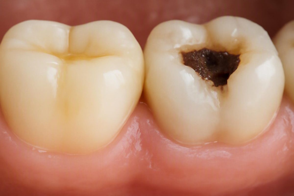
The short answer: how a stain differs from a cavity
A tooth stain primarily changes color, either on the surface or within the tooth. A cavity involves decay-related damage to tooth structure. The distinction sounds simple, but stains and cavities can look alike. White, brown, gray, or black coloring cannot establish the cause by itself.
At home, you can look for patterns that make staining or decay more plausible, but you cannot reliably diagnose a tooth from its color, texture, location, or symptoms. Early decay may be painless, while decay between teeth or beneath dental work may not be visible in a mirror. A medically reviewed stain-versus-cavity overview likewise notes that the two can be difficult to distinguish by appearance alone.
Here is a practical clue-not-diagnosis comparison:
| Feature | More consistent with staining | More concerning for decay | Important limitation |
|---|---|---|---|
| Surface integrity | Enamel appears flat and intact | Localized pit, opening, chipped-looking area, or missing structure | Early or hidden decay may have no visible hole |
| Texture | Appears smooth | Appears rough, irregular, or pitted | Do not scrape or probe the tooth to test it |
| Distribution | Broad discoloration or similar coloring on several teeth | Concentrated mark on one area of one tooth | Either pattern can have other causes |
| Symptoms | Often painless and not sensitive | Sensitivity, biting discomfort, or toothache | A painless spot may still be decay |
| Progression | May remain similar or change with pigment exposure | Appears to enlarge, deepen, darken, or catch food | Lighting and memory make home comparisons unreliable |
| Common locations | Exposed flat surfaces, deposits near the gumline, or several teeth | Molar grooves, between teeth, near the gumline, or around restorations | Location alone cannot confirm the cause |
| Response to cleaning | Superficial pigment may improve after professional cleaning | Structural damage remains | Failure to brush away a mark does not prove decay |
| Likely next step | Discuss cleaning or cosmetic options after identifying the cause | Dental examination and treatment based on the findings | Any persistent unexplained spot deserves assessment |
A smooth, intact, broadly distributed, painless discoloration is more consistent with staining than an isolated area of structural change. None of those reassuring features rules out decay.
Concern is greater when a mark appears rough or enlarging, has an apparent pit or opening, repeatedly catches food, or is accompanied by sensitivity, pain when biting, or an unexplained toothache. These are reasons to arrange an assessment, not proof that a cavity is present. Conversely, the absence of pain or a visible hole does not prove that the tooth is healthy.
Editorial disclosure: Decay Guide provides general education and cannot diagnose an individual tooth. It is written by a health writer rather than a dentist, and its articles are not clinician-reviewed. Its content does not replace a dental examination. Read more about how Decay Guide is written.
Why color cannot diagnose a tooth spot
The same color can arise from different processes. Brown or black areas may represent external pigment, tartar, a stained groove, an old restoration, a crack, trauma-related change, or decay. Early mineral loss may instead appear chalky white or off-white before an obvious surface opening develops. White spots can also have causes unrelated to active decay.
In practical terms:
- A dark spot is not automatically a cavity.
- A light spot is not automatically harmless.
- A gray tooth is not necessarily affected by surface staining.
- A brown groove cannot be diagnosed from color alone.
Distribution offers a tendency, not a rule. Pigment from coffee, tea, red wine, tobacco, or dark foods may affect broad surfaces or several teeth, while decay is often more localized. But pigment can collect in a single deep groove, and mineral or developmental changes can involve several teeth. A single spot should not be labeled decay solely because it is isolated, and widespread discoloration should not automatically be dismissed as cosmetic.
Location provides useful context. Decay can develop in the grooves of back teeth, between adjacent teeth, near the gumline, and around or beneath an existing filling or other restoration. Decay between teeth or under dental work may not be visible from the outside, and imaging may sometimes be considered after an examination when the suspected area cannot be assessed adequately from the visible surface. A dental-practice comparison of cavities and stains describes both symptom-free early decay and cavities hidden between teeth or beneath fillings.
Three common assumptions are especially unreliable:
- Myth: “Black means cavity.” Black can reflect decay, but it may also be pigment, tartar, a restoration-related shadow, or another change.
- Myth: “A cavity always looks like a hole.” Decay may begin beneath an apparently intact surface or in a place you cannot see.
- Myth: “No pain means no decay.” Early and hidden cavities may cause no pain or sensitivity.
Online photographs cannot resolve these uncertainties. Any photographs used in dental education should be treated as illustrative examples, not diagnostic references. Two similar-looking marks may have different causes, while the same condition may look different from one tooth to another.
Clues that favor staining—and clues that raise concern for decay
The most useful way to consider a stain vs cavity is through four lenses: surface and texture, symptoms, location and distribution, and change over time. Each supplies a clue; no single lens supplies a diagnosis.
Surface and texture
A flat or apparently smooth mark on intact enamel is more consistent with discoloration than with an established opening. That is particularly true when the coloring follows repeated exposure to coffee, tea, red wine, tobacco, dark sauces, or other pigment-producing substances.
A rough-looking area, localized pit, visible opening, chipped contour, or apparent loss of structure raises more concern. Food repeatedly lodging in the same place may also suggest a contour change, although food can collect for reasons other than decay.
Do not press, scrape, pick, or probe the area with a fingernail, toothpick, dental instrument, or another object. Do not bite hard food to test whether the tooth hurts.
Symptoms
Surface stains ordinarily affect appearance rather than sensation. Possible cavity clues include:
- Sensitivity to sweet foods or drinks
- Sensitivity to heat or cold
- Pain or discomfort when biting
- An unexplained toothache
- Localized tenderness or repeated food catching
These symptoms are not proof of decay.
Just as importantly, an early cavity may cause no pain or sensitivity. Possible cavity signs include pits, color changes, temperature or sweet sensitivity, toothache, and biting pain, but symptoms vary with the location and extent of decay. Aquafresh’s cavity-and-stain article also notes that early cavities may have no symptoms.
Location and distribution
Broad, fairly even pigmentation across several teeth favors an exposure-related stain. A localized mark in one molar groove, between two teeth, beside the gumline, or next to a restoration deserves closer attention.
These remain tendencies. A molar groove may simply be deeply pigmented, while a dark area near the gumline may be tartar rather than a cavity. Decay can also develop beneath the contact point between teeth without producing an obvious external mark.
When comparing teeth, look rather than manipulate. You may note whether similar coloring appears on neighboring teeth, whether the mark is close to a filling, or whether the tooth was previously injured. Those observations can help during a dental consultation, but they cannot determine the diagnosis or treatment.
Change over time
A mark that appears to enlarge, deepen, darken, become rough, begin catching food, or develop symptoms deserves assessment. However, lighting, wetness, viewing angle, and heightened attention can make a spot appear different. A photograph may help you describe what you noticed, but it is not a substitute for an examination.
Improvement after professional cleaning supports the explanation that at least some of the color was on the surface. Brushing and whitening should not be treated as diagnostic tests. Pigment may remain because it is within tooth structure, trapped in a groove, incorporated into tartar, or associated with a restoration. Staining and decay may also coexist.
A mark that remains after ordinary brushing could represent:
- Intrinsic discoloration
- Tartar
- A naturally or externally stained groove
- A shadow associated with an existing restoration
- Trauma-related change
- A crack or chip
- Early mineral loss
- Active or inactive decay
Failure to brush away the mark does not reveal which explanation applies. Early cavities may remain completely painless, so waiting for discomfort is not a reliable way to screen a tooth.
Not every tooth spot fits the stain-or-cavity binary
“Stain or cavity” is often the first question, but tooth color can change for several reasons. It helps to distinguish surface pigment, color arising within the tooth, hardened deposits, structural damage, and decay.
Extrinsic staining occurs on the outer surface. Common contributors include coffee, tea, red wine, tobacco, pigmented foods, plaque, and tartar. The appearance varies with the exposure and tooth surface. Surface pigment is generally more likely than intrinsic discoloration to improve with professional cleaning or whitening.
Intrinsic discoloration arises within tooth structure. It may be associated with trauma, medication exposure, fluorosis, aging, developmental changes, or processes occurring inside the tooth. Because the color is not simply sitting on the surface, it may not respond like an ordinary external stain.
Tartar is a hardened deposit that may appear yellow, brown, or black, especially near the gumline. It generally requires professional removal rather than harder brushing. Existing dental work can also affect appearance: an older silver-colored restoration may make nearby tooth structure appear gray or dark, while a chip or crack may expose darker material or create a shadow. A clinical overview of dark tooth spots identifies tartar, surface pigment, cracks, trauma, old silver restorations, and decay as possible explanations.
Patterned white or brown discoloration across multiple teeth is one possible presentation of fluorosis, but the pattern is not diagnostic. Multiple affected teeth can have other explanations.
One tooth becoming gray or markedly darker after an injury warrants dental assessment, even if it does not hurt. The change may arise within the tooth rather than from ordinary surface pigment, but color alone cannot establish the condition of the internal tissues.
Another possibility is arrested decay, meaning a lesion considered inactive or stable rather than currently progressing. Such a lesion may be dark and can resemble a stained groove. Professional assessment is needed to distinguish inactive decay from active decay, ordinary pigmentation, or another defect; a reader should not independently decide that a dark, painless mark is safe to monitor. These alternative explanations and their limits are described in a dentist-authored review of dark tooth spots.
Early mineral loss is not the same as an established hole
Tooth decay is better understood as a continuum than as a switch that suddenly changes from “no cavity” to “cavity.” The process may begin with mineral loss from enamel. If it continues, the surface can break down, structural damage can deepen, and inner tooth layers may become involved.
Early mineral loss may appear as a chalky white or off-white patch and may cause no pain. At this point, the surface can still be intact even though the enamel has changed.
Terminology can be confusing because consumer sources may use “early cavity” for several points along the continuum. Important distinctions include:
- Non-cavitated mineral loss: The enamel has lost minerals, but there is no established opening or missing structure.
- Cavitation: The surface has broken down and tooth structure has been lost.
- Active lesion: Findings suggest that the process is continuing.
- Arrested lesion: Findings suggest that the lesion is inactive or stable.
These distinctions can affect management. Fluoride may sometimes help restore minerals or arrest early enamel damage when the surface remains intact and care is guided by a dental professional. That does not mean every early lesion can be reversed. Once an actual hole or structural loss is present, fluoride, brushing, and whitening cannot rebuild the missing tooth; treatment depends on a professional assessment of the location and extent of damage. The distinction between early fluoride-responsive damage and established cavities is summarized in the Aquafresh cavity guide.
An article cannot determine whether a particular white, brown, or dark mark is non-cavitated, structurally cavitated, active, or arrested. Those terms should not be used for self-diagnosis.
How a dentist distinguishes discoloration from decay
A dental professional does not ordinarily rely on one visual rule. Assessment combines the history of the mark with its location, surface integrity, surrounding enamel, symptoms, and relationship to existing dental work.
Be ready to mention:
- When you first noticed the mark
- Whether it seems to have changed
- Whether the tooth was ever hit or injured
- Whether food catches in the area
- Any sensitivity, toothache, or biting discomfort
- Frequent coffee, tea, wine, tobacco, or other pigment exposure
- Existing fillings, crowns, bonding, or other restorations
- Recent whitening or changes in oral-care products
During the examination, the dentist may consider whether the surface appears intact, whether there is a pit or defect, how the area relates to the tooth’s natural anatomy, and whether nearby teeth or gums show related changes. Existing restorations may also be examined because discoloration can occur beside dental work and decay can develop at or beneath a restoration.
An X-ray is not automatically required for every dark or white mark. Imaging is selected by the dentist after the examination according to the suspected location and clinical need. It may contribute useful information when an area between teeth, beneath a restoration, or elsewhere cannot be assessed adequately from the visible surface. Professional assessment therefore uses history, location, surface condition, surrounding structures, dental work, and imaging when indicated rather than treating any one test as conclusive, as outlined in this overview of professional stain-versus-cavity evaluation.
Stained grooves, early lesions, restorations, and overlapping conditions can require clinical judgment or follow-up. Home inspection, online photographs, and symptom checklists cannot establish the condition of a particular tooth.
Treatment depends on whether the problem is pigment or structural damage
Treatment should follow identification of the cause. The response to removable pigment differs from treatment for hardened deposits, intrinsic discoloration, early mineral loss, or missing tooth structure.
For superficial stains, options may include professional cleaning or polishing, reducing the relevant exposure, or whitening. Not every color change responds, and cleaning may remove surface deposits without altering the tooth’s underlying shade.
For tartar, professional cleaning is generally needed. More forceful brushing will not substitute for professional removal, and the deposit should not be treated as something to scrape off at home.
For intrinsic discoloration, management depends on why the tooth changed color and whether it is otherwise healthy. Options discussed after examination may include professional whitening, bonding, veneers, or another approach. No single cosmetic treatment is suitable for every cause or every tooth.
For early non-cavitated mineral damage, a dentist may recommend fluoride and measures intended to control the conditions that allow decay to remain active. An established cavity generally requires restoration. Depending on depth, location, severity, and whether the tooth can be restored, treatment may include a filling, crown, root canal treatment, or extraction. A dental-practice treatment comparison outlines the difference between stain management and restorative treatment for structural decay.
Whitening changes color; it does not remove decay, repair a crack, remove tartar, or replace missing tooth structure. Do not start whitening solely to discover whether an unexplained spot is a stain, particularly if the tooth is already sensitive. Whitening products can cause tooth sensitivity or gum irritation, so professional advice is appropriate when the cause of a localized color change is uncertain.
What to do next: safe care and reasons to seek help
Arrange a dental examination for a mark that is:
- New or unexplained
- Persistent
- Changing, enlarging, or darkening
- Rough or associated with an apparent pit
- Catching food
- Sensitive to sweets, heat, or cold
- Painful during biting
- Associated with an unexplained toothache
These features do not prove that decay is present, but they justify assessment rather than waiting for the mark to hurt. Swelling around a tooth or gum also requires attention. Severe or escalating pain, or gum or facial swelling, warrants urgent dental assessment rather than waiting for a routine cleaning. Pain, swelling, a visible opening, or an enlarging or darkening mark are among the warning signs identified in guidance on dark tooth spots.
While waiting for an appointment:
- Continue gentle, regular brushing with fluoride toothpaste.
- Continue your normal interdental cleaning routine.
- Do not scrape, probe, press, or pick the mark with a fingernail, toothpick, or other object.
- Do not brush aggressively in an attempt to remove it.
- Do not begin whitening solely as a diagnostic test.
- Note when you first noticed the spot, any symptoms, and whether the tooth has been injured or restored.
General preventive habits overlap for staining and decay: brush regularly with fluoride toothpaste, clean between teeth, limit frequent sugary or acidic foods and drinks, drink or rinse with water after pigment-producing exposures, and obtain regular dental care.
The central decision rule is simple: color cannot settle the stain-versus-cavity question. Smoothness, distribution, symptoms, surface damage, and change over time are clues only. Continue normal fluoride brushing, avoid scraping or whitening an unexplained spot as a test, and seek professional assessment for a persistent, changing, rough, painful, or sensitive mark—or one associated with a pit, food catching, or swelling. Only an examination can determine whether the cause is staining, tartar, early mineral loss, active decay, inactive decay, or something else. Decay Guide is educational and cannot diagnose an individual tooth.
Frequently asked questions
Can a cavity be present without pain or a visible hole?
Yes. Early decay may cause no symptoms, while decay between teeth or beneath a restoration may not be visible in a mirror. The absence of pain does not rule out decay. A dentist may need to assess the visible surfaces and decide whether imaging would provide useful information for a hidden area. A cavity-and-stain comparison from Ashley Harrison, DDS describes both painless early decay and decay hidden between teeth or beneath fillings.
Can a tooth stain turn into a cavity?
A stain does not directly transform into a cavity. Staining is a color change, whereas decay is a process that damages tooth structure. The two can coexist, and some habits—such as frequently consuming certain sugary or acidic drinks—may contribute both to discoloration and to conditions associated with decay.
Will brushing remove a stain or cavity?
Ordinary brushing may reduce some recent surface pigment and helps control plaque, but many stains will not disappear through brushing alone. Tartar and intrinsic discoloration also remain after brushing for different reasons.
An established cavity cannot be brushed away because tooth structure has already been damaged or lost. A mark remaining after brushing is not automatically a cavity; it could be intrinsic color, tartar, a restoration-related shadow, a stained groove, trauma-related change, or decay.
Does every dark tooth spot require an X-ray?
No. Imaging is selected by the dentist according to the examination, suspected location, and clinical need. It may be useful when a possible problem lies between teeth, beneath a restoration, or in another area that cannot be assessed adequately from the visible surface. An X-ray contributes information but does not replace the rest of the examination.
Is it safe to whiten a tooth when I do not know what caused the spot?
Do not use whitening as a test for an unexplained spot. Whitening may alter some surface or internal discoloration, but it cannot treat decay, repair a crack, remove tartar, or replace missing tooth structure. Whitening products can also cause temporary sensitivity or gum irritation.
Have a new or unexplained localized mark assessed first, especially if the tooth is sensitive, painful, damaged, or noticeably different from neighboring teeth. A dentist can evaluate the likely cause and discuss whether whitening or another approach is appropriate.