
Is the Tooth Temporarily Injured or No Longer Vital?
A bruised tooth may retain living pulp and recover; a dead tooth cannot regain vitality. Pain and color alone cannot confirm either.
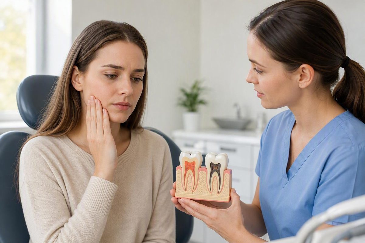
A “bruised tooth” may still contain living pulp and recover. A dead, or non-vital, tooth has lost pulp vitality and cannot become vital again. However, pain, tenderness, looseness, swelling, and discoloration can occur after several types of dental injury. A dentist needs to assess both the pulp and the tissues supporting the tooth (overview of bruised and non-vital teeth).
The difference at a glance
“Bruised tooth” is an informal term most often used for irritation or injury of the periodontal ligament—the supporting tissue between a tooth’s root and socket—after trauma or excessive bite pressure. “Dead tooth,” by contrast, describes pulp status: the tissue inside the tooth is non-vital.
| Feature | Bruised tooth | Dead or non-vital tooth |
|---|---|---|
| Primary tissue involved | Usually the periodontal ligament or nearby supporting tissue | Pulp inside the tooth |
| Pulp status | May remain alive and capable of recovery | No longer vital and cannot return to a vital state |
| Common causes | Impact, biting something hard, grinding, clenching, uneven bite pressure, or recent dental work | Trauma, deep decay, cracks, repeated dental procedures, or infection |
| Possible symptoms | Biting tenderness and localized soreness; broader trauma may also cause swelling, mobility, discoloration, or temperature symptoms | May be painless or painful if inflammation or infection affects tissues around the root; swelling, drainage, or mobility may occur |
| Temperature sensitivity | Can occur after trauma, particularly if the pulp is also affected | A fully non-vital pulp does not itself respond normally to temperature, but a person may report sensitivity from evolving pulp injury, another tooth, or a concurrent condition |
| Discoloration | Color change can follow trauma but may indicate injury beyond an isolated ligament strain | May become gray or dark, but color alone does not establish pulp death |
| Likely course | A limited ligament injury may settle if the cause is removed and the pulp remains healthy | Non-vital pulp does not recover; treatment depends on infection, surrounding tissues, and whether the tooth can be restored |
| Infection concern | An isolated ligament injury is not necessarily infected | Necrotic tissue may become infected or be associated with disease around the root |
| Professional assessment | History, examination, bite and mobility checks, pulp and root-area assessment, and sometimes X-rays | The same combined assessment, with treatment based on the professional pulpal and periapical diagnosis |
| Possible treatment | Monitoring, relieving excessive pressure, protecting the tooth, or splinting when indicated | Root canal treatment may be indicated if the tooth can be retained; extraction is considered only when the tooth’s condition warrants it |
The key distinction is therefore not whether the tooth hurts. It is whether the pulp remains vital.
What can be injured even when the tooth looks intact
The periodontal ligament connects the tooth’s root to its socket and helps cushion biting forces.
The pulp is different. It is the soft tissue inside the tooth containing blood vessels, nerves, and connective tissue. Trauma can damage it even when the enamel has no visible chip or crack, according to the American Association of Endodontists’ explanation of pulp injury.
Possible triggers for tenderness after dental injury include:
- A blow, fall, or sports injury
- Biting a hard food or object
- Grinding or clenching
- An uneven filling or other restoration
- Some dental procedures
These events do not establish that a painful tooth is merely “bruised.” A crack, decay, infection, gum disease, or another dental problem may produce similar symptoms. Even when the timing strongly suggests trauma or excessive pressure, the responsible tissues still need to be identified.
An isolated ligament strain is most closely associated with soreness or tenderness under pressure. Swelling, discoloration, or temperature symptoms warrant assessment for other injury too.
Why pain and tooth color cannot settle the question
Several findings can occur after either limited supporting-tissue trauma or a more complicated dental injury:
- Tenderness while biting
- Localized soreness
- Reported sensitivity to hot or cold
- Gum swelling
- Tooth mobility
- Pink, gray, or darker discoloration
A non-vital tooth may be painless. It may also hurt when inflammation or infection develops in tissues around the root. Pain therefore does not prove that the pulp is alive, while fading pain does not prove either recovery or pulp death.
Temperature symptoms require similar caution. Living but injured or severely inflamed pulp may respond to hot or cold. Trying hot or cold substances at home is not a dependable vitality test.
Gray or dark discoloration after trauma raises concern about internal injury, but it is not proof of necrosis. Pink discoloration is likewise not proof that the tooth is healing. Published dental-practice guidance describes both pink and gray changes as findings that warrant professional evaluation rather than interpretation by color alone (guidance on post-trauma tooth discoloration).
Pain, sensitivity, swelling, and discoloration can also occur with decay, cracks, gum problems, or infection. The overlap is why the dentist must assess the entire clinical picture rather than assign pulp status from one symptom.
Clue, not conclusion: Pain, lack of pain, temperature sensitivity, swelling, looseness, and visible color changes can guide a dental investigation. None can confirm pulp vitality at home.
How a dentist assesses the injury
Assessment may combine several types of information:
- History: What happened, when it happened, whether the bite changed, and how symptoms have developed.
- Clinical examination: Checking the tooth and gums for decay, fractures, displacement, swelling, drainage, or other damage.
- Bite and mobility checks: Determining whether the tooth contacts too heavily, has moved from its normal position, or is more mobile than expected.
- Pulp and root-area assessment: Evaluating the pulp and the tissues surrounding the root.
- Dental X-rays: Looking for fractures, displacement, infection, or changes around the root.
These findings are considered together. The resulting pulpal and periapical diagnoses help determine whether observation, pulp-preserving treatment, root canal treatment, or another approach is appropriate.
Follow-up can be particularly important after significant trauma because pulp status and other complications may evolve. Loose teeth, displaced teeth, and adult teeth that were knocked out and reinserted need continuing dental care, including assessment of vitality. Cambridge University Hospitals also notes that injured teeth may be splinted to hold them in position and limit excess movement while healing occurs (dental-injury aftercare guidance).
Follow-up is not evidence that the tooth is already dead or certain to need a root canal. It allows the dentist to identify changes that were not apparent at the first examination and adjust treatment if necessary.
Treatment depends on which tissues can recover
Treatment should follow the diagnosis rather than the tooth’s color or pain level.
If the pulp appears vital and the injury is limited: The dentist may monitor the tooth and address excessive pressure. Depending on the cause, this may involve protecting the tooth, temporarily avoiding force on it, or correcting a bite problem.
If the tooth is loose or displaced: It may need to be repositioned and splinted. A splint holds the tooth in position and reduces excess movement while supporting tissues heal.
If living pulp can be preserved: Selected injuries caused by trauma, decay, or restorative treatment may be managed with a vital-pulp procedure. The aim is to retain functioning living pulp. Suitability depends on the pulpal and periapical diagnoses, infection, associated fractures or displacement, and the tooth’s stage of development.
If the pulp is non-vital or infected: The dentist will assess whether root canal treatment can retain the tooth. Treatment choices require an examination; this symptom comparison cannot establish which procedure is appropriate.
During root canal treatment, inflamed or infected pulp is removed. The interior of the tooth is then cleaned, disinfected, filled, and sealed before the tooth receives an appropriate protective restoration. A fully developed tooth can continue functioning without pulp because surrounding tissues continue to nourish it.
If the tooth cannot or should not be retained: Extraction may be considered based on structural damage, infection, supporting tissues, restorability, and the broader treatment plan. It is not the automatic outcome for every painful, loose, or discolored tooth.
What to do while arranging care—and when it is an emergency
The appropriate response depends on whether symptoms are mild and stable, persistent or worsening, or accompanied by signs that infection may be spreading.
1. Conservative interim protection
- Choose soft foods.
- Avoid chewing on the affected tooth.
- Clean the area gently rather than stopping oral hygiene.
- Avoid sports and other activities that could cause another impact.
- If using an over-the-counter pain medicine, follow the product label and use it only when medically appropriate for you.
These measures may limit further irritation, but they cannot determine pulp vitality or treat non-vital or infected tissue.
2. Arrange prompt dental assessment
Contact a dentist promptly for:
- Persistent or worsening pain
- Increasing discoloration
- New or increasing looseness
- A changed bite
- Worsening tenderness
- Swelling
- Pus or other signs of an abscess
- An unpleasant taste
- Fever
Significant trauma also needs prompt assessment, especially if a tooth moved from its normal position or was knocked out and reinserted. Do not rely solely on home observation when symptoms persist or worsen.
3. Seek emergency medical assessment
Go to an emergency department or use your local emergency service if mouth or facial swelling:
- Makes breathing or swallowing difficult
- Extends toward the eye
- Prevents you from opening your mouth fully
These are warning signs of potentially spreading infection that require emergency assessment. NHS Wales lists the same features as reasons to attend emergency care for mouth or facial swelling (mouth and facial swelling guidance).
There is no dependable universal healing deadline for a “bruised tooth.” Recovery varies with injury severity and whether the periodontal ligament, pulp, tooth structure, or several tissues are involved. A limited injury with vital pulp may recover, while confirmed non-vital or infected pulp requires professional treatment planning.
Decay Guide provides general educational information only. It is not a dental practice, and this information is not a diagnosis or individualized treatment advice.