
What a Gum Bump Might Mean—and When You Should Get Help
It can drain, shrink and become less painful without proving that the underlying source of the problem has resolved.
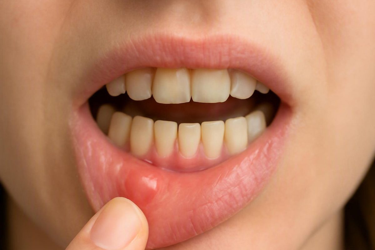
A bump on your gums cannot be diagnosed reliably from its color, firmness, location, pain level, or a photograph. Abscesses, cysts, irritation fibromas, canker sores, injuries, and other oral lesions can overlap in appearance. Use the bump’s symptoms and behavior to decide how quickly to seek care rather than trying to name it yourself.
First, decide how urgently the bump needs attention
Not every gum bump requires emergency medical care. The clearest distinction is between signs of a potentially spreading infection, symptoms that warrant prompt contact with a dentist, and an apparently minor irritation that is already improving.
Seek emergency medical care now if you have:
- Difficulty breathing or swallowing
- Rapidly worsening swelling of the face, jaw, or neck
- Increasing difficulty opening your mouth
- Swelling accompanied by fever, chills, or feeling seriously unwell
These symptoms may indicate that an oral infection is spreading or interfering with normal breathing or swallowing. Dental-practice emergency guidance identifies facial or jaw swelling, fever, chills, restricted mouth opening, and breathing or swallowing problems as escalation signs; breathing or swallowing difficulty should not be monitored at home (dental infection warning signs).
Contact a dentist promptly if the bump occurs with:
- Pus or drainage
- A foul or unusual taste
- Significant, throbbing, or worsening pain
- Warmth or swelling around one tooth
- Sensitivity to pressure, biting, hot, or cold
- Fever or swollen lymph nodes
- Pain spreading toward the jaw, ear, or neck
- A pimple-like bump beside a tooth
- A bump that drains, shrinks, and then returns
These features may indicate an infection, but no single symptom proves that the bump is an abscess. A dentist must examine the gums and nearby teeth to identify the source.
The supplied evidence does not establish a universal rule that every person with these symptoms must be seen within a particular number of hours. Contact the dental office, describe the symptoms clearly, and let the clinician determine the appropriate timing. Emergency warning signs override that process.
Arrange a dental examination if the bump:
- Persists without a clear explanation
- Is enlarging or changing
- Bleeds without an obvious recent injury
- Repeatedly returns
- Remains after an apparent irritant has been removed
- Is firm or painless but does not go away
- Occurs with a persistent red or white area
- Interferes with chewing
- Is concerning or difficult to interpret
A painless lesion is not automatically safe to ignore. Cysts, fibromas, and other lesions may initially produce little or no discomfort.
Brief monitoring may be reasonable when there is an obvious minor trigger—such as sharp food, forceful brushing, or rubbing from an appliance—and the area is already improving after the trigger stops. Stop monitoring and contact a dentist if the bump grows, drains, becomes more painful, returns, or occurs with fever or spreading swelling.
There is no diagnostic one-, two-, or three-week cutoff. One supplied dental-practice source recommends assessment after more than one week, while another uses persistence beyond two weeks as a reason to seek care (one-week dental-assessment threshold).
A commercial oral-care article advises contacting a dentist when a presumed ulcer has not healed within three weeks. Because the supplied sources conflict, roughly two weeks is best treated as a cautious examination point for an otherwise stable, unexplained lesion—not as permission to wait when symptoms are severe, infectious, or worsening (mouth-ulcer and abscess guidance).
This article is educational, not diagnostic. The clinical evidence supplied for it consists largely of commercial dental-practice and oral-care articles rather than formal clinical guidelines. Decay Guide also states that its material is written by a health writer, not a dentist, that no article has been clinician-reviewed, and that its content cannot replace an examination (Decay Guide’s editorial limitations).
What the bump’s features can—and cannot—tell you
Pain, drainage, texture, growth, and duration can help determine urgency. They cannot establish a diagnosis by themselves.
| Possible explanation | Commonly described features | Typical course | Appropriate next step |
|---|---|---|---|
| Dental or gum abscess | Raised swelling that may feel warm, tender, or soft; possible pus, drainage, bad taste, throbbing pain, tooth sensitivity, or nearby swelling | May worsen, drain temporarily, shrink, or recur; drainage does not prove that the infection has cleared | Contact a dentist promptly; seek emergency medical care for breathing or swallowing difficulty or rapidly spreading swelling |
| Canker sore | Usually a shallow, painful ulcer rather than a deep lump; may have a white, yellow, or gray center with a red border | Commonly improves and heals within approximately one to two weeks | Protect it from irritation; arrange an examination if it persists, grows, becomes unusually painful, or repeatedly returns |
| Local irritation or trauma | Tender, scraped, ulcerated, or swollen area near sharp food, forceful brushing, braces, dentures, or another appliance | May improve after the trigger is removed | Monitor briefly only if it is clearly improving; seek care if it persists or develops infection signs |
| Dental cyst | May produce a slow-growing, initially painless swelling; discomfort can develop if it enlarges or becomes infected | May remain unnoticed and does not have a dependable self-resolution pattern | Arrange a dental examination, particularly if the area persists or changes |
| Irritation fibroma | Often firm, smooth, and painless; may occur where tissue is repeatedly rubbed | May remain present rather than behaving like a short-lived sore | Have a persistent lump examined; do not assume that lack of pain makes it harmless |
| Gum disease-related inflammation | Broader redness, swelling, or bleeding; a localized infection may create a more distinct swelling | May persist or progress without treatment of the gum problem | Arrange a dental and gum assessment |
| Suspicious or unexplained lesion | Persistent growth, unexplained bleeding, change in color or texture, red or white patch, loose tooth, or difficulty chewing or swallowing | Persists, enlarges, changes, or remains unexplained | Seek prompt examination; referral or biopsy may be considered after assessment |
A canker sore is usually described as a surface defect: a shallow ulcer with a pale center and red rim. An abscess is more often described as a raised swelling, sometimes with warmth, redness, pus, throbbing pain, drainage, fever, swollen lymph nodes, or a bad taste. Canker sores commonly heal in 7 to 14 days, whereas an abscess is an infection requiring professional dental assessment (canker sore and abscess comparison).
The distinction is not foolproof. A sore can look raised because the surrounding tissue is swollen. An abscess can appear white or yellow where material lies close to the surface. Irritated tissue may be red and painful, while a draining infection may become less painful.
Descriptions such as “hard,” “soft,” “white,” “red,” “painful,” and “painless” are therefore not diagnoses:
- A white area might be the center of an ulcer, material beneath a swelling, or another tissue change.
-
Redness can accompany irritation, inflammation, infection, or repeated trauma.
-
Softness may accompany swelling or fluid but does not establish what the fluid is.
- Pain may arise from a sore, injury, inflammation, or infection.
- Lack of pain does not rule out a cyst, fibroma, chronic draining pathway, or another persistent lesion.
An abscess may rupture or drain, reducing pressure and pain. That improvement does not show that the source in a tooth, root, or periodontal tissue has healed. A repeatedly appearing “gum pimple” may act as an outlet from an unresolved problem underneath.
To make an appointment more useful, record:
- When you first noticed the bump
- Whether it is growing, shrinking, or changing
- Whether it has appeared before in the same place
- Whether it drains and whether the drainage has a bad taste
- Whether a nearby tooth is sensitive to temperature, pressure, or biting
- Whether you have fever, facial swelling, or swollen lymph nodes
- Any recent biting injury, sharp food, forceful brushing, or appliance rubbing
- Any recent toothache, cracked tooth, cavity, or gum problem
A dated photograph can document change, but it cannot confirm the diagnosis.
Possible causes of a bump on the gums
The following is a limited differential, not a ranking of what is most likely. The supplied evidence does not establish which cause is most common among adults with a new gum bump.
Dental or periodontal abscess
A dental abscess is a pocket of pus that forms in or around a tooth or the nearby gums because of a bacterial infection Dental Cyst Vs Abscess: How to Tell the Difference - Spring Dental Arts. It may be associated with decay, a crack or injury that allows bacteria into a tooth, infection around a tooth root, or gum disease. Possible symptoms include a pimple-like swelling, tenderness, throbbing pain, tooth sensitivity, warmth, drainage, bad taste, fever, or facial swelling.
Some abscesses are intensely painful. Others may hurt less after they drain, so pain intensity alone cannot show whether the source has resolved.
Canker sore
A canker sore is a shallow, noncontagious mouth ulcer that is often painful, particularly when touched by food or a toothbrush. It may have a white, yellow, or gray center and a red border. These sores commonly heal within approximately one to two weeks.
A canker sore is generally ulcerated rather than a deep, pus-filled swelling. Surrounding inflammation can nevertheless make it feel raised, which is one reason visual self-diagnosis is unreliable.
Local irritation or trauma
Gum tissue can become swollen or ulcerated after:
- Rubbing from braces, aligners, dentures, retainers, or another oral appliance
- Contact with sharp or hard food
- Biting or scraping the tissue
- Aggressive brushing
- Repeated friction in one spot
An irritation-related area may begin to improve when the trigger stops. A persistent lump beneath a denture or beside an appliance still deserves examination, particularly if the appliance continues to rub or no longer fits properly.
Braces, dentures, sharp food, and aggressive brushing are all identified in the supplied evidence as possible causes of gum irritation; the same source associates gingivitis with broader redness, swelling, and easy bleeding and more advanced gum disease with localized infection-related swelling (possible causes of gum bumps and swelling).
Dental cyst
A dental cyst is generally described as a fluid-filled lesion around a tooth root or within the jaw. It may grow slowly and initially cause no pain. If it enlarges or becomes infected, it can produce swelling, pressure, tenderness, or other symptoms.
Cysts and abscesses can be difficult to distinguish without an examination and imaging. Management varies according to what the lesion is, its size and location, and its relationship to teeth and surrounding structures; it should not be assumed that every cyst requires the same procedure (dental cyst and abscess comparison).
Irritation fibroma
An irritation fibroma is a noncancerous growth associated with repeated friction or injury to oral tissue. It is commonly described as firm, smooth, and usually painless. Dentures and other oral devices may contribute by repeatedly rubbing one area.
A fibroma is not the same as temporary swelling after a single minor injury. It may remain present, and other lesions can resemble it. The supplied evidence describes oral fibromas as painless growths associated with irritated tissue but does not support assuming that they will reliably disappear without assessment (oral cyst, fibroma, abscess, and canker-sore features).
Gum disease
Gingivitis may cause more generalized redness, swelling, tenderness, and bleeding rather than one isolated bump. With more advanced gum disease, deeper spaces around teeth may become infected, and localized swelling or a periodontal abscess may develop.
Bleeding while brushing does not identify the cause by itself. Recurrent swelling, gum recession, tooth mobility, persistent bad breath, or pus around a tooth should be examined.
Less common suspicious lesion
A persistent, enlarging, changing, bleeding, or otherwise unexplained lesion may require assessment for oral cancer or another disorder. Warning features described in the supplied evidence include a nonhealing or bleeding sore, a red or white patch, localized thickening, unexplained loose teeth, and difficulty chewing or swallowing.
These signs have several possible causes and do not mean cancer is the likely explanation. They do mean that a reassuring diagnosis should not be assigned at home. A dentist may consider referral or biopsy when examination findings are suspicious or inconclusive (persistent oral-lesion warning signs).
When a pimple-like bump may be an abscess
A “pimple” on the gum may be an opening through which an underlying infection drains. Features that raise concern for an abscess or another infection include:
- Throbbing or progressively worsening pain
- Warmth, redness, or localized swelling
- Pus-like material or drainage
- A foul taste or persistent bad breath
- Sensitivity in a nearby tooth
- Pain when biting
- Fever, chills, fatigue, or swollen lymph nodes
- Pain extending toward the jaw, ear, or neck
- Facial swelling
- A bump that repeatedly drains, shrinks, and returns
These symptoms must be interpreted together. Pus or a bad taste raises concern for infection, but no single feature conclusively identifies the lesion.
Dentists use general location categories for dental abscesses. A gingival abscess occurs in gum tissue; a periodontal abscess involves tissues supporting a tooth; and a periapical abscess occurs near the end of a tooth root. These categories help clinicians locate the source but are not labels a reader can reliably assign in a mirror.
A recurrent pimple-like outlet—sometimes called a gum boil or parulis—may drain an underlying infection. When it opens, pressure may fall and pain may improve. If the source remains in a tooth or periodontal tissue, however, the outlet can close and return.
A suspected abscess warrants prompt dental assessment because infection may spread into surrounding tissues. Difficulty breathing or swallowing, rapidly worsening facial or neck swelling, or increasing difficulty opening the mouth requires emergency medical evaluation. Rupture may cause foul-tasting fluid and temporary pain relief, but professional treatment is still needed (gum abscess symptoms and escalation signs).
Treatment is selected only after the source has been identified. General possibilities include:
- Draining an infected collection
- Cleaning and treating an affected gum or periodontal area
- Treating the inside of an infected tooth with root-canal treatment
- Removing a tooth that cannot appropriately be treated or retained
- Using antibiotics when the clinical findings indicate they are needed
This list is not a prediction of what any individual will require. The supplied evidence describes antibiotics as something a dentist may use “when needed,” alongside drainage and treatment of the affected tooth. It does not support presenting antibiotics as necessary for every abscess or as a substitute for identifying and treating the source (dental abscess assessment and treatment options).
Canker sore, irritation, cyst, or fibroma?
The first useful distinction is often between an ulcerated surface and a raised swelling.
An ulcer is a break in the surface tissue. A canker sore is commonly described as shallow, round or oval, painful, and white, yellow, or gray in the center with a red border. Acidic, spicy, salty, or hard foods may make it sting. Canker sores typically heal within one to two weeks (dentist-reviewed canker-sore and gum-abscess comparison).
A raised swelling projects from or beneath the tissue. It may reflect inflammation, fluid, infection, a cyst, a fibroma, or another lesion. This distinction narrows the possibilities but cannot confirm them: an ulcer may have swollen edges, and a draining abscess may have an ulcerated opening.
Irritation or trauma becomes more plausible when the area developed directly after biting the gum, eating sharp food, brushing aggressively, or experiencing repeated rubbing from braces, aligners, dentures, or another appliance. It should show a trend toward improvement once the trigger stops.
If an appliance seems responsible, contact the dentist or orthodontist who manages it.
A cyst may remain painless while growing slowly. Symptoms can emerge if it becomes larger, infected, or begins affecting nearby structures. Because it may lie beneath the visible surface, its size and relationship to teeth or bone may not be apparent without imaging.
A fibroma is more often firm, smooth, and painless and may occur where tissue is repeatedly irritated. It does not behave like a short-lived ulcer. Because other lesions can resemble it, a persistent lump should be examined rather than assumed to be harmless.
Arrange an examination when a presumed sore or irritation:
- Is not clearly improving
- Remains present for roughly two weeks
- Repeatedly returns
- Becomes unusually large or painful
- Develops pus, warmth, or spreading swelling
- Grows or changes
- Bleeds without an obvious recent injury
- Interferes with eating, chewing, or appliance use
Persistent red or white patches, unexplained bleeding, loose teeth, a changing lump, or difficulty chewing or swallowing also warrant prompt assessment. These findings do not mean the bump is cancer; irritation, infection, gum disease, and other conditions can produce overlapping symptoms. The purpose of examination is to avoid assigning a reassuring diagnosis when a lesion persists or changes.
What to do—and what not to do—while waiting for care
Do not pop, squeeze, puncture, cut, or try to drain the bump. Even if fluid comes out, home drainage cannot treat a source inside a tooth or below the gumline.
Continue oral hygiene, but be gentle:
- Brush nearby teeth carefully rather than abandoning cleaning.
- Avoid aggressively scrubbing a painful ulcer or swelling.
- Do not probe the bump with a fingernail, toothpick, or another object.
- Follow the instructions already provided for cleaning around an appliance.
A warm salt-water rinse may provide temporary comfort for irritated tissue or a canker sore. It does not eliminate an abscess or repair the source of a dental infection. Supplied dental-practice guidance similarly recommends gentle hygiene and salt-water rinses while warning against popping or draining a bump at home (temporary care for a gum bump).
Avoid an obvious source of irritation when practical. For example, stop chewing sharp food on that side and avoid repeatedly rubbing the area with your tongue. If a removable appliance is causing friction, contact the clinician who provided it rather than altering it yourself.
For an ulcerated area, avoid foods that clearly increase discomfort. Hard, spicy, acidic, or very salty foods may aggravate exposed tissue. Softer foods may be more comfortable while the area heals.
Nonprescription pain relief may be considered in general terms, but follow the product label and ask a pharmacist, dentist, or clinician if you are unsure whether a product is suitable for you. Do not exceed label directions.
Topical numbing products, rinses, and pain relievers may reduce symptoms without changing the cause. Spontaneous drainage may likewise reduce pressure. Neither response proves that an infection has resolved.
While awaiting care, watch for:
- Increasing size
- New or worsening drainage
- Fever or chills
- Facial, jaw, or neck swelling
- Increasing pain
- New tooth sensitivity
- Difficulty opening the mouth
- Difficulty swallowing
- Difficulty breathing
Contact the dental office if symptoms worsen before the scheduled appointment. Seek emergency medical care for breathing or swallowing difficulty or rapidly spreading facial or neck swelling.
What a dentist may check and how treatment is chosen
Before the appointment, prepare a concise history that includes:
- When the bump began
- Whether its size, color, or texture has changed
- Whether it is painful and what triggers the pain
- Whether it has drained or caused a bad taste
- Whether you have had fever, chills, or facial swelling
- Whether a nearby tooth is sensitive
- Whether the bump has appeared before
- Any injury, sharp food, or forceful brushing
- Use of braces, aligners, dentures, retainers, or other appliances
- Recent cavities, cracked teeth, gum problems, or dental treatment
The assessment may include looking for decay or cracks, evaluating the tissues around nearby teeth, and using dental imaging when necessary to investigate a tooth-root, bone, cystic, or periodontal problem.
The bump’s color is only one part of that assessment. Its history, growth pattern, drainage, texture, nearby tooth findings, gum health, and imaging findings may all affect the decision.
Depending on the diagnosis, care might involve:
- Removing or correcting a source of irritation
- Monitoring documented healing
- Addressing an ulcer or recurring trigger
- Draining an infection
- Treating an affected tooth
- Cleaning or treating an infected periodontal area
- Assessing and managing a cyst
- Removing a persistent lesion
- Referring to an oral surgeon, periodontist, oral medicine clinician, or another professional
If a lesion persists or has suspicious features, the dentist may recommend referral or biopsy.
There is no single treatment for every gum bump. An ulcer, fibroma, cyst, abscess, irritation injury, and suspicious lesion have different causes and management. Even similar-looking infections may require different treatment depending on whether the source lies in a tooth, gum tissue, or supporting structures.
Examination is particularly valuable for a painless, recurrent, or repeatedly draining bump. Those features can make a problem easy to postpone even when something persists beneath the surface.
Can a gum abscess drain and then go away on its own?
It can drain, shrink, and become less painful, but that does not establish that the underlying source has resolved. Drainage may release pressure while infection remains in a tooth, around a root, or in periodontal tissue. A bump that drains and returns should be assessed promptly.
Is a painless or hard bump on the gum still worth showing a dentist?
Yes—particularly if it persists, grows, changes, returns, bleeds, or has no clear explanation. Cysts and irritation fibromas may initially be painless, and other lesions can also produce a firm lump.
A hard or painless bump does not automatically require emergency care. Its behavior and accompanying symptoms determine whether routine or more prompt assessment is appropriate.
How long should I wait before having a gum bump examined?
Do not wait for a monitoring deadline if there is worsening pain, pus, drainage, tooth sensitivity, fever, facial swelling, rapid growth, or another infection sign.
For an apparently irritation-related area that is already improving after the trigger has stopped, brief monitoring may be reasonable. For an unexplained or nonhealing lesion, roughly two weeks is a cautious point for arranging an examination. It is not a diagnostic cutoff, and worsening symptoms should always override it.
When does a gum bump require emergency medical care rather than a dental appointment?
Seek emergency medical help for difficulty breathing or swallowing, rapidly worsening facial or neck swelling, increasing inability to open the mouth, or swelling accompanied by fever, chills, or serious illness.
Without those emergency features, a suspected localized infection generally calls for prompt contact with a dentist, who can determine how quickly you need to be seen.
Should I pop or squeeze a pimple-like bump on my gum?
No. Do not squeeze, puncture, cut, or attempt to drain it yourself. Manipulation may injure the tissue or contribute to infection spread, and drainage would not treat a possible source inside a tooth or below the gumline.
The practical rule is simple: do not diagnose or pop a gum bump yourself. Seek prompt dental care for pain, pus, drainage, recurrence, growth, unexplained bleeding, or failure to heal. Seek emergency medical help for breathing or swallowing difficulty or rapidly spreading facial or neck swelling. A bump that drains or stops hurting can still have an unresolved source, while a painless bump can still warrant examination if it persists or changes.