
What That Pimple-Like Bump on Your Gum Could Mean
Do not pop or puncture it. Seek emergency care for breathing or swallowing difficulty, substantial facial or neck swelling, or restricted mouth opening.
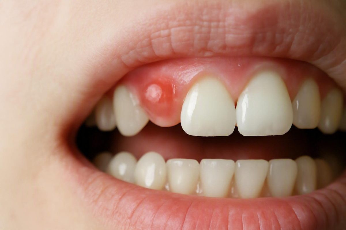
The short answer: a gum pimple may be an infection, but appearance cannot confirm it
A pimple-like bump on your gum is not ordinary acne. Gum tissue does not contain the oil glands involved in clogged-pore skin pimples. One important possibility is a parulis, also called a gum boil or dental sinus tract: a small opening through which an infection deeper around a tooth or its supporting tissues drains to the surface (Aspen Dental’s gum-pimple and abscess guide).
The visible bump may therefore be an exit point rather than the place where the problem began. It can appear near an infected tooth root, beside a periodontal pocket, or elsewhere along a drainage route.
However, not every gum bump is an abscess. A shallow mouth ulcer, dental cyst, irritation-related growth, eruption cyst over a developing tooth, trauma-related swelling, or benign bony lump can look similar.
If you have found an unexplained pimple-like bump on your gums:
- Do not squeeze, lance, puncture, or repeatedly press it.
- Arrange a dental evaluation, particularly if it is painful, draining, recurring, enlarging, changing, or persistent.
- Seek emergency medical care for difficulty breathing or swallowing, substantial or rapidly increasing facial or neck swelling, marked difficulty opening your mouth, or rapidly worsening illness. These can be warning signs that an infection or swelling needs urgent assessment (Cleveland Clinic’s medically reviewed periodontal-abscess guidance).
Editorial disclosure: Decay Guide is an educational publication written by a health writer, not a dentist. Its articles are not clinician-reviewed, cannot diagnose your bump, and do not replace a dental examination. Read more about how Decay Guide is written.
Possible causes of a pimple-like bump on the gums
The possibilities below are not ranked by likelihood. Their features overlap, and every visual clue is suggestive rather than diagnostic.
| Possible cause | Typical clues—suggestive, not diagnostic | What may be happening beneath the surface | Appropriate next step |
|---|---|---|---|
| Periapical abscess or draining sinus tract | A white, yellow, or red bump near a tooth; possible pus, unpleasant taste, toothache, chewing pain, or temperature sensitivity. It may also be painless. | Infection may have developed around the root tip after decay reached the pulp or following a crack, injury, or failed dental work. The bump may be a drainage route rather than the original infection site (Gentle Dental’s gum-boil overview). | Arrange prompt dental assessment. Seek emergency medical care for airway symptoms, substantial swelling, restricted mouth opening, or rapidly worsening illness. |
| Periodontal abscess | A localized swollen bump beside a tooth, gum tenderness, bad taste, chewing pain, drainage, or a loose tooth. Pain may be absent. | A pus-filled infection has begun in the gums and supporting tissues, often within a periodontal pocket (Cleveland Clinic’s periodontal-abscess guide). | Obtain prompt dental assessment, including evaluation of the gums and pocket around the tooth. |
| Gingival abscess | A localized, tender bump in the surface gum, sometimes following food trapping or irritation. | Food or another foreign object may be trapped in superficial gum tissue (Gentle Dental’s explanation of gingival abscesses). | Have a dentist assess the tissue and remove any trapped material when necessary. Do not dig into the gum yourself. |
| Canker sore | Usually a shallow, painful ulcer with a white or yellow center and red border rather than a raised, pus-filled bump (Duo-Lase’s comparison of oral bumps). | A surface ulcer has formed rather than a drainage tract from a tooth or gum infection. | Avoid further irritation and monitor it, but seek assessment if it is unexplained, worsening, recurrent, or not healing. |
| Dental cyst | A smooth or firm swelling that may be painless and slow to become noticeable. | A fluid-filled sac may be developing near a tooth root or another dental structure (For Beautiful Smiles’ gum-bump overview). | Arrange a dental examination; imaging may be needed to identify its location and relationship to nearby teeth. |
| Irritation fibroma | A smooth, firm, usually painless lump near an area exposed to repeated rubbing or trauma. | Tissue may have overgrown in response to chronic irritation from biting, an appliance, or a rough restoration (For Beautiful Smiles’ discussion of oral fibromas). | Seek dental review to identify the lesion and address a possible irritant. |
| Bony torus | A very hard, slow-growing lump, commonly without pain or inflammation. | A benign bony prominence may be present rather than a soft-tissue infection (The 247 Dentist’s painless-lump guide). | Arrange non-emergency assessment if it is unexplained, particularly if it grows, changes, ulcerates, or interferes with function. |
| Eruption cyst | In a child, a soft blue, purple, clear, or translucent bump directly over an emerging tooth; often painless. | Fluid has collected over a tooth that is coming through the gum. | Seek pediatric dental advice if the diagnosis is uncertain or the bump is large, painful, persistent, or accompanied by signs of infection. |
| Other persistent oral lesion | Firmness, enlargement, ulceration, unexplained bleeding, or a change in color, shape, surface, or texture. | Several benign and less common conditions can produce a growth that does not fit the patterns above. | Obtain dental or specialist assessment. An unusual lesion may occasionally require biopsy to identify it. |
Periapical abscess
A periapical abscess develops around the tip of a tooth root. One pathway begins when decay reaches the pulp—the inner tissue containing nerves and blood vessels—and bacteria travel through the root system. A crack, traumatic injury, deep or failing restoration, or previous dental treatment can also be relevant.
The bump may not sit directly over the responsible tooth. Infection can travel through surrounding tissue and drain at another point on the gum. That is one reason a surface photograph cannot identify the source reliably.
Periodontal abscess
A periodontal abscess begins in the gums and supporting tissues rather than inside the tooth. Pus may collect in a periodontal pocket beside a tooth, causing localized swelling, tenderness, drainage, chewing discomfort, or tooth mobility. It may be painful or painless.
Periodontal and periapical abscesses can produce overlapping symptoms. Determining which tissue is involved matters because the source and definitive treatment differ.
Gingival abscess
A gingival abscess is a localized infection of the surface gum. It may occur when food or another foreign object becomes lodged in the tissue.
Even if a fragment appears visible, do not probe below the gum with a pin, fingernail, toothpick, or other implement. Doing so can injure the tissue or push material farther beneath the surface.
Canker sore
A canker sore is usually an ulcer, meaning there is a shallow break in the surface rather than a raised pocket filled with pus. It commonly has a white or yellow center surrounded by a red border and may hurt sharply when touched by food or a toothbrush.
That distinction is helpful but not definitive. Do not attempt to diagnose a lesion solely by its center color or border.
Dental cyst
A dental cyst is a fluid-filled sac that may occur near a tooth root. It can remain painless, particularly while small, and may be discovered only after swelling develops or an X-ray is taken.
A dentist must distinguish a possible cyst from an abscess and other jaw or soft-tissue changes before discussing treatment. Appearance alone cannot show whether a lump contains fluid, pus, solid tissue, or bone.
Irritation fibroma and bony tori
An irritation fibroma is generally a smooth, firm overgrowth associated with repeated rubbing or trauma. A rough filling, denture edge, brace, biting habit, or another local irritant may be relevant. Correcting the irritant may form part of management, but a persistent lump still needs examination to establish what it is.
A torus is made of bone and typically feels much harder than an abscess, cyst, or fibroma. Tori are generally benign and slow-growing. Even so, a new or changing hard lump should not automatically be ignored; a dental professional can determine whether its location and characteristics are consistent with a torus.
Signs that make a dental infection more concerning
No single symptom confirms an abscess. Concern rises when a gum bump occurs with several local or whole-body signs.
Local signs that can accompany infection include:
- Pus or other drainage
- A foul, unpleasant, or salty taste
- Persistent bad breath
- Red or swollen gums
- Toothache or throbbing pain
- Pain when biting or chewing
- Sensitivity to hot or cold
- Tenderness around a tooth or the gum
- Gum bleeding
- A tooth that feels loose
Broader symptoms can include:
- Fever or chills
- Swollen lymph nodes
- Fatigue or increasing weakness
- Nausea or vomiting
- Swelling of the face, jaw, or neck
- Feeling increasingly unwell
These symptoms can occur with tooth or periodontal abscesses, although their presence and intensity vary. Alberta’s reviewed patient-care guidance lists red or swollen gums, throbbing or chewing pain, bad taste, fever, and jaw swelling among possible signs of an abscessed tooth (MyHealth Alberta’s abscessed-tooth guidance).
An abscess can be painful or painless. Lack of pain does not establish that a bump is harmless. If infection has created an opening through which fluid escapes, the reduction in pressure may make the tooth or gum feel better. That is symptom relief, not evidence that bacteria or damaged tissue at the source are gone.
Color is similarly unreliable. An infection-related bump may look white or yellow when fluid lies near the surface, or red when surrounding tissue is inflamed. Ulcers, cysts, irritated tissue, and eruption-related lesions can share some of those colors.
A bump that shrinks, ruptures, temporarily disappears, and then returns may be draining intermittently. The cycle can resemble healing because pressure and discomfort decline after drainage. Recurrence instead suggests that the source may still be present and needs investigation.
When to see a dentist—and when to seek emergency medical care
Judge urgency by symptoms and progression rather than trying to name the bump at home.
Emergency medical assessment
Seek emergency medical help rather than waiting for a routine dental appointment if you have:
- Difficulty breathing
- Difficulty swallowing
- Substantial or rapidly increasing swelling of the face or neck
- Marked difficulty opening your mouth
- Rapidly worsening illness
- Severe systemic symptoms, such as repeated vomiting, significant weakness, or feeling acutely unwell
These signs may indicate that swelling or infection is affecting spaces beyond the original tooth or gum. Serious spread is uncommon, but it is possible. Breathing difficulty requires emergency care, while worsening pain, swelling, redness, pus drainage, or fever warrants immediate clinical advice or assessment (MyHealth Alberta’s reviewed care instructions).
Prompt dental evaluation
Contact a dental professional promptly for a bump accompanied by:
- Pus or foul-tasting drainage
- Toothache or throbbing pain
- Pain when biting or chewing
- Lingering sensitivity to hot or cold
- Localized gum or jaw swelling
- Fever
- A bump that repeatedly returns
- Symptoms that are becoming more intense or spreading
Explain the full pattern when calling, including whether the bump has drained, disappeared, or returned. The dental office can determine whether immediate, same-day, or otherwise prompt assessment is appropriate based on your symptoms and health history.
Non-emergency but necessary evaluation
A painless, unexplained lump without fever or spreading swelling may not require an emergency department. It should still be examined, particularly if it:
- Persists rather than settling
- Enlarges
- Changes color, shape, surface, or texture
- Becomes firm
- Develops an ulcer
- Bleeds without an obvious injury
- Returns repeatedly
There is no rigid number of days that proves a lesion is a canker sore, cyst, abscess, or something else. Oral lesions have overlapping timelines, and the available evidence does not support one exact appointment deadline for every symptom combination. Contact a dental professional for case-specific triage rather than using a fixed waiting period to rule infection in or out.
How a dentist finds the source of the bump
The location of the visible bump may not show where the problem began. A drainage tract can connect the gum surface to infection around a root, while a periodontal abscess may begin inside a pocket beside a tooth. The nearest-looking tooth is not necessarily responsible.
A dental evaluation may include:
- Symptom history. The dentist may ask when the bump appeared, whether it drained or returned, what the fluid tasted like, and whether you have pain, sensitivity, fever, swelling, trauma, or previous treatment in that area.
- Inspection of the lesion. Its location, surface, consistency, drainage, bleeding, surrounding redness, and relationship to nearby teeth can provide clues.
- Examination of nearby teeth. The dentist may look for decay, cracks, tenderness, mobility, damage, large fillings, crowns, or other restorations.
- Assessment of the gums. Periodontal pocket measurements can help identify a possible gum-tissue source.
- Dental imaging. X-rays may be used to look for changes around a root, supporting bone, a possible cyst, or another hidden source.
- Further investigation when needed. A persistent or unusual growth may require specialist assessment and, in some cases, biopsy. A biopsy is used to identify a lesion; recommending one does not mean the lesion is probably cancerous.
Treatment depends on whether the source is the tooth, gums, or another lesion
There is no universal “gum pimple treatment.” Appropriate care depends on where the lesion started and what the examination and imaging show.
If the source is inside or around a tooth root
Treatment may involve draining accumulated fluid and addressing infection within or around the tooth. Depending on the diagnosis and whether the tooth can be retained, possible care includes:
- Root-canal treatment
- Treatment of a damaged or failing restoration
- Repair planning for a cracked or injured tooth
- Extraction when the tooth cannot reasonably be retained
Drainage may relieve pressure, while root-canal treatment or extraction addresses the underlying source. These are related but distinct goals.
If the source is the gums and supporting tissues
A periodontal abscess may require drainage and treatment of the infected periodontal pocket. Care can include scaling and root planing or, in some cases, gum surgery.
The choice depends on factors found during professional evaluation, including the abscess location, severity, supporting bone, tooth condition, and broader periodontal diagnosis.
If the source is superficial gum tissue
For a localized gingival abscess associated with trapped food or another object, a dentist may remove the material and clean or treat the affected tissue.
This differs from digging into the area at home. A clinician can inspect the tissue, use controlled instruments, and determine whether the apparent surface problem has a deeper source.
If the lesion is a cyst, fibroma, or another growth
Management may range from observation or correction of a chronic irritant to surgical removal or specialist care. A cyst associated with a tooth may require treatment of both the sac and the dental source. A suspected irritation fibroma is managed differently from a bony torus or unexplained ulcerated lesion.
Where antibiotics fit
Antibiotics are not automatically required for every localized dental abscess. When they are used, they do not replace treatment of the source. They cannot by themselves remove infected tissue enclosed within a tooth, clean a periodontal pocket, repair a crack, or remove trapped material. Professional treatment may involve drainage, periodontal therapy, root-canal care, restoration-related treatment, or extraction, depending on the diagnosis (Medical News Today’s dentist-reviewed gum-boil guide).
It helps to separate four concepts:
- Symptom relief: less pain or pressure
- Surface drainage: fluid escaping through the bump
- Temporary infection control: reducing bacterial activity or spread
- Definitive source control: treating the infected tooth, periodontal pocket, foreign object, cyst, or other cause
Only definitive source control addresses why the bump developed. No procedure or antibiotic can be recommended for an individual reader without an examination and, where indicated, imaging.
What to do—and what not to do—while waiting for care
Home care may make the area more comfortable, but it cannot diagnose the bump or cure an established abscess.
Safer temporary measures
- Rinse gently with warm salt water. Avoid forceful swishing that repeatedly disturbs the bump.
- Continue gentle brushing and flossing. Keeping the mouth clean is generally preferable to abandoning oral hygiene unless a clinician has given different instructions for the affected area.
- Use a wrapped cold pack outside the cheek for temporary discomfort or swelling. Do not place ice directly on the skin or gum.
- Choose foods that do not aggravate the area. Chewing on the other side and avoiding very hot, sharp, or irritating foods may be more comfortable.
- Use over-the-counter pain medicine only if it is suitable for you. Follow the product label rather than improvising a dose.
Medication suitability depends on the individual. Ask a pharmacist, dentist, or medical clinician if you are uncertain.
Do not manipulate the bump
Do not:
- Pop or squeeze it
- Lance it with a needle
- Puncture it
- Repeatedly press it to check for drainage
- Dig below the gum for suspected food or a foreign object
- Apply caustic or unproven substances in an attempt to draw out infection
Attempting to drain a gum bump at home does not treat an infected tooth or periodontal pocket and may injure the surrounding tissue. Commercial dental-provider guidance likewise advises against squeezing, popping, or draining a suspected abscess yourself (Aspen Dental’s gum-abscess guidance).
If the bump bursts on its own
If you notice fluid, blood, or a foul or salty taste:
- Spit out the drainage rather than pressing for more.
- Rinse gently with warm salt water.
- Do not insert anything into the opening.
- Note whether you have tooth pain, fever, swelling, unpleasant taste, or recurring drainage.
- Keep or arrange the dental appointment even if the bump flattens and the pain improves.
Rupture may reduce pressure, but it does not demonstrate that the underlying source has healed.
Painless, recurring, or post-treatment gum bumps still need attention
A drainage tract can open and close. While it is open, infection-related fluid may escape and pressure can fall, causing the bump and pain to diminish. If the opening closes while the source remains, the bump may fill or become visible again.
That is why a painless or recurring gum lesion is not automatically benign. Report the pattern—not just how the bump looks on the appointment day. Useful details include:
- How often it appears
- Whether it drains
- Whether it becomes smaller after drainage
- Any foul or salty taste
- Sensitivity or chewing pain
- Previous swelling or fever
- Whether it always appears in the same place
A recurring bump beside a root-canal-treated tooth needs reassessment. Bacteria may persist or re-enter, including through a crack, but the bump alone cannot determine which treatment is appropriate (Avalon Dental Group’s gum-boil overview).
Also tell the dentist about:
- A damaged or cracked tooth
- Previous trauma, even if it happened long ago
- A crown near the bump
- A large filling
- A restoration that feels loose, broken, or different
- Previous root-canal treatment or dental surgery
These details can help identify possible pathways but do not establish a diagnosis.
Recurrence is not always caused by infection. Persistent firmness, enlargement, ulceration, spontaneous bleeding, or changes in color, shape, or texture warrant investigation for cysts, irritation-related growths, and other oral lesions. A dentist may recommend specialist assessment or biopsy when examination and imaging cannot confidently identify an unusual growth.
A pimple on a child’s gums: infection versus an eruption cyst
Children can develop several of the same gum bumps as adults, including abscesses associated with cavities, gum infection, or trauma. They can also develop an eruption cyst over a tooth that is coming through.
An eruption cyst is generally a soft, fluid-filled bump directly over an emerging tooth. It may look bluish, purple, clear, or translucent and is often painless. Many uncomplicated eruption cysts resolve as the tooth breaks through the gum, while a large, painful, persistent, or uncertain lesion should be assessed by a pediatric dentist.
Color alone cannot distinguish an eruption cyst from trauma or infection. Seek prompt pediatric dental care if the child has:
- Pain or significant tenderness
- Pus or repeated drainage
- Unexplained bleeding
- Foul odor or taste
- Fever
- Facial or jaw swelling
- General illness
- A bump that persists or enlarges
- A large cavity or history of injury near the affected tooth
Do not pop or drain either an apparent eruption cyst or a suspected abscess. Manipulating the area can cause pain, bleeding, and tissue injury without identifying or treating the source.
For discomfort, use only medicine labeled for the child’s age and circumstances. Ask a pediatric dentist, pharmacist, or medical clinician if the child has allergies, health conditions, takes other medicines, or if you are uncertain which product is appropriate.
Fluoride toothpaste, age-appropriate brushing and flossing, regular dental care, and limiting frequent sugary snacks may reduce decay- and gum-related infections. These measures cannot prevent every bump because eruption cysts, injuries, tooth cracks, and some other causes are not preventable through oral hygiene alone.
Frequently asked questions
Can a pimple on the gums be painless and still be an abscess?
Yes. A periodontal abscess or drainage opening from a deeper infection can be painless. Drainage may lower pressure and reduce discomfort even though the tooth or gum source remains.
A painless bump may also be a cyst, fibroma, torus, or another lesion. Lack of pain therefore neither confirms nor excludes infection. Arrange dental evaluation for an unexplained bump, particularly if it drains, recurs, changes, enlarges, or appears beside a damaged or previously treated tooth.
What should I do if the gum bump bursts or drains on its own?
Spit out the drainage, rinse gently with warm salt water, and avoid squeezing or probing the opening. Note any foul or salty taste, tooth pain, fever, swelling, or return of the bump.
Keep or arrange a dental appointment because rupture and temporary pain relief do not prove that the source has resolved. Seek emergency medical help if you also develop breathing or swallowing difficulty, substantial facial or neck swelling, restricted mouth opening, or rapidly worsening illness.
Should I pop a white pimple-like bump on my gums?
No. White coloring does not confirm that the bump is a simple pocket that can be safely emptied. It could be a drainage tract, ulcer, cyst, eruption-related lesion, or another growth.
Popping or puncturing it may damage tissue and will not treat an infected tooth or periodontal pocket. Leave it alone, continue gentle oral hygiene, and arrange a dental assessment.
Will antibiotics make a gum abscess go away?
Not necessarily. Antibiotics may be used in selected circumstances, but they are not automatically required for every localized abscess and do not replace treatment of the source.
Even when antibiotics help control bacterial infection, they cannot remove infected tissue inside a tooth, clean a deep periodontal pocket, repair a crack, or remove a trapped foreign object. Depending on the diagnosis, definitive care may involve drainage, root-canal treatment, periodontal care, restoration-related treatment, or extraction.
Is a gum abscess contagious?
The abscess itself is not generally considered contagious. You cannot catch another person’s pocket of infection in the way you catch a respiratory virus.
Action plan: Do not pop or puncture a pimple-like bump on your gums. Seek emergency medical help for breathing or swallowing difficulty, substantial facial or neck swelling, restricted mouth opening, or rapidly worsening illness. Otherwise, arrange dental evaluation for an unexplained bump—especially one that is painful, draining, recurring, enlarging, changing, or persistent. Temporary drainage and lack of pain do not rule out infection; only an examination, often supported by dental X-rays, can identify the cause and appropriate treatment.