
Why Dental X-Rays Are Based on Risk, Not a Fixed Schedule
Dental X-ray timing depends on decay risk, exam findings and earlier images—not simply the calendar. Learn what can shorten or lengthen the interval.
How this site is written
Decay Guide is written by a health writer, not by a dentist, and no article here has been reviewed by a clinician. We work from public patient-education sources — the NHS, the CDC, the American Dental Association and hospital patient guides — and link to them so you can check what we say. Figures such as pocket depths are quoted as the educational benchmarks those sources use, not as thresholds you can apply to yourself. Nothing here replaces an examination.
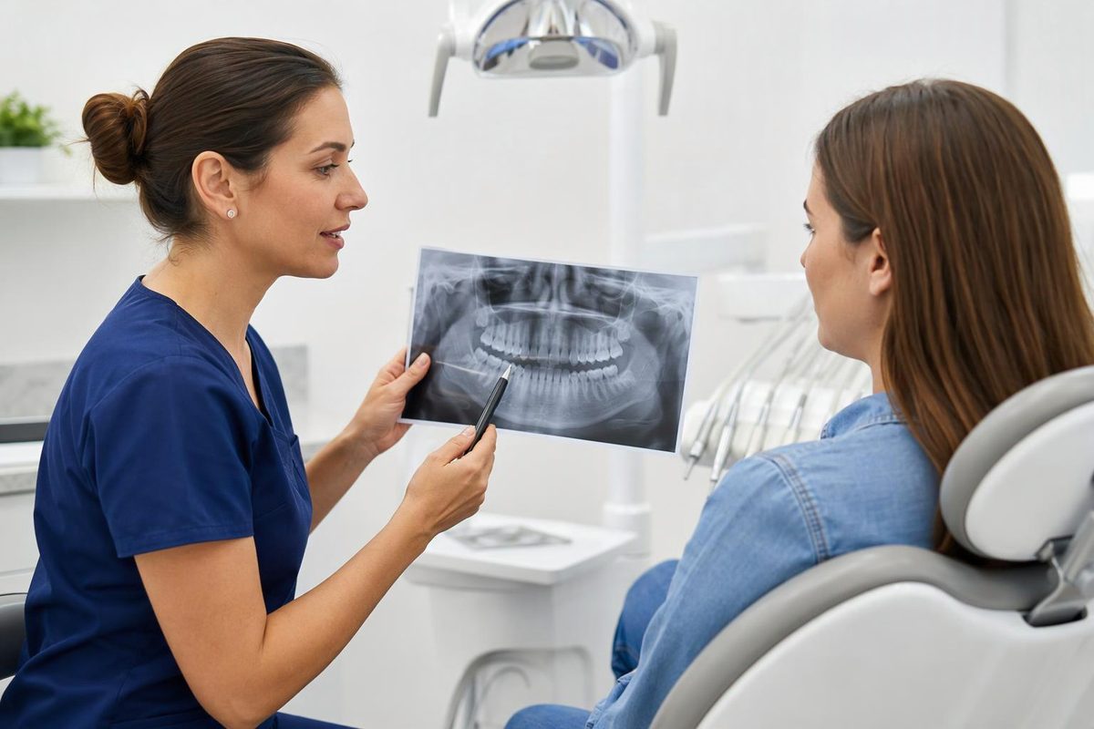
Dental X-rays should be timed according to what they are likely to reveal and whether that information will help guide care, not simply because another year has passed. Current oral health, disease risk, age, symptoms and examination findings all affect that decision. The American Dental Association’s updated 2026 patient-selection recommendations emphasize reviewing the history and examining the patient before ordering images. ADA recommendations summary
That means annual X-rays can be appropriate for some people without being necessary for everyone. It also means a new concern can justify imaging before a previously planned interval has elapsed.
Why the same interval does not fit everyone
X-rays can reveal disease that is not visible during an ordinary examination. Bitewings—a type of dental X-ray—can help detect decay on the touching side surfaces of neighboring teeth. Their accuracy varies with the stage of decay. ADA caries guidance Whether those surfaces can be examined directly also affects the decision to take images. ADA’s 2026 recommendations summary
For someone developing new decay, repeat images may provide useful information sooner. For someone with stable findings and low decay risk, taking the same images as often may add little information. The aim is to avoid both unnecessary exposure and an interval that leaves clinically important changes unchecked.
This is why a checkup schedule and an X-ray schedule are separate decisions. An examination can reassess whether imaging is needed without automatically including it. The ADA’s patient information explicitly notes that an X-ray may not be needed at every dental visit. MouthHealthy
What changes the risk assessment?
“Risk” means the likelihood of disease, not a judgment about how hard someone tries to care for their teeth. The ADA’s caries-risk assessment considers factors including:
- Recent decay: new lesions or recent restorations placed because of decay.
- Dry mouth or low saliva flow: severe dry mouth or visibly inadequate saliva flow can indicate higher decay risk.
- Frequent sugar exposure: repeated or prolonged sugary food and drink exposures between meals matter.
- Difficulty carrying out oral care in children: the assessment considers health conditions or disabilities that limit effective care by the child or a caregiver.
These factors are considered alongside the examination rather than used as an automatic X-ray checklist. Brushing regularly, by itself, does not establish low risk. ADA risk-assessment guidance
Age and dental development also matter. Children and teenagers may need images more often than adults because their teeth and jaws are developing, but age alone does not dictate a universal schedule. MouthHealthy
Not all imaging is about cavities. For periodontal disease, which affects the tissues supporting teeth, the 2026 recommendations say repeat-imaging frequency depends on clinical findings and treatment response. A cavity-monitoring interval cannot simply be applied to every other dental problem. ADA’s 2026 recommendations summary
Earlier images and the type of X-ray matter
Changing dentists does not automatically make existing images unusable. A new dentist may obtain previous X-rays rather than repeat them, depending on whether they supply the information needed now. Forwarding earlier images can also help establish what has changed. MouthHealthy
Likewise, “dental X-rays” are not one interchangeable test. The image should fit the question being investigated. A 3-D cone-beam CT scan is not a routine replacement for small 2-D images; ADA safety guidance recommends using it only when lower-exposure options cannot provide the necessary diagnostic information. ADA radiography guidance
Questions that make the recommendation clearer
A useful discussion is more specific than “Are X-rays due?” Consider asking:
- What are these images intended to detect or monitor?
- Which findings or risk factors make them useful now?
- Can my previous images answer that question?
- What would make the next imaging interval shorter or longer?
Dental imaging generally contributes a small amount of radiation exposure, but a small exposure still needs a clinical reason. Risk-based timing is not “never take X-rays unless something hurts.” It is take the appropriate images when their expected benefit justifies the exposure. ADA guidance
Decay Guide is an independent information publisher, not a dental practice. This article explains general principles, not an individual imaging schedule.