
What a Pale Layer in a Healing Tooth Socket May Mean
It may appear around days 2-3 and remain through the first week or longer. Improving pain and swelling are more informative than color alone.
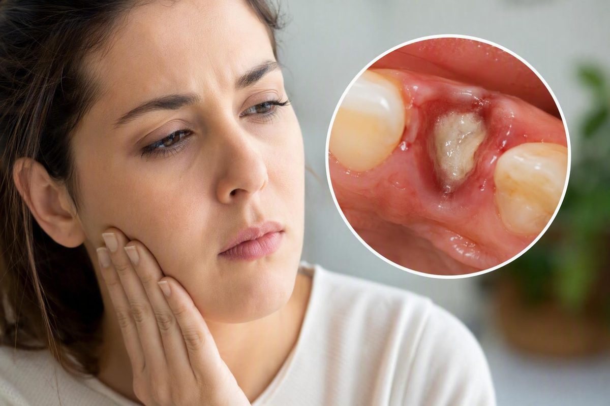
The short answer: a pale film can be normal, but color cannot diagnose it
A white, cream, or pale-yellow covering can appear during the early healing of a tooth-extraction socket. It may reflect changes in the blood clot, a fibrin-rich surface, developing granulation tissue, or another normal feature of repair. A pale layer is therefore not automatically pus, infection, exposed bone, or proof that the clot has been lost.
The appearance is more reassuring when:
- Soreness is becoming milder rather than stronger.
- Swelling is stable or decreasing.
- There is no persistent foul taste or odor.
- There is no thick discharge, fever, chills, or general feeling of illness.
- The surface appears stable rather than actively draining.
The most useful rule is to focus on symptom direction rather than color. A socket that looks unusual but feels progressively better is generally more compatible with routine healing. Increasing pain, expanding swelling, discharge, foul odor, fever, or other symptoms moving in the wrong direction deserve professional assessment. Patient guidance on extraction-site healing likewise emphasizes the recovery trend rather than what a photograph appears to show when evaluating pale repair tissue.
A mirror inspection or photograph cannot confirm whether the material is:
- Fibrin
- Granulation tissue
- Food or plaque
- Pus
- Exposed bone
- Collagen or graft material
- Packing, a membrane, or a surgical dressing
These possibilities can overlap in color and texture. Even visible bone does not, by itself, establish dry socket.
Do not test the material. Avoid touching, picking, scraping, pulling, wiping, directly brushing, or forcefully rinsing it. Those actions may disturb a protective clot, fragile repair tissue, stitches, a membrane, a dressing, or graft material deliberately placed during the procedure.
This article provides general reference information, not an assessment of an individual extraction site. Decay Guide is not a dental practice and does not diagnose or treat patients. The dentist or oral surgeon who performed the extraction is best placed to identify what was used during surgery and examine the socket directly.
Fibrin and granulation tissue: related healing terms, not interchangeable labels
Patient-facing explanations sometimes use fibrin and granulation tissue for a similar-looking pale surface. The terms are related to healing, but they describe different things.
Fibrin is a fibrous protein mesh produced during coagulation. It helps reinforce and stabilize the initial blood clot after tissue and blood vessels have been injured. One dental-clinic explanation describes fibrin as forming when fibrinogen is converted into insoluble fibrin during coagulation, creating a network that strengthens the clot during early wound healing.
Granulation tissue is temporary repair tissue that develops as healing progresses. It contains cells, collagen, connective tissue, and small developing blood vessels. It helps occupy and repair the wound bed while the socket moves toward gum coverage and longer-term healing. Dental patient education describes this tissue as forming after the clot and gradually giving way to healthier-looking gum tissue as surface repair continues.
A simplified healing continuum is:
- Bleeding occurs after the tooth is removed.
- A blood clot forms and begins protecting the socket.
- Fibrin helps stabilize that clot.
- Cells and small blood vessels enter the wound area.
- Granulation tissue develops and begins filling the socket.
- Gum tissue advances across the surface while deeper repair continues.
This is a biological model, not a visual diagnostic tool. During early healing, the visible surface may include clot material and fibrin while repair tissue is developing beneath or within it. Later, granulation tissue may become more prominent. A person looking in a mirror cannot identify that microscopic transition precisely.
Terminology in patient resources is inconsistent. Some sources call a pale coating fibrin; others call a similar white or pale-yellow appearance granulation tissue. Some use both labels without clearly separating them. That disagreement is one reason not to treat the appearance as proof of either material.
The practical distinction is:
- Fibrin participates in coagulation and clot stabilization.
- Granulation tissue is developing repair tissue.
- Both can be associated with normal healing.
- A pale surface could also be something unrelated to either.
- Color and texture cannot establish which material is present.
No particular shade, texture, or postoperative day proves that a layer is fibrin or granulation tissue. “White fibrin film” may be a convenient description of what a patient sees, but it is not a diagnosis based on appearance alone.
An approximate healing timeline from blood clot to pinker gum tissue
Socket healing follows a general sequence, but it does not run on a universal calendar. The extraction type, socket depth, surgical difficulty, suturing, grafting, health conditions, medications, smoking, and postoperative care can all affect what is visible and how the recovery progresses.
The stages below are approximate observations rather than deadlines.
First 24 hours: the clot forms
A dark red or purple blood clot generally forms during the first day. It helps control bleeding, covers the underlying bone and nerves, and provides a foundation for early repair. Protecting the clot is one reason postoperative instructions warn against disturbing the socket during this period after tooth extraction.
The clot does not necessarily remain dark red throughout healing. Its visible surface may change as coagulation and repair proceed. A lighter appearance does not automatically mean the clot has disappeared.
Around days 2–3: the surface may become pale
A whitish, cream, or pale-yellow area may become noticeable as the clot changes and early repair develops. Some patient-education sources describe the visible layer as fibrin, while others call it early granulation tissue. Reports commonly place this transition at approximately the second or third day, but the timing cannot identify the material or determine whether an individual socket is healthy.
A pale appearance during this period is more reassuring when discomfort and swelling are improving. It is less reassuring when accompanied by increasing pain, thick drainage, spreading redness, fever, or a persistent foul taste or odor.
Days 3–7: pale repair material may remain visible
During the remainder of the first week, the socket may still contain pale fibrin-rich material, developing granulation tissue, or both. The site may look moist, uneven, cream-colored, yellowish, pinkish, partly red, or visibly open.
In an uncomplicated recovery, pain and swelling should generally trend downward rather than intensify. The important distinction is not whether every day feels identical, but whether the overall recovery is improving or worsening.
Following weeks: the surface generally becomes pinker
As gum tissue advances over the site, the visible surface usually becomes more like the surrounding pink tissue. The opening may narrow gradually rather than closing all at once.
A socket can still look like a hole while healthy repair is occurring underneath. Conversely, a surface that appears almost closed does not mean deeper bone repair has finished. The visible surface cannot show the condition of the bottom or sides of the socket.
There is no universal day by which every white or yellowish film must disappear. Patient guidance describes pale repair tissue as changing during the first week and sometimes remaining visible into the following weeks, with timing varying by the procedure and the individual healing course. Persistence without concerning symptoms does not by itself establish a complication. If the layer remains alongside worsening pain, swelling, discharge, odor, bleeding, numbness, or uncertainty about surgical material, contact the treating office.
What else can look white or yellow inside an extraction socket?
A pale extraction site has several possible explanations. Considering them is not a way to diagnose the material at home; it explains why color alone is insufficient.
| Possible material | Typical context | Associated symptoms or clues | Safest next step |
|---|---|---|---|
| Fibrin-rich healing surface | Early clot stabilization and repair | Stable pale coating with improving pain and swelling | Leave it undisturbed and follow postoperative instructions |
| Granulation tissue | Temporary repair tissue developing after the clot | Pale, cream, pink-white, moist, or uneven surface with an improving recovery | Do not touch or directly brush it |
| Food or plaque | Debris collects near the socket while normal cleaning is limited | May be pale or yellowish; appearance is inconclusive | Do not probe; use only the cleaning method authorized by the dentist |
| Collagen, membrane, dressing, or packing | Material deliberately placed during surgery | May look soft, stringy, folded, sponge-like, or gauze-like | Contact the treating office before cleaning or removing it |
| Bone-graft particles | Socket preservation or grafting was performed | May appear chalky, sandy, granular, or off-white | Do not pick out particles; ask the treating office what was placed |
| Pus-like drainage | Possible infection | Thick or opaque drainage with increasing pain, swelling, warmth, redness, foul taste or odor, fever, chills, or illness | Arrange prompt dental assessment |
| Exposed bone | Covering tissue or clot may be absent or disrupted | Hard-looking white or off-white surface, especially with severe or escalating pain | Seek prompt dental assessment rather than testing it |
Normal repair material
A fibrin-rich surface or developing granulation tissue can appear white, off-white, cream, or pale yellow. Temporary repair material does not have to match the surrounding pink gum.
These colors are not exclusive to healing tissue. Food, plaque, surgical products, discharge, and bone can produce an overlapping appearance.
Food or plaque
Food may collect near an open socket after eating resumes. Plaque can also accumulate around the area while brushing is restricted or uncomfortable. Either may resemble a pale patch.
Do not insert a fingernail, cotton swab, toothpick, toothbrush, or irrigation device to find out whether the material moves. Even if it is food, physical testing may disturb the healing surface. If your clinician did not provide a socket-cleaning plan, ask before attempting irrigation.
Surgical material
Following a surgical extraction, the clinician may place a collagen membrane, dressing, packing, gauze-like product, or bone graft. Depending on the material, it may appear:
- Chalky or sandy
- Soft or sponge-like
- Stringy or folded
- White, cream, or off-white
- Granular at the surface
Bone-graft particles and collagen products may therefore be mistaken for food or abnormal tissue. Dental aftercare material specifically notes that intentionally placed graft particles or collagen membranes can have pale, chalky, sandy, soft, or stringy appearances at the extraction site.
If grafting, packing, or a membrane was used—or you are unsure whether it was—contact the treating office before trying to clean or remove anything.
Pus-like drainage
Pus-like material is more concerning when it is thick, opaque, and actively draining rather than forming a stable surface. It may appear yellow, yellow-green, gray, or whitish, but color remains insufficient by itself.
Increasing pain or swelling, spreading warmth or redness, persistent foul taste or odor, fever, chills, swollen lymph nodes, or feeling generally unwell can support concern about infection and warrant prompt dental assessment.
Exposed bone
Bone may look white or off-white, but it should not be touched to test whether it feels hard. Exposed bone can occur with dry socket, although neither visible bone nor a white-looking socket establishes that diagnosis. Severe, escalating, throbbing, or radiating pain is a more important clue.
Normal healing, possible infection, and dry socket compared
Routine healing, possible infection, and dry socket are better compared through the pattern and direction of symptoms than through socket color.
| Pattern | Typical symptom course | Appropriate response |
|---|---|---|
| Routine healing | Mild or manageable soreness and swelling that improve; a stable pale surface; no thick discharge, persistent foul odor, unusual swelling, fever, or systemic illness | Continue the clinician’s aftercare plan and leave visible material alone |
| Possible infection | Increasing pain or swelling; spreading redness or warmth; thick pus-like drainage; persistent foul taste or odor; fever, chills, swollen lymph nodes, or feeling unwell | Contact the treating dentist promptly |
| Possible dry socket | Severe throbbing pain that begins or intensifies several days after extraction; pain may radiate toward the ear or temple; the socket may look empty or show bone; foul taste or odor may occur | Arrange urgent dental review rather than trying to replace the clot |
Routine healing is an improving pattern
A stable pale surface can fit routine healing when soreness and swelling are becoming less noticeable and no new warning signs are appearing. The socket does not have to look neat, pink, or closed during early recovery.
Mild symptoms can remain while the surface looks white or yellowish. What is reassuring is the combination of a stable appearance and a recovery that continues to move in the right direction.
Infection is often inflammation- or discharge-led
Possible infection is more likely to involve a cluster of inflammatory or systemic signs: increasing swelling, warmth, redness, pus-like drainage, fever, chills, swollen lymph nodes, foul taste or odor, or feeling unwell. Increasing pain may occur as part of that pattern.
No single sign is conclusive:
- Yellow does not automatically mean pus.
- White does not automatically mean healthy tissue.
- Absence of fever does not rule out infection.
- A photograph cannot assess warmth, tenderness, drainage depth, or systemic symptoms.
Dry socket is primarily pain-led
Dry socket is a different pattern from infection. It is associated with severe throbbing pain that begins or becomes significantly worse several days after extraction. Pain may travel toward the ear or temple. The socket may look empty, the clot may appear absent, or bone may be visible; an unpleasant taste or odor may also occur. These pain and appearance patterns are described in patient guidance on post-extraction dry socket.
The visual findings are secondary to the pain pattern. A white area without severe or escalating pain does not establish dry socket. Conversely, a photograph that appears normal cannot rule it out when the pain pattern is strongly concerning.
Infection and dry socket are not the same condition and may require different professional management. Neither should be self-treated by scraping the socket, inserting substances, packing the opening, or trying to replace the clot. Prompt dental assessment is the appropriate next step for either concerning pattern.
Leave the film alone: safe care around a healing socket
The preservation rule is straightforward:
Do not pick, scrape, pull, wipe, or directly brush visible material inside the socket.
Attempted removal could disturb:
- The blood clot
- A fibrin-rich surface
- Fragile granulation tissue
- Sutures
- A collagen membrane
- A surgical dressing or packing
- Bone-graft particles
You do not need to identify the material before deciding to preserve it. Leaving it undisturbed is the safer response across several plausible explanations.
Rinsing should be gentle and procedure-specific
General extraction aftercare commonly advises avoiding rinsing during the first 24 hours, followed by gentle rinsing after that period when consistent with the treating clinician’s instructions. Vigorous swishing and forceful spitting may disturb the clot or delicate tissue. This approach is described in extraction aftercare guidance on protecting the healing socket.
“Gentle” does not mean repeatedly rinsing to see whether the pale material comes off.
Brush the rest of the mouth without testing the socket
Continue oral hygiene according to the instructions you received, but do not use a toothbrush to investigate visible socket material. Cleaning nearby teeth and brushing inside an extraction wound are different actions.
If you were told to avoid the area temporarily, follow that direction. If you received a specific brushing or irrigation schedule, that procedure-specific plan takes priority over general online information.
If you think food is trapped
Do not probe the socket to confirm the suspicion. Food, healing tissue, collagen, and graft particles may look similar, particularly in a deep or difficult-to-see site.
If your clinician supplied an irrigation syringe or another cleaning device, use it only at the time and in the manner prescribed. If no method was provided, contact the office rather than inventing one. The treating team can account for the extraction type, socket depth, stitches, grafting, and other procedural details.
After grafting, packing, or membrane placement
Be especially cautious if the procedure included socket preservation, a bone graft, a collagen membrane, medicated packing, or a dressing. Material that looks loose or foreign may have been intentionally placed. Contact the treating office before attempting to move, rinse away, or remove it.
When to monitor, call a dentist, or seek emergency care
Use a tiered response based on the complete recovery pattern rather than one color or an exact postoperative day.
Reasonable to monitor
Continued monitoring while following your postoperative instructions may be reasonable when:
- The pale layer is stable.
- Soreness is becoming milder.
- Swelling is stable or decreasing.
- There is no thick or active drainage.
- There is no persistent foul odor or bad taste.
- There is no fever, chills, or feeling of illness.
- No other part of the recovery is moving in the wrong direction.
You may document the appearance if useful, but do not manipulate the socket to obtain a better view. How symptoms change over time is more informative than whether the site immediately looks like normal gum.
Contact the treating dentist promptly
Call the dentist or oral surgeon for:
- Severe or escalating pain
- Pain that begins improving and then becomes substantially worse
- Severe throbbing or pain radiating toward the ear or temple
- Worsening or expanding swelling
- Spreading redness or warmth
- Thick or pus-like discharge
- Persistent foul taste or odor
- Fever, chills, swollen lymph nodes, or feeling unwell
- Persistent or recurrent bleeding
- Persistent numbness or a concerning change in sensation
- A suspected missing clot
- An empty-looking socket associated with significant pain
- Unidentified material after grafting, packing, or membrane placement
- Any overall recovery trend moving in the wrong direction
Severe throbbing or radiating pain that develops several days after extraction is compatible with dry socket and warrants urgent dental review. Increasing swelling, discharge, fever, or systemic symptoms may instead suggest infection. These patterns cannot be reliably separated through color or a photograph alone.
Do not wait for every listed symptom to appear. Absence of fever does not make severe pain, discharge, or worsening swelling safe to ignore.
Seek emergency care
Difficulty breathing or swallowing requires emergency evaluation. It should not be managed by monitoring the socket or waiting for a routine appointment. Post-extraction patient guidance identifies trouble breathing or swallowing as an emergency warning sign requiring immediate care.
This article does not use rigid at-home thresholds for fever, bleeding, numbness, or pain because the appropriate response depends on the procedure and the complete clinical picture. The clinician who performed the extraction knows which tooth was removed, how the procedure went, whether stitches or grafts were used, and what materials were placed. That context—and a direct examination—is more useful than the color of the socket.
Frequently asked questions
Can a yellowish layer be normal if pain and swelling are improving?
Yes. A cream or pale-yellow layer can be compatible with routine healing when pain and swelling are steadily improving and there is no thick discharge, persistent foul odor or taste, fever, chills, or other concerning change.
Yellow is not synonymous with pus. Healing material, food, collagen, and graft particles can all appear yellowish or off-white. Leave the layer undisturbed and judge the recovery by its overall direction.
If the material is thick or actively draining—or it accompanies increasing pain, swelling, warmth, redness, odor, fever, or illness—contact the treating dentist promptly.
How long can a white or yellow film remain visible after tooth extraction?
A pale layer may become noticeable around the second or third day and remain visible through the first week or longer. Some sockets retain pale repair tissue during part of the following weeks as the surface gradually becomes pinker.
These are approximate observations, not a deadline. Timing varies with the extraction, socket depth, health conditions, medications, smoking, grafting, and aftercare.
Persistence alone does not prove a complication. Contact the treating office if the layer remains alongside worsening pain, swelling, discharge, odor, fever, bleeding, numbness, or uncertainty about surgical material.
Does a white socket mean the blood clot has fallen out or I have dry socket?
No. A socket can become pale as the clot changes and early repair develops. White or off-white material may also be fibrin, granulation tissue, food, plaque, surgical material, graft particles, or bone.
Dry socket cannot be diagnosed from whiteness, visible bone, or an empty-looking area alone. A more characteristic clue is severe throbbing pain that begins or intensifies several days after extraction and may radiate toward the ear or temple. A foul taste or odor and an apparently absent clot may also occur.
If that pain pattern develops, arrange urgent dental review. Do not try to replace the clot or pack the socket yourself.
What should I do if I think the white material is trapped food or bone-graft material?
Do not probe, scrape, brush, pull, or forcefully irrigate it. Food and graft material can be difficult to distinguish visually, and attempted removal may disturb healing tissue or deliberately placed material.
Follow the cleaning instructions supplied by the dentist or oral surgeon. If you received an irrigation device, use it only when and how the clinician authorized. If grafting, packing, a membrane, or a dressing may have been used, contact the treating office before attempting removal.
Can a photograph tell whether the layer is fibrin, granulation tissue, or pus?
No. A photograph may document a change, but it cannot reliably distinguish fibrin, granulation tissue, food, plaque, pus, exposed bone, or surgical material.
A photograph also cannot assess warmth, tenderness, tissue depth, odor, drainage consistency, or the full pain pattern. Those details—and whether symptoms are improving or worsening—are essential to clinical assessment.
The bottom line
A stable white, cream, or pale-yellow film accompanied by steadily improving symptoms is often compatible with healing, but its identity cannot be confirmed from appearance alone. Leave it undisturbed and follow the treating dentist’s instructions.
Request prompt dental assessment if pain, swelling, discharge, odor, fever, bleeding, or numbness worsens or persists. Seek emergency care for difficulty breathing or swallowing.