
A Dead Tooth May Still Be Saved—But It Needs a Dental Diagnosis
A dentist assesses remaining structure, bone support, fractures and canal access to decide whether the tooth can be saved and restored.
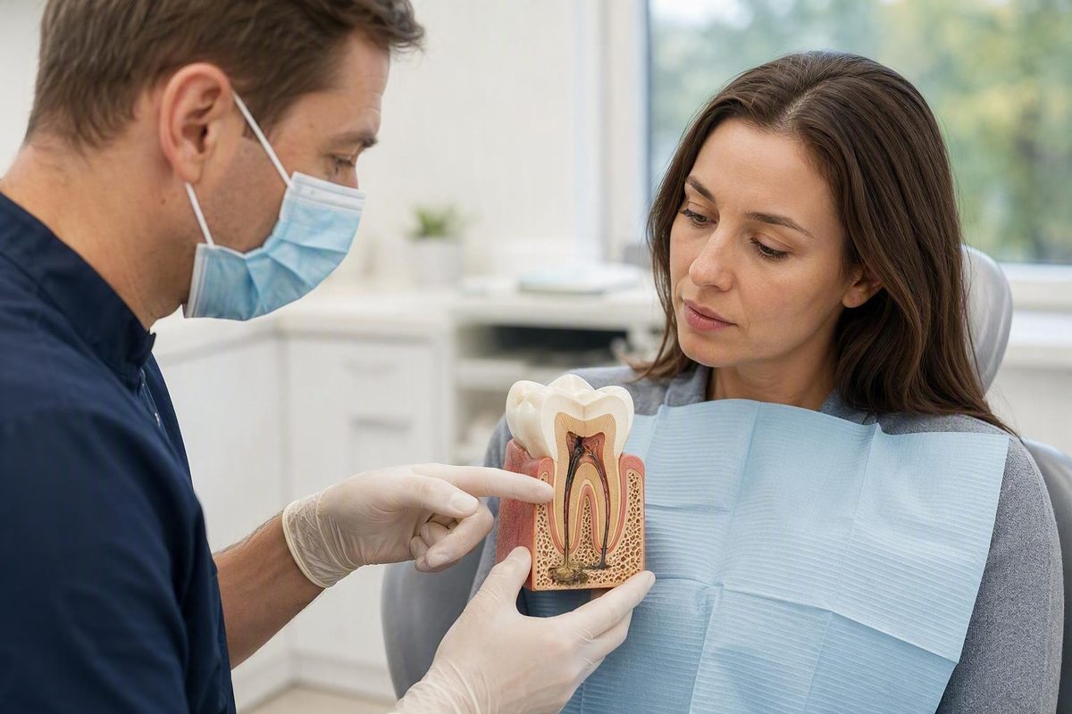
A dead tooth cannot be revived at home. If a dentist confirms that the pulp has died, the usual tooth-preserving treatment is root canal therapy followed by a permanent restoration. Extraction may be necessary when the tooth is too damaged, poorly supported or otherwise unsuitable for predictable restoration. Pain stopping does not mean the problem has resolved: symptoms may disappear as the pulp dies even though infection continues (NHS 111 Wales).
The short answer: arrange a dental assessment
Dead dental pulp does not naturally regenerate. Brushing, rinses and other home measures cannot restore its blood supply or clean and seal the root-canal system. A dentist must confirm whether the pulp is dead, look for infection and determine whether the remaining tooth can be saved. Root canal treatment may preserve a structurally sound tooth; extraction is the alternative when preservation is not practical (Leeds Teaching Hospitals).
A painless tooth still deserves prompt assessment. Pulp damage can develop quietly, and severe pain may disappear as the pulp dies. That change does not establish that infection has gone away. There is no dependable timetable for when a painless dead tooth will cause complications.
Use this urgency ladder:
- Arrange a prompt dental appointment if a tooth has darkened, stopped responding normally or changed after trauma—even if it does not hurt.
- Seek urgent dental care for worsening pain, pus, persistent or increasing swelling, fever, or swelling of the face or jaw.
- Seek emergency medical care for spreading infection, facial swelling accompanied by fever, or difficulty breathing. These escalation signs are identified in Leeds Teaching Hospitals’ pediatric root-canal guidance; adults with these signs should also seek immediate professional assessment rather than relying on this article (Leeds Teaching Hospitals).
Important: Decay Guide provides general information. It is not a dental practice and cannot determine whether your tooth is dead, infected or savable. That requires an examination by a qualified dental professional, as explained in Decay Guide’s editorial and clinical limitations.
Why symptoms and tooth color cannot confirm pulp death
A “dead” or non-vital tooth is one whose pulp—the soft tissue containing nerves, connective tissue and blood vessels—has died after losing its viable blood supply. Possible causes include deep decay, trauma, cracks and damage associated with previous or extensive dental work. These are risk factors, not a diagnosis.
Possible signs include:
- Gray, brown or otherwise dark discoloration
- Tenderness when biting
- Gum swelling near the tooth
- A pimple-like bump that drains on the gum
- Pus or an unpleasant taste
- Persistent bad breath
- Pressure or throbbing
- No noticeable symptoms at all
These signs are associated with pulp damage and root-canal infection, but none confirms pulp death by itself. A darkened tooth is not automatically necrotic, while a tooth that looks normal may have internal damage (Delta Dental).
One nonresponsive pulp test is also inconclusive. Recent trauma, calcification and testing limitations can produce a negative response without establishing necrosis. Diagnosis therefore combines medical and dental history, previous symptoms or trauma, visual and periodontal examination, X-rays, and comparison with nearby teeth using pulp and periapical tests. The American Association of Endodontists emphasizes that treatment should follow a complete pulpal and periapical diagnosis rather than a single symptom or test result (AAE guidance on endodontic diagnosis).
It also helps to distinguish three related problems:
- Pulp necrosis: the tissue inside the tooth has died.
- Root-canal infection: bacteria have entered or persisted within the canal system.
- Inflammation or infection around the root: living tissues outside the tooth react, sometimes producing biting pain, swelling, drainage or an abscess.
Pulp necrosis alone does not cause inflammation around the root unless the root canal is infected. This also explains how a tooth with dead internal nerves can still hurt: the ligament, gum and bone surrounding the root remain alive and can become painful or swollen.
Root canal or extraction? How the decision is made
The practical question is not simply whether the pulp is dead. It is whether the tooth can be cleaned, sealed and restored well enough to remain useful.
| Clinical situation | Likely treatment path | What happens next |
|---|---|---|
| Enough usable tooth structure, adequate periodontal and bone support, no unmanageable fracture, and canals that can be treated | Root canal treatment | The canals are cleaned, disinfected, filled and sealed; the tooth then receives an appropriate permanent restoration |
| Severe decay, fracture below the gumline, too little remaining structure, inadequate bone support, or canals that cannot be treated adequately | Extraction may be necessary | The tooth is removed; healing and possible replacement options are discussed |
| Infection or symptoms after a previous root canal | Reassessment for retreatment or root-end surgery | The existing treatment, canal anatomy, restoration and fracture status are evaluated before extraction is assumed necessary |
These pathways reflect the structural and technical factors described by the American Association of Endodontists and Leeds Teaching Hospitals. A complete diagnosis or confirmation that treatment is feasible may sometimes be possible only after the tooth has been opened.
The final recommendation depends on restorability, gum and bone support, fracture location and depth, canal accessibility, expected prognosis, biting forces, and the patient’s circumstances and goals. Saving a natural tooth is not always possible. Cost, appointment time, possible grafting, replacement needs and long-term maintenance are case-specific and should be compared as complete treatment pathways with the treating dentist.
When earlier root canal treatment has not healed, extraction is not necessarily the immediate next step. Depending on the cause, an endodontist may consider nonsurgical retreatment or surgery at the root end. Surgical treatment is generally assessed after conventional treatment or retreatment, and it cannot guarantee that the tooth will be retained.
What happens during root canal treatment
A normal filling repairs a defect in the visible part of a tooth. It cannot remove dead or infected material located inside the root canals. Root canal treatment reaches that internal space.
The procedure generally follows this sequence:
- Local anaesthetic is given to numb the tooth and surrounding area.
- The tooth is isolated, commonly with a rubber dam to keep it dry and reduce contamination from saliva.
- An access opening is made through the tooth into the pulp chamber.
- Dead or infected tissue is removed.
- The canals are cleaned, shaped and disinfected with fine instruments and irrigating solutions.
- The prepared canals are filled with root-filling material to reduce the space in which bacteria could persist or return.
- The access opening is sealed, either temporarily when another visit is needed or with the appropriate permanent restoration.
If an abscess is present, it may also be drained during treatment.
Root canal treatment usually takes 2 or more appointments Root canal treatment - NHS. Medication and a temporary filling may be placed inside the tooth between visits. X-rays may be used to assess canal length and the completed root filling. Leeds Teaching Hospitals describes isolation, canal preparation, disinfection, filling and final restoration as the core sequence.
Local anaesthetic is normally used to control procedural pain, but a painless experience cannot be guaranteed. Pressure or discomfort may occur, and the tissues around the tooth can be sore afterward.
Reported success figures vary because studies examine different teeth, patient groups, follow-up periods and definitions of success. X-ray healing, symptom relief and retention of a functional tooth are related but different outcomes, so no single percentage predicts what will happen to an individual tooth.
The final restoration is part of the treatment
Cleaning and filling the canals addresses the internal problem, but the tooth still needs a durable external seal and enough strength to function. The permanent restoration may be a filling, onlay or crown. The quality and timing of that final seal affect both reinfection and fracture risk.
A crown is common for a weakened back tooth because molars and premolars carry substantial chewing forces and may already have lost considerable structure to decay, cracks, large fillings or the treatment access opening. However, not every root-canal-treated tooth automatically needs a crown. A front tooth with substantial sound structure may have different restorative needs.
The choice depends mainly on:
- Whether the tooth is at the front or back of the mouth
- How much healthy tooth structure remains
- The size and condition of existing restorations
- Whether cracks are present
- The tooth’s role in the bite
- Grinding, clenching and other fracture risks
Follow the treating dentist’s restoration schedule rather than relying on a universal deadline. Until the permanent restoration is complete, avoid heavy chewing or hard foods on that tooth. A temporary filling does not provide the finished restoration’s long-term seal or fracture protection.
What to do while waiting—and why antibiotics are not a cure
There is no home treatment that can revive dead pulp or properly clean, fill and seal root canals. Brushing, salt-water rinses, mouthwash, clove oil and other home remedies cannot replace definitive treatment.
While waiting for an appointment:
- Avoid chewing hard or sticky foods on the affected tooth.
- Keep the area gently clean without pressing or picking at a swelling.
- Monitor for increasing pain, drainage, fever or facial swelling.
- Seek faster care if symptoms worsen.
Nonprescription pain relief may be suitable for some people, but the choice depends on age, pregnancy, allergies, medical conditions, other medicines and the product instructions. Follow the label or advice from a pharmacist, dentist or doctor rather than using an individualized dose from a general article.
Antibiotics are not a substitute for root canal treatment or extraction. They do not remove necrotic tissue, clean canal walls or create a lasting seal inside the tooth.
Recovery, follow-up, and treatment that does not heal as expected
Mild soreness or biting tenderness can occur for several days after treatment and should generally improve. Contact the treating provider if pain is severe, swelling persists or increases, the temporary filling comes out, or symptoms worsen rather than settle.
Follow-up examinations and X-rays may be used to monitor the tissues around the root. Healing is not judged by symptoms alone: a tooth can feel comfortable before radiographic changes have fully resolved.
Reinfection or incomplete healing can occur because of:
- New decay
- Leakage around a filling, onlay or crown
- A loose or failed restoration
- Trauma or a new crack
- Complex, curved, blocked or previously untreated canals
- Persistent bacteria in anatomy that was difficult to clean
The usual escalation path is reassessment, correction of any restoration problem, nonsurgical retreatment where appropriate, and possible root-end surgery. Extraction remains an option if the tooth cannot be restored, has a poor structural prognosis or does not respond to preservation attempts.
Long-term outcome depends on the tooth’s initial condition, remaining structure, periodontal support, canal anatomy, final seal, oral hygiene and biting forces. Root canal therapy treats disease inside the tooth; it cannot make a severely cracked or poorly supported tooth structurally normal.
If extraction is necessary: replacement, appearance, and special cases
After extraction, possible replacement approaches include:
- A bridge: a fixed replacement supported by surrounding teeth.
- A dental implant: a fixture placed in the jaw that supports a crown or another restoration.
- A removable partial denture: an appliance that replaces one or more teeth and can be removed for cleaning.
No option is universally best. The choice depends on the gap’s location, nearby teeth, available bone, gum health, medical circumstances, treatment time, maintenance preferences and cost. The NHS dental treatment guide explains the basic differences among bridges, implants and removable dentures.
If a saved tooth remains dark, appearance is a secondary issue. Internal bleaching, a veneer or a crown may sometimes be considered, but only after infection has been controlled and the tooth’s structural treatment has been planned.
Children and adolescents with immature permanent teeth may require specialist management because their roots may still be developing. Depending on the tooth, treatment may involve an open-root barrier or another pediatric endodontic approach rather than the standard adult sequence. NHS pediatric guidance describes specialist assessment and different management for teeth with open roots (St George’s University Hospitals).
A tooth affected by a fall, sports injury or blow to the face should be assessed promptly, even if it initially causes little or no pain. Trauma can disrupt the pulp’s blood supply, and the consequences may only become apparent later.
The practical sequence is straightforward: do not try to revive or sterilize the tooth at home. Arrange a dental examination, have the pulpal and root diagnosis confirmed, and determine whether the tooth is restorable. If it can be saved, complete both the root canal and final restoration. If it cannot, discuss extraction and replacement choices. Lack of pain is not reassurance; fever, facial or spreading swelling, or breathing difficulty requires urgent escalation.